<?xml version="1.0" encoding="ISO-8859-1"?><article xmlns:mml="http://www.w3.org/1998/Math/MathML" xmlns:xlink="http://www.w3.org/1999/xlink" xmlns:xsi="http://www.w3.org/2001/XMLSchema-instance">
<front>
<journal-meta>
<journal-id>0036-3634</journal-id>
<journal-title><![CDATA[Salud Pública de México]]></journal-title>
<abbrev-journal-title><![CDATA[Salud pública Méx]]></abbrev-journal-title>
<issn>0036-3634</issn>
<publisher>
<publisher-name><![CDATA[Instituto Nacional de Salud Pública]]></publisher-name>
</publisher>
</journal-meta>
<article-meta>
<article-id>S0036-36342003000900020</article-id>
<title-group>
<article-title xml:lang="en"><![CDATA[Recommendations for cervical cancer screening programs in developing countries: the need for equity and technological development]]></article-title>
<article-title xml:lang="es"><![CDATA[Recomendaciones a programas de detección oportuna de cáncer cervical en países en desarrollo: necesidad de equidad y desarrollo tecnológico]]></article-title>
</title-group>
<contrib-group>
<contrib contrib-type="author">
<name>
<surname><![CDATA[Lazcano-Ponce]]></surname>
<given-names><![CDATA[Eduardo]]></given-names>
</name>
<xref ref-type="aff" rid="A01"/>
</contrib>
<contrib contrib-type="author">
<name>
<surname><![CDATA[Alonso]]></surname>
<given-names><![CDATA[Patricia]]></given-names>
</name>
<xref ref-type="aff" rid="A02"/>
</contrib>
<contrib contrib-type="author">
<name>
<surname><![CDATA[Ruiz-Moreno]]></surname>
<given-names><![CDATA[José Antonio]]></given-names>
</name>
<xref ref-type="aff" rid="A03"/>
</contrib>
<contrib contrib-type="author">
<name>
<surname><![CDATA[Hernández-Avila]]></surname>
<given-names><![CDATA[Mauricio]]></given-names>
</name>
<xref ref-type="aff" rid="A01"/>
</contrib>
</contrib-group>
<aff id="A01">
<institution><![CDATA[,Instituto Nacional de Salud Pública Centro de Investigación en Salud Poblacional ]]></institution>
<addr-line><![CDATA[ ]]></addr-line>
</aff>
<aff id="A02">
<institution><![CDATA[,Universidad Nacional Autónoma de México Facultad de Medicina Hospital General de México]]></institution>
<addr-line><![CDATA[México DF]]></addr-line>
<country>México</country>
</aff>
<aff id="A03">
<institution><![CDATA[,Secretaria de la Defensa Nacional Escuela Militar de Graduados de Sanidad ]]></institution>
<addr-line><![CDATA[México DF]]></addr-line>
<country>México</country>
</aff>
<pub-date pub-type="pub">
<day>00</day>
<month>00</month>
<year>2003</year>
</pub-date>
<pub-date pub-type="epub">
<day>00</day>
<month>00</month>
<year>2003</year>
</pub-date>
<volume>45</volume>
<fpage>449</fpage>
<lpage>462</lpage>
<copyright-statement/>
<copyright-year/>
<self-uri xlink:href="http://www.scielo.org.mx/scielo.php?script=sci_arttext&amp;pid=S0036-36342003000900020&amp;lng=en&amp;nrm=iso"></self-uri><self-uri xlink:href="http://www.scielo.org.mx/scielo.php?script=sci_abstract&amp;pid=S0036-36342003000900020&amp;lng=en&amp;nrm=iso"></self-uri><self-uri xlink:href="http://www.scielo.org.mx/scielo.php?script=sci_pdf&amp;pid=S0036-36342003000900020&amp;lng=en&amp;nrm=iso"></self-uri><abstract abstract-type="short" xml:lang="en"><p><![CDATA[The cervical cancer screening programs (CCSP) have not been very efficient in the developing countries. This explains the need to foster changes on policies, standards, quality control mechanisms, evaluation and integration of new screening alternatives considered as low and high cost, as well as to regulate colposcopy practices and the foundation of HPV laboratories. Cervical cancer (CC) is a disease most frequently found in poverty-stricken communities and reflecting a problem of equity at both levels gender and regional, and this, is not only due to social and economic development inequalities, but to the infrastructure and human resources necessary for primary care. For this reason, the CCSP program must be restructured, a) to primarily address unprivileged rural and urban areas; b) to foster actions aimed at ensuring extensive coverage as well as a similar quality of that coverage in every region; c) to use screening strategies in keeping with the availability of health care services. In countries with a great regional heterogeneity, a variety of screening procedures must be regulated and standardized, including a combination of assisted visual inspection, cervical cytology and HPV detection; d) regional community intervention must be set up to assess the effectiveness of using HPV detection as an strategy in addition to cervical cytology (pap smear); e) the practice of colposcopy must be regulated to prevent the use of it in healthy women at a population level, thus preventing unnecessary diagnosis and treatment which not only are expensive but also causes unnecessary anxiety to women at risk; f) the operation of those clinical laboratories using HPV as a detection strategy must likewise be accredited and regulated and g) the CCSP program for assuring health care quality should meet the expectations of its beneficiaries, and increase the knowledge in cervical cancer related matters. Finally, though a variety of clinical tests on prophylactic and therapeutic vaccines against HPV are recently being developed worldwide; it will take at least from 5 to 10-years time to have them available in the market. For this reason, it will be necessary to intensify the CCSP programs. All these reasons lay emphasis on the need to reinforce actions for CCSP programs.]]></p></abstract>
<abstract abstract-type="short" xml:lang="es"><p><![CDATA[Los programas poblacionales de detección oportuna de cáncer cervical (DOC) han sido poco eficientes en países en desarrollo. Por esta razón, es necesario impulsar cambios en las políticas, normatividad, mecanismos de control de calidad, evaluación e integración de nuevas alternativas de tamizaje consideradas de bajo y alto costo, así como la regulación de la práctica de colposcopía y de la futura integración de laboratorios para el VPH. El cáncer cervicouterino (CC) es una enfermedad de la pobreza que refleja un problema de equidad de género y de equidad regional, no sólo en cuanto a diferencias de desarrollo económico y social, sino en cuanto a la infraestructura física y de recursos humanos que otorgan servicios de atención primaria. Por esta razón, un programa de DOC debe reorganizarse para: a) dirigirse predominantemente a regiones geográficas rurales y urbanas marginadas, b) impulsar acciones que garanticen una cobertura ampliada y calidad similar en todas las regiones, c) utilizar estrategias de tamizaje acordes con la disponibilidad de servicios de atención médica. En países con gran heterogeneidad regional deben regularse y normarse diversos escenarios de tamizaje que incluyan la combinación de imagen visual asistida, citología cervical y determinación del VPH, d) se deben iniciar intervenciones comunitarias regionales que evalúen la efectividad de utilizar el VPH como estrategia adicional a la citología cervical (Pap), e) se debe regular la práctica de la colposcopía para evitar su utilización en mujeres sanas a nivel poblacional, y así, evitar sobre-diagnóstico y sobre-tratamiento, que no sólo tienen implicaciones de elevados costos, sino que producen ansiedad innecesaria en las mujeres en riesgo, f) debe acreditarse y regularse la práctica de los laboratorios clínicos que determinen el VPH como estrategia de detección, y g) el programa de DOC para garantizar calidad, también deberá satisfacer las expectativas de las usuarias, así como aumentar el conocimiento sobre cáncer cervical. Finalmente, a pesar de que diversos ensayos clínicos de vacunas profilácticas y terapéuticas contra el VPH se desarrollan actualmente a nivel mundial al menos durante un periodo de entre 5 y 10 años las vacunas contra este virus no estarán disponibles comercialmente. Por esta razón, será necesario reforzar las acciones de los programas de DOC.]]></p></abstract>
<kwd-group>
<kwd lng="en"><![CDATA[cervical cancer]]></kwd>
<kwd lng="en"><![CDATA[developing countries]]></kwd>
<kwd lng="en"><![CDATA[screening]]></kwd>
<kwd lng="en"><![CDATA[cervical cytology]]></kwd>
<kwd lng="en"><![CDATA[colposcopy]]></kwd>
<kwd lng="en"><![CDATA[HPV]]></kwd>
<kwd lng="en"><![CDATA[vaccine]]></kwd>
<kwd lng="es"><![CDATA[cáncer cervical]]></kwd>
<kwd lng="es"><![CDATA[países en desarrollo]]></kwd>
<kwd lng="es"><![CDATA[detección oportuna de cáncer]]></kwd>
<kwd lng="es"><![CDATA[citología cervical]]></kwd>
<kwd lng="es"><![CDATA[colposcopia]]></kwd>
<kwd lng="es"><![CDATA[VPH]]></kwd>
<kwd lng="es"><![CDATA[vacunas]]></kwd>
</kwd-group>
</article-meta>
</front><body><![CDATA[ <p align="right"><font face="Verdana" size="2"><b>ARTICLE    </b> ARTÍCULOS</font></p>     <p>&nbsp;</p>     <p><font face="Verdana" size="4"><b>Recommendations    for cervical cancer screening programs in developing countries. The need for    equity and technological development </b></font></p>     <p>&nbsp;</p>     <p><font face="Verdana" size="3"><b>Recomendaciones    a programas de detecci&oacute;n oportuna de c&aacute;ncer cervical en pa&iacute;ses    en desarrollo. Necesidad de equidad y desarrollo tecnol&oacute;gico</b></font></p>     <p>&nbsp;</p>     <p>&nbsp;</p>     <p><font face="Verdana" size="2"><b>Eduardo Lazcano-Ponce,    MD, Dr Sc<sup>I</sup>; Patricia Alonso, MD<sup>II</sup>; Jos&eacute; Antonio    Ruiz-Moreno, MD<sup>III</sup>; Mauricio Hern&aacute;ndez-Avila MD, Dr Sc<sup>I</sup>    </b></font></p>     <p><font face="Verdana" size="2"><sup>I</sup>Centro    de Investigaci&oacute;n en Salud Poblacional. Instituto Nacional de Salud P&uacute;blica,    Cuernavaca, Morelos, M&eacute;xico    <br>   <sup>II</sup>Unidad de Patolog&iacute;a. Facultad de Medicina de la Universidad    Nacional Aut&oacute;noma de M&eacute;xico-Hospital General de M&eacute;xico.    M&eacute;xico, DF, M&eacute;xico    ]]></body>
<body><![CDATA[<br>   <sup>III</sup>Escuela Militar de Graduados de Sanidad. Secretaria de la Defensa    Nacional. M&eacute;xico, DF, M&eacute;xico</font></p>     <p>&nbsp;</p>     <p>&nbsp;</p> <hr size="1" noshade>     <p><font face="Verdana" size="2"><b>ABSTRACT </b></font></p>     <p><font face="Verdana" size="2">The cervical cancer screening programs (CCSP)    have not been very efficient in the developing countries. This explains the    need to foster changes on policies, standards, quality control mechanisms, evaluation    and integration of new screening alternatives considered as low and high cost,    as well as to regulate colposcopy practices and the foundation of HPV laboratories.    Cervical cancer (CC) is a disease most frequently found in poverty-stricken    communities and reflecting a problem of equity at both levels gender and regional,    and this, is not only due to social and economic development inequalities, but    to the infrastructure and human resources necessary for primary care. For this    reason, the CCSP program must be restructured, a) to primarily address unprivileged    rural and urban areas; b) to foster actions aimed at ensuring extensive coverage    as well as a similar quality of that coverage in every region; c) to use screening    strategies in keeping with the availability of health care services. In countries    with a great regional heterogeneity, a variety of screening procedures must    be regulated and standardized, including a combination of assisted visual inspection,    cervical cytology and HPV detection; d) regional community intervention must    be set up to assess the effectiveness of using HPV detection as an strategy    in addition to cervical cytology (pap smear); e) the practice of colposcopy    must be regulated to prevent the use of it in healthy women at a population    level, thus preventing unnecessary diagnosis and treatment which not only are    expensive but also causes unnecessary anxiety to women at risk; f) the operation    of those clinical laboratories using HPV as a detection strategy must likewise    be accredited and regulated and g) the CCSP program for assuring health care    quality should meet the expectations of its beneficiaries, and increase the    knowledge in cervical cancer related matters. Finally, though a variety of clinical    tests on prophylactic and therapeutic vaccines against HPV are recently being    developed worldwide; it will take at least from 5 to 10-years time to have them    available in the market. For this reason, it will be necessary to intensify    the CCSP programs. All these reasons lay emphasis on the need to reinforce actions    for CCSP programs. This paper is available too at: <a href="http://www.insp.mx/salud/index.html">http://www.insp.mx/salud/index.html</a></font></p>     <p><font face="Verdana" size="2"><b>Keywords:</b>    cervical cancer; developing countries; screening; cervical cytology; colposcopy;    HPV; vaccine </font></p> <hr size="1" noshade>     <p><font face="Verdana" size="2"><b>RESUMEN</b></font></p>     <p><font face="Verdana" size="2">Los programas poblacionales    de detecci&oacute;n oportuna de c&aacute;ncer cervical (DOC) han sido poco eficientes    en pa&iacute;ses en desarrollo. Por esta raz&oacute;n, es necesario impulsar    cambios en las pol&iacute;ticas, normatividad, mecanismos de control de calidad,    evaluaci&oacute;n e integraci&oacute;n de nuevas alternativas de tamizaje consideradas    de bajo y alto costo, as&iacute; como la regulaci&oacute;n de la pr&aacute;ctica    de colposcop&iacute;a y de la futura integraci&oacute;n de laboratorios para    el VPH. El c&aacute;ncer cervicouterino (CC) es una enfermedad de la pobreza    que refleja un problema de equidad de g&eacute;nero y de equidad regional, no    s&oacute;lo en cuanto a diferencias de desarrollo econ&oacute;mico y social,    sino en cuanto a la infraestructura f&iacute;sica y de recursos humanos que    otorgan servicios de atenci&oacute;n primaria. Por esta raz&oacute;n, un programa    de DOC debe reorganizarse para: a) dirigirse predominantemente a regiones geogr&aacute;ficas    rurales y urbanas marginadas, b) impulsar acciones que garanticen una cobertura    ampliada y calidad similar en todas las regiones, c) utilizar estrategias de    tamizaje acordes con la disponibilidad de servicios de atenci&oacute;n m&eacute;dica.    En pa&iacute;ses con gran heterogeneidad regional deben regularse y normarse    diversos escenarios de tamizaje que incluyan la combinaci&oacute;n de imagen    visual asistida, citolog&iacute;a cervical y determinaci&oacute;n del VPH, d)    se deben iniciar intervenciones comunitarias regionales que eval&uacute;en la    efectividad de utilizar el VPH como estrategia adicional a la citolog&iacute;a    cervical (Pap), e) se debe regular la pr&aacute;ctica de la colposcop&iacute;a    para evitar su utilizaci&oacute;n en mujeres sanas a nivel poblacional, y as&iacute;,    evitar sobre-diagn&oacute;stico y sobre-tratamiento, que no s&oacute;lo tienen    implicaciones de elevados costos, sino que producen ansiedad innecesaria en    las mujeres en riesgo, f) debe acreditarse y regularse la pr&aacute;ctica de    los laboratorios cl&iacute;nicos que determinen el VPH como estrategia de detecci&oacute;n,    y g) el programa de DOC para garantizar calidad, tambi&eacute;n deber&aacute;    satisfacer las expectativas de las usuarias, as&iacute; como aumentar el conocimiento    sobre c&aacute;ncer cervical. Finalmente, a pesar de que diversos ensayos cl&iacute;nicos    de vacunas profil&aacute;cticas y terap&eacute;uticas contra el VPH se desarrollan    actualmente a nivel mundial al menos durante un periodo de entre 5 y 10 a&ntilde;os    las vacunas contra este virus no estar&aacute;n disponibles comercialmente.    Por esta raz&oacute;n, ser&aacute; necesario reforzar las acciones de los programas    de DOC. Este art&iacute;culo tambi&eacute;n est&aacute; disponible en: <a href="http://www.insp.mx/salud/index.html">http://www.insp.mx/salud/index.html</a>    </font></p>     <p><font face="Verdana" size="2"><b>Palabras clave:</b>    c&aacute;ncer cervical; pa&iacute;ses en desarrollo; detecci&oacute;n oportuna    de c&aacute;ncer; citolog&iacute;a cervical; colposcopia; VPH; vacunas </font></p> <hr size="1" noshade>     <p>&nbsp;</p>     ]]></body>
<body><![CDATA[<p>&nbsp;</p>     <p><font face="Verdana" size="2">The exfoliative    cytology test (PAP) as a medical intervention at a population-level has proven    to be a very effective early detection technique for reducing mortality from    cervical cancer (CC) in developed countries, when is used regularly.<SUP>1</SUP>    </font></p>     <p><font face="Verdana" size="2"> Unfortunately,    the impact of CC detection in developing countries<SUP>2</SUP> is low due to    the fact that detection coverage is at its lowest and the quality of the service    provided is poor: among the responsible factors we find high-risk population    low coverage, poor quality of the health care process and, in some countries,<SUP>3</SUP>    the unreliability of PAP diagnoses due to the absence of quality control systems    and to the lack of ongoing training for the staff in charge. It must be considered    that in order to be able to develop a cancer detection program based on conventional    cytology, a high level of training and education is required for each and everyone    that is involved in the process, especially the health professional collecting    the specimens and the cytology technologists involved in the readings, as well    as for the colposcopists that round out the diagnose and treatment. </font></p>     <p><font face="Verdana" size="2"> In the face of    the situation in developing countries, actions must be implemented to help the    cytopathology laboratories improve their efficiency, such as setting up standards    to collect specimens correctly,<SUP>4</SUP> as well as guarantee the reproducibility    and the diagnostic validity of the PAP test<SUP>5</SUP> throughout ongoing monitoring,    total quality control management at the gynecological cytology reading centers<SUP>6</SUP>    including: a) a well thought-out quality of the cytopathology laboratory throughout    the use of criteria and standards; b) identifying problems, and c) propose corrective    actions in resources and accountability, functions, procedures, education and    incentives, in order to meet the action's main objective, that of decreasing    the number of false negative results. </font></p>     <p><font face="Verdana" size="2"> The quality of    the PAP diagnosis is directly related to adequate training on cytopathology,    to the consistency of the criteria used in the diagnoses, efficient nomenclature    and adequate quality-control mechanisms, for specimen collection as well for    diagnosis accuracy.<SUP>7</SUP> </font></p>     <p><font face="Verdana" size="2"> Furthermore, the    CCSP programs in poor countries are highly heterogeneous and rely on the social    and economic development of the different geographical areas; in the poorest    ones, the common denominator is the short supply in laboratory equipment available    to manage high volumes of cytological specimens. It is therefore necessary to    modify and to increase the possibilities of use of the conventional cytological    test along with new technological alternatives in accordance with existing technical    infrastructure and human resources.
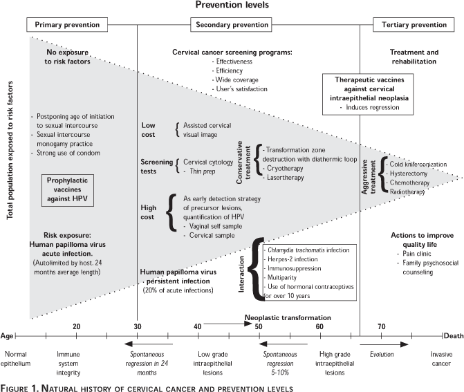
 </font></p>     <p><font face="Verdana" size="2"> Based on the above    information, <a href="/img/revistas/spm/v45s3/3a22f01.gif">Figure 1</a> shows the prevention    strategies for CC for each prevention level based on the current knowledge of    CC's natural history, according to which, based on extensive epidemiological<SUP>8</SUP>    and laboratory<SUP>9</SUP> research, a causal relationship has been shown between    the human papilloma virus (HPV) and CC<SUP>10</SUP> and on the basis of these    findings, prevention measures are currently under investigation. In that respect,    HPV infection is one of the most common sexually-transmitted infections, and    it is estimated that at least 50% of sexually active adults have suffered from    a HPV genital infection.<SUP>11</SUP> A majority of such infections, however,    can be effectively controlled by the host's immune response.<SUP>12</SUP> Finally,    the HPV detection tests plays a major role in the research on the natural history    of the infection and on the potential development of prophylactic vaccines,    a primary prevention measure likely to decrease to a great extent the incidence    of CC as well as the mortality from the disease. In this order of ideas, listed    below are the factors to be considered for the improvement of CCSP programs    in poor countries. </font></p>     <p><font face="Verdana" size="2"><b>Promoting equity    in health care </b></font></p>     <p><font face="Verdana" size="2">The definition    of inequity may be a notorious difference in the quality and/or quantity of    a health care service between two or more communities, with a differential medical    coverage depending on ethnic origin, gender, schooling, site of residence or    other criteria.<SUP>13</SUP> </font></p>     <p><font face="Verdana" size="2"> A primary goal    of a National Health System is eliminating disparities, and this is why a CCSP    program must be promoted and implemented with the same quality in both urban    and rural areas, mainly in rural geographical areas and underprivileged urban    communities. In order to assess the conditions of equality in CC detection,    indicators must be set up to evaluate the CCSP program coverage and the mortality    figures, stratifying by urban and rural regional areas, for the North, Center    and South regions of each country. </font></p>     ]]></body>
<body><![CDATA[<p><font face="Verdana" size="2"><b>Promoting actions    that ensure extensive coverage </b></font></p>     <p><font face="Verdana" size="2">One of the great    benefits of CC screening is involving women at risk, especially those at very    high risk, in a CCSP program. In that respect, an extended coverage and adhesion    to it are paramount for the effectiveness of a CCSP program. CC cannot be eradicated    without an early detection of the precursor lesions by means of a quality PAP    test. </font></p>     <p><font face="Verdana" size="2"><b>Using screening    strategies in accordance with health care services availability </b></font></p>     <p><font face="Verdana" size="2"><I>The screening    test settings scenarios in developing countries</I> </font></p>     <p><font face="Verdana" size="2">In countries with    a great regional heterogeneity, a variety of screening settings must be regulated    and standardized to include a combination of assisted visual inspection, PAP    test and HPV detection. </font></p>     <p><font face="Verdana" size="2"><I>
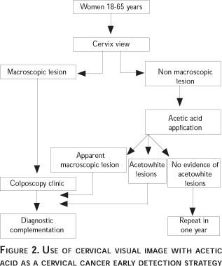
CC detection    by means of acetic acid visual inspection</I>. Prior to the advent of PAP in    1944, visual inspection of the cervix was the only strategy available for CC    detection. The magnification of the cervix by means of the colposcope, a device    invented in 1925 by Hans Hinselmann, revealed tiny invasive cancer cells, invisible    to the naked eye.<SUP>14</SUP> Back in those times, the carcinoma <I>in situ    </I>in the cervix was known, although nobody paid attention to it because its    precursor-of-cancer nature was unknown. The image of an exophytic tumor, a necrotic    area, or a deep ulceration prompted to carry out a biopsy in order to obtain    an accurate diagnosis. Unassisted or colposcopic visual inspection permitted    a high number of early diagnoses and the patients treated with the available    means (radical surgery or radiotherapy) survived to the disease with an acceptable    quality of life. The use of acetic acid, a fortuitous finding by Hinselmann,    improved the detection outcomes by permitting a clearer image of the cervical    surface. Acetic acid dissolves mucus and other detritus, causes a degree of    vasoconstriction with the effect of bringing out the images and showing lesions    invisible to the naked eye and qualified as acetowhite for that matter, because    they are rich in keratin which precipitates under the action of the acid, with    the effect of turning them white and more readily identifiable. Unfortunately,    the method's good results were conditioned to the identification of early cases,    which today would be classified as Stage IB1 invasive cancer (tumor of less    than 4 cm in its larger dimension), because the treatment outcomes depended    on how advanced the lesion was. The late diagnosis of CC, which visual inspection    or colposcopy did not contribute to, offered no benefit in terms of the use    of health facilities, costs, and caused suffering for the women and their families.    A majority of developed countries currently base their CC detection strategy    on the PAP, which has proven useful to that effect.<SUP>15</SUP> This however    is not always feasible in developing countries or certain underprivileged geographic    areas whereas there is neither facilities infrastructure nor human resources    at least to carry out a PAP test, a low cost alternative being the visual inspection    of the cervix with acetic acid. </font></p>     <p><font face="Verdana" size="2"> This strategy    must be implemented as follows: women without an abnormal bleeding history are    placed in gynecological position and a vaginal speculum is introduced (<a href="#fig2">Figure    2</a>) to permit visual inspection of the cervix. The surface is carefully scrutinized    under appropriate lighting. If a macroscopic lesion (exophytic tumor, a necrotic    area or a deep ulceration) is found, the patient must be referred to the Colposcopy    Clinic, for a complementation of the diagnosis. In the absence of a lesion,    the cervix is embrocated with a swab drenched in a 5% concentration of acetic    acid. Two to three minutes later, the swab is removed and the cervix examined.    In the presence of macroscopic lesions, as already mentioned, the patient will    be referred to the Colposcopy Clinic, likewise in the presence of acetowhite    lesions (invisible to the naked eye and visible under the action of acetic acid).    Unfortunately, acetowhite lesions are not restricted to potential cervical neoplasia,    but are also common in the presence of a metaplasia, some inflammatory process,    repair tissue, and atrophy. This constitutes a serious limitation for the method,    because it is likely to unnecessarily saturate the colposcopy facilities with    false positive outcomes which give the test very little specificity and a low    predictive value. In a geographical area where the detection strategy is based    on visual inspection with acetic acid, the number and distribution of colposcopy    facilities must be in keeping with this need, rather than with those generated    by a cervical cytology detection program. </font></p>     <p><a name="fig2"></a></p>     <p>&nbsp;</p>     <p align="center"><img src="/img/revistas/spm/v45s3/3a22f02.gif"></p>     ]]></body>
<body><![CDATA[<p>&nbsp;</p>     <p><font face="Verdana" size="2">In the absence    of macroscopic and acetowhite lesions, the patient may be reasonably assumed    free of invasive neoplasia and precursor lesions, and a repeated exploration    after 12 months is recommended. </font></p>     <p><font face="Verdana" size="2"> As for women with    a history of abnormal bleeding, it is preferable to carry out a gentle vaginal    recognition and in the event of finding an exophitic cervical tumor, without    even using the vaginal speculum; the patient should be referred to a Gynecological    Oncology clinic. In that respect, the patient's assessment is obviously incomplete,    but the idea is to avoid forcing the health care first level units to eventually    face the problem of a severe vaginal hemorrhage, the truth being that they might    not have the material resources to do it. </font></p>     <p><font face="Verdana" size="2"> The visual inspection    of the cervix with or without the application of acetic acid is so easy a proceeding    that, in many countries, it is carried out by nurses specially trained to that    effect.<SUP>16,17</SUP> The outcomes of epidemiological studies with the acetic    acid test are diverse. Case-sensitivity varies between 49.4% and 90%; so does    specificity at a 48.5% to 92% ratio; positive predictive value (PPV) is between    17% and 18.9%; and negative predictive value (NPV) fluctuates between 79.8%    and 97%. In those studies comparing the cervical visual inspection and other    methods, the PAP, the cervicography or de HPV-DNA demonstration yielded superior    outcomes; the cervical visual inspection test, however, may be quite useful    in geographical areas lacking health infrastructure. Finally, with the specificity    and HPV figures, it can be maintained that the women with negative acetic acid    readings are not likely to show, for the time being, an invasive cancer or high    grade precursor lesions. The test may be performed yearly to maintain its reliability.    </font></p>     <p><font face="Verdana" size="2"><I>CCSP programs    based on cervical cytology. </I>The PAP was described in 1944 as a CC diagnosis    method. It soon proved to be able to identify not only the invasive cancer,    but also precursor lesions, the treatment of which was and still is simpler,    cheaper, and more effective. In those countries where the cytological study    has been implemented as a detection strategy in communities, the CC frequency    decreased considerably and has remained stable for the past 50 years.<SUP>18,19</SUP>    </font></p>     <p><font face="Verdana" size="2"> A very significant    change introduced to improve the PAP's diagnostic reliability has been the appropriate    collection of specimens. The smears performed with material accumulated at the    bottom of the vaginal pouch, as recommended by Papanicolaou, proved to be inadequate    for the diagnosis because the cells had exfoliated some time before and had    been exposed to the action of environmental aggressive agents such as the vagina's    physiological acidity and the normal resident flora. On the contrary, if the    smears include "live" cells, collected directly from the precursor    lesion site, the outcomes improve notoriously. Thus, the cytology for the detection    of precursor lesions of cervical-uterine cancer stopped being vaginal or cervical-uterine    in nature to become specifically cervical. </font></p>     <p><font face="Verdana" size="2"> The programs implemented    in developed countries state that the person in charge of collecting specimens    must make sure that the whole transformation zone has been duly sampled. Therefore,    the cervix must be visualized at the moment of performing the smear. The laboratory    staff can never be sure that the whole circumference of the cervix has been    sampled, whatever the nature or the content of the cells in the smear. For the    pathologist, a smear collected on one half of the cervix will look just the    same as one collected on its whole circumference.<SUP>20</SUP> Ideally, a PAP    smear should include a combined sample (one specimen of endocervical and exocervical    cells). This is the reason why the sample collectors must be duly trained in    smear performance, and continued medical updating and training is recommended,    that is, an improvement process to permit the obtainment of advanced knowledge    as well as the development of specific skills and abilities. Provisions should    likewise be made for training inexperienced health staff involved for the first    time in smear collection. For the specimen collector to be able to evaluate    the quality of a cervical smear, and basically, to ensure such quality, the    laboratory must mention in the results report the existence or inexistence of    indicators of the potential sampling of the transformation zone based on the    presence of metaplastic squamous endocervical cells. </font></p>     <p><font face="Verdana" size="2"> The lack of reliable    indicators for the transformation zone sampling in atrophic smears, explains    why no additional comments are needed in reports on that kind of smears. Likewise,    the sampling of the transformation zone may be difficult in the case of women    who have received treatment for pre-malignant lesions. In such circumstances,    the use of an endocervical brush may be needed in addition to a spatula, and    if it is not possible to obtain material from the area, the patient may be referred    for a colposcopic evaluation. Primary screening should not be performed only    with an endocervical brush. Such smears may contain only endocervical cells    and possible do not sample mature squamous cells or epithelium from the transformation    zone. </font></p>     <p><font face="Verdana" size="2"> If the sample    collector wishes to replicate a smear not showing cytological evidence for the    sampling of the transformation zone, he must notify it immediately explaining    the reasons for his request. Such a proceeding should be considered as a part    of the screening process.<SUP>21</SUP> </font></p>     <p><font face="Verdana" size="2"> The proceeding    for PAP specimen collection is the following: in cases without a history of    abnormal bleeding, the patient is placed in gynecological position and the vaginal    speculum is introduced (<a href="#fig3">Figure 3</a>), permitting the visual    inspection of the cervix; under adequate lighting conditions, cervix surface    is carefully inspected. If a macroscopic lesion (exophytic tumor, a necrotic    area or a deep ulceration) is found, the patient must be referred to the Colposcopy    Clinic, for a complementation of the diagnosis. In the absence of a lesion,    the PAP is collected, beginning with the ectocervical sample with an Ayre spatula    or any modified version of it; the ectocervical surface is gently scraped in    a 360&ordm; circular motion. A wooden spatula is needed, making sure to avoid    the use of a plastic one, for this material decreases the cells' ability to    adhere to the slide. The scrapings are quickly deposited in a linear extended    fashion on the distal half of a slide previously marked on one end for correct    identification. To avoid the drying out of the ectocervical material, the endocervical    sample is taken immediately after with a brush gently introduced in the endocervical    canal and rotated in a 180&ordm; motion in a likewise gentle fashion. The material    is immediately fixated with the indicated aerosol. The fixator's recipient must    be at a 30 cm distance from the plate, in order for the fluid to fall as fine    rain on the slide. </font></p>     ]]></body>
<body><![CDATA[<p><a name="fig3"></a></p>     <p>&nbsp;</p>     <p align="center"><img src="/img/revistas/spm/v45s3/3a22f03.gif"></p>     <p>&nbsp;</p>     <p><font face="Verdana" size="2"> It is paramount    to stick to the order specified for the collection of samples; the endocervical    sampling may cause light bleeding even with gentle motions due to the very characteristics    of the glandular epithelium, there being a vessels network situated immediately    below the epithelial layer. Bleeding is more likely in the presence of a certain    amount of inflammation, which happens frequently. If the endocervical sample    is collected first, the blood contaminates the exocervical surface and goes    together with the material on that site, causing a distortion of the cells collected    upon performing the smear and making the interpretation more difficult. It is    recommended in some texts to perform the PAP with a sole tool used to collect    both endocervical and ectocervical material at one time. We do not recommend    doing so. Ample evidence exists for the convenience of collecting two different    samples, in particular, for collecting the endocervical sample with a brush.<SUP>22,23</SUP>    </font></p>     <p><font face="Verdana" size="2"> If the cervix    is coated with a normal cervicovaginal secretion, it must be cleaned with a    swab soaked in physiological solution previous to the visual inspection and    PAP collection. If the secretion's aspect suggests the presence of a vaginal    or cervical infection, the patient should be referred to a first level care    unit, where the infectious process shall be diagnosed and treated. Only after    the treatment and the elimination of the remains of whatever topic medication,    can the PAP be performed. </font></p>     <p><font face="Verdana" size="2"> In women with    a history of abnormal bleeding, it is recommended to first perform a vaginal    palpation without using lubricant, or only wearing a glove moistened with saline    solution. If the palpation reveals a cervical exophitic tumor, the patient shall    be referred to an Oncological Gynecology clinic, without even introducing the    vaginal speculum. If nothing of the sort is found, proceed as mentioned. </font></p>     <p><font face="Verdana" size="2"> It has been said    that cytology yields a high ratio of false negative outcomes, which puts a limitation    on its usefulness. False negative outcomes should not be found in more than    10% of studies and a way of achieving this is carrying out carefully every step    described. A false negative outcome has no impact on a woman's health if the    study is replicated in reasonable amount of time, hence the necessity of her    having her first two negative cervical cytology outcomes obtained at a one year    interval, in order to later increase the screening periodicity. Furthermore,    false negative outcomes are more frequent in low grade intraepithelial lesions    (LSIL), whose natural history shows a very high tendency (in 80% of cases) to    disappear.<SUP>24</SUP> </font></p>     <p><font face="Verdana" size="2"> The age for starting    a cytological study for precursor lesions detection is important, due to the    needed balance between infrastructure and demand. The first cytological study    should be carried out at age 35, or when a woman is considered at risk for having    more than two sexual partners. Otherwise, the facilities would be saturated    with too many inconsequent LSIL's. The natural progression of CC carcinogenesis    is slow enough, so that a PAP test carried out every three years allows sufficient    time for an early detection. </font></p>     <p><font face="Verdana" size="2"> Technological    variations have been recently introduced in the classical PAP technique, such    as thin prep cytology or automated readings. The high costs of both do not warrant    their application in a community program, no matter what their usefulness which    as yet is not sufficiently documented at that level.<SUP>25</SUP> </font></p>     ]]></body>
<body><![CDATA[<p><font face="Verdana" size="2"><I>HPV- DNA as    a strategy for the detection of CC precursor lesions. </I>Since it became possible    to identify the HPV- DNA in secretions collected from the internal part of the    vagina or the cervix, this test has been used as a strategy for the detection    of CC precursor lesions (high grade intraepithelial squamous lesion &#91;HSIL) in    population programs. The test, as most detection devices, has both advantages    and limitations. </font></p>     <p><font face="Verdana" size="2"> The determination    of HPV as a screening strategy is likely to be used at a large scale through    the hybrid capture technique (Hybrid capture II) because it can be implemented    in a semi-automated fashion. The hybrid capture technique allows identifying    over 95% of the HPV types linked to CC, including types 16, 18, 31, 33, 35,    39, 45, 51, 52, 56, 58, 59, and 68.<SUP>26</SUP> </font></p>     <p><font face="Verdana" size="2"> The age-group    most appropriate for using the HPV as a CCSP strategy comprises women over 35    years-old, for whom the prevalence of false positive outcomes is very low. The    HPV may play other roles, the most obvious of which is improving the treatment    of women with low grade or "borderline" lesions. In that context,    using the HPV may help identify those women in need of an immediate referral    to colposcopy. The HPV must also be used in the post-treatment follow-up in    order to be able to monitor complete excisions. The preliminary outcomes of    HPV studies as a screening strategy are quite promising because they increase    considerably the likehood of identifying high grade lesions undiagnosed by conventional    cytology. Especially en poor countries, such a strategy has, however, many limitations,    such as: </font></p> <ul>       <li><font face="Verdana" size="2">In order to      be able to evidence the presence of a HSIL, a CC detection test must be administered      to a very large population of healthy or apparently healthy women. In that      respect, if the PAP has not reached the intended coverage in the national      program, the DNA-HPV will hardly have an extensive coverage. </font></li>       <li><font face="Verdana" size="2">A CC detection      test must sort out between women with HSIL, the healthy ones, and those suffering      another type of HPV-caused disease. In this respect, a positive DNA-HPV test      identifies those women suffering a HPV genital infection of the latent type      (healthy carriers with negative cytology and colposcopy), those with acute      (productive), or chronic (transformative), and those with HSIL and invasive      cancer. The positiveness, which does not define the type of disease suffered,      will cause the referral of the women to a Colposcopy Clinic where their cases      shall be valuated, and the group they belong to will be determined. While      awaiting these results, a majority of women will experience unnecessary anxiety      thinking they might have cancer, when they really are healthy carriers or      only show a transitory LSIL likely to disappear within a few months without      leaving any consequences. The only relevant women for a detection program      are those with HSIL and an invasive cancer. Taking into account the fact that      15% to 30% of the women are healthy carriers,<SUP>27</SUP> these clinics'      population will surely increase, entailing their saturation, which will decrease      the quality of the services, provided and increase the detection program's      costs. </font></li>       <li><font face="Verdana" size="2">The DNA and      HPV tests yield a percentage of false negative results and may leave undetected      cases of HSIL or cancer. However, as compared with other detection methods,      the false negative ratio is much lower, which definitely is an advantage.      Other methods such as cytology may decrease the false negative ratio if every      step in the program is carried out satisfactorily; and the irreducible false      negative ratio (approximately 10%) can be decreased by a replication of the      test, which is effective and does not contribute to a major increase in the      program's cost, for individual studies are quite cheap. </font></li>       <li><font face="Verdana" size="2">In poor countries,      an excessive marketing activity is carried out to convince the clinics of      the usefulness of HPV-DNA determination; however, there does not seem to be      an accurate clinical awareness of the usefulness of the test in such countries,      resulting in inaccurate diagnoses and over-treatment (usually a resection      of the transformation zone with a diathermic loop); an ethically objectionable      and potentially hazardous proceeding). </font></li>       <li><font face="Verdana" size="2">Among other      potential limitations, it must be mentioned that: Amendments to approved CCSP      programs would be very costly and require an extensive training program; presently,      the unitary cost of HPV is higher than PAP's; the HPV screening is likely      to create a financial dependency; and finally, there is no evidence that the      HPV screening would decrease the high frequency and mortality rates from CC.      </font></li>     </ul>     <p><font face="Verdana" size="2"> On the other hand,    the test does offer certain advantages, among which we find: </font></p>     ]]></body>
<body><![CDATA[<p><font face="Verdana" size="2"> The HPV is a potential    candidate for screening as a cancer prevention measure.<SUP>28</SUP> However,    this is still at an early stage and HPV could only be beneficial if it were    to prevent CC through the early detection of asymptomatic precursor lesions.    Many of these potential tests use probes systems amplifying many genotypes and    based on a cocktail of specific tests. There are presently three probes systems    (the MY09/11 and the GP5+/6+), and type II hybrid capture.<SUP>29</SUP> </font></p>     <p><font face="Verdana" size="2"> All three methods    are highly sensitive and specific for the detection of oncogenic viruses and    have a potential for automation. Current technological developments should improve    specificity without substantially decreasing sensitivity. This possibility should    be relevant in terms of public health for it has been shown that HPV-DNA detection    in the absence of cytological abnormalities may reveal the presence of high    grade cervical intraepithelial neoplasia, which cannot be diagnosed through    cytology.<SUP>22</SUP> At any rate, every study suggests that adding HPV test    to PAP test in CC screening may increase the identification of CC precursor    lesions by up to 50-100%. Further studies and prediction schemes will be needed    to evaluate the usefulness as well as the cost-effectiveness of the HPV test    as a screening strategy in developing countries, and how it could also be used    in combination with other diagnosis methodologies. </font></p>     <p><font face="Verdana" size="2"> Another potential    advantage is that the sample needed for the detection may be self-collected.    In that respect, one of the limitations of the CCSP national program is that    the patients refrain from coming to the PAP detection facilities for personal    or family reasons having to do with the very process of sample collection. The    possibility of self-collection and its proven effectiveness may help going round    this difficulty. </font></p>     <p><font face="Verdana" size="2"> <a href="/img/revistas/spm/v45s3/3a22f04.gif">Figure    4</a> shows the algorithm suggested for the implementation of this proposal,    considering that regional community interventions must be initiated to valuate    the convenience of using HPV additionally to PAP. The HPV-DNA is useful for    the valuation of uncertain cytological diagnoses, such as ASC-US. In that respect,    the ASC-US cytological diagnosis, as listed in the Bethesda System 1988, was    an uncertain diagnosis in need of an urgent revaluation, since it could as well    correspond to a healthy woman, or to the carrier of such a serious lesion as    an invasive cancer. Under such conditions, the HPV-DNA determination pointed    out the group of women at risk, who were immediately referred to colposcopy.    The ASC-US diagnosis is no longer found in the revised System Bethesda 2001,    and two diagnosis alternatives are suggested instead, that is, ASC-US and ASC-H.    The latter obviously must be immediately referred to colposcopy, because the    probabilities of it corresponding to a HSIL are high. The former, on the contrary,    is rarely associated to a HSIL, and monitoring through repeated cytologies is    sufficient.<SUP>30,31</SUP> </font></p>     <p><font face="Verdana" size="2"><I>The usefulness    of HPV-DNA for post-treatment control</I> </font></p>     <p><font face="Verdana" size="2">In that respect,    if after treatment for HSIL or a more advanced problem, a patient is HPV-DNA    negative, the check-up appointments may be at larger intervals.<SUP>32</SUP>    If a four-monthly evaluation is recommended during the first two years of monitoring,    this evaluation may later be appointed annually on the basis of negative HPV-DNA    outcomes. </font></p>     <p><font face="Verdana" size="2"><I>The relevance    of HPV-DNA for revaluation</I> </font></p>     <p><font face="Verdana" size="2">In addition to    the harshly criticized ALTS study, many research studies have been conducted    to establish the relevance of HPV-DNA in the clinical management of patients.    These outcomes are necessary to be able to determine that relevance. </font></p>     <p><font face="Verdana" size="2"><b>Management of    abnormal cervical cytology outcomes </b> </font></p>     <p><font face="Verdana" size="2">Since Papanicolaou's    time to this date, the way the results of a cervicovaginal cytology study are    reported has changed notoriously. From a terminology based on the combination    of minus and plus signs and Roman numerals from I to V, one is now used to describe,    interpret and diagnose in terms identical to those used in pathology. Such a    development has permitted a more select management of each case, for the specified    purpose of preventing the overload of colposcopy facilities, which is the next    step in the evaluation of most cytological abnormalities. It must be pointed    out that the imprecision in Papanicolaou's nomenclature was not relevant in    those times, for it was feasible that every woman having from Negative III (an    "all-inclusive" category where just about anything would fit) to Positive    V (invasive cancer) outcomes would go to her gynecologist who, with or without    a colposcope, would perform a biopsy in order to be able to make a precise diagnosis.    Biopsy could vary from a small fragment obtained with a nipper, to a knife cone,    in the operating room. A treatment was determined on the basis of the pathological    diagnosis, resulting in an actual decrease in mortality from CC. This would    not be feasible now because detection reaches a greater number of women every    day and the check-up facilities are overloaded with work. </font></p>     ]]></body>
<body><![CDATA[<p><font face="Verdana" size="2"> Cytological nomenclature    advanced gradually: the nomenclature proposed by Richart<SUP>33</SUP> including    term: 1, 2, and 3 grade Cervical Intraepithelial Neoplasia (CIN) replaced such    terms as mild, moderate, and severe dysplasia, and carcinoma in situ used in    the WHO's recommendations and, finally, there was the Bethesda System, created    in 1988<SUP>34</SUP> and revised in 2001.<SUP>35</SUP> Despite the differences    in appellations and the various ways of classifying the precursor lesions, the    terms are entirely equivalent. The recommendations made in this article include    those derived from the Bethesda System 2001 classification. </font></p>     <p><font face="Verdana" size="2"> In this context,    it is estimated that very high-quality detection programs<SUP>31</SUP> of cytological    studies result abnormal in a 7%, as where is unorganized programs it goes from    10% to 12%. More importantly, many of such abnormalities can and must be managed,    at least temporarily, at the detection site or health care alternate facilities    other than the Colposcopy Clinics. A community program for CC detection must    take care that the Colposcopy Clinics do not become overloaded, for this would    impact negatively on the quality of the service provided, which would be detrimental    for both the patients and the program. </font></p>     <p><font face="Verdana" size="2"> The Bethesda System    2001 cytological diagnoses belong to two main categories: </font></p> <ol>       <li><font face="Verdana" size="2"> Diverse cell      abnormalities unrelated to precursor lesions or cancer, and </font></li>       <li><font face="Verdana" size="2"> Squamous and      glandular cell alterations linked to such diagnoses. The management of each      potential cytological diagnosis will be analyzed </font></li>     </ol>     <p><font face="Verdana" size="2"><I>Non-neoplastic    alterations (<a href="/img/revistas/spm/v45s3/3a22f05.gif">Figure 5</a>) </I></font></p>     <p><font face="Verdana" size="2"><I>The presence    of microorganisms. </i>Evidence of a microorganism in the lower genital tract<I>    (Tricomonas vaginalis, Candida </I>or of some other organisms such as<I> Gardnerella    vaginalis, etc.) </I>will call for a specific treatment. Cervical cytology must    be performed three months later, and later treatment will depend on the outcome.    </font></p>     <p><font face="Verdana" size="2"><I>Other non-neoplastic    findings. Reactive cellular changes. </I>These changes are important because    they often complicate the cytological diagnosis. This is why it is important    to obtain clinical data concerning radiation history and the use of an intrauterine    device. In both instances, the cytological abnormalities are irrelevant. </font></p>     <p><font face="Verdana" size="2"> The inflammation-linked    reactive cellular changes include the metaplastic cells which may be immature,    nevertheless reflecting a physiological process. They may also evidence an unbalance    in the vaginal flora, usually associated to the action of external agents such    as vaginal douches, spermicides, lubricant jellies, intercourse with a partner    using a condom or the vaginal application of medicines. If the inflammation    is mild (graded from + to ++) the possibilities of a cytological diagnosis for    the detection of precursor cells or CC are not hindered, and there is no need    to recommend any changes. However, in the presence of a severe inflammation    (graded from +++ or ++++) the patient must be referred to a specialized facility.    The PAP shall be replicated six months later, and the outcome shall determine    the management of patient. </font></p>     ]]></body>
<body><![CDATA[<p><font face="Verdana" size="2"> A cytological    atrophy picture corresponds to a physiological state in certain stages of life.    The cytological diagnosis may prove difficult to the laboratory, although it    does not have any particular implications for the physician. </font></p>     <p><font face="Verdana" size="2"> The presence of    post-hysterectomy glandular cells shows that the cervix resection was incomplete,    that part of the squamocolumnar junction remains, that there is metaplasia and    therefore, transformation zone. These patients must participate in the CCSP    program just as any other woman. An incomplete hysterectomy does not provide    any protection. </font></p>     <p><font face="Verdana" size="2"> The presence of    normal endometrial cells in post-menopausal women, even in the absence of symptoms,    may be the first indication of a glandular precursor lesion of the endometrium    or an endometrial adenocarcinoma, and a hysteroscopy or curettage is recommended.    </font></p>     <p><font face="Verdana" size="2"><I>Neoplastic cytological    alterations (<a href="/img/revistas/spm/v45s3/3a22f06.gif">Figure 6</a>) </I></font></p>     <p><font face="Verdana" size="2"><I>Atypical squamous    cells with undetermined significance (ASCUS).</i> This category, proposed on    the basis of the Bethesda System 1988 nomenclature, was divided into two sections    for the Bethesda System 2001 revised version: a) <I>atypical squamous cells    with undetermined significance (ASC-US)</I>, favoring a reactive lesion, and    b) <I>atypical squamous cells cannot exclude HSIL (ASC-H)</I>. It is recommended    that category a) be managed as a LSIL and category b) be managed as HSIL, the    frequency of HSIL diagnosed through biopsy is between 5-17% in the former, and    24-94% in the latter.<SUP>36</SUP> </font></p>     <p><font face="Verdana" size="2"><I>ASC-US/LSIL</I>.    LSIL natural history shows its disappearance within 24 months, which actually    occurs in 80% of cases.<SUP>37 </SUP>Therefore, in the presence of ASC-US and    LSIL diagnoses, it is recommended to repeat the cytology study 6 and 12 months    later. If both studies evidence a disappearance, repeat the PAP twice and go    through the detection process every three years thereafter; if both studies    evidence persistence, the patient must be referred to CC, just as well as if    a more severe diagnosis were made. </font></p>     <p><font face="Verdana" size="2"><I>ASC-US under    special circumstances</I>. a) Post-menopausal woman: recommend vaginal estrogens    for seven days and a PAP test five days after the conclusion of treatment. Some    SIL cases not visible due to atrophy thus become apparent. If the cytology turns    out negative after the estrogen treatment, perform it again six months after    a new epithelial maturation. If preferred, oral estrogens may be prescribed.    Two negative results send the patient again to a four-monthly control; b) woman    with immunosuppression: immediately refer the patient to the colposcopy clinic,    and c) pregnant woman: must be monitored normally and revaluated after the puerperium.    </font></p>     <p><font face="Verdana" size="2"><I>ASC-H/HSIL</I>.    Immediately refer to the colposcopy clinic. If the colposcopy of a patient with    ASC-H turns out negative and the woman is post-menopausal, immediately use the    epithelial maturation with estrogens; if she is still menstruating, repeat the    cervical cytology six months later and manage according to the results obtained.    </font></p>     <p><font face="Verdana" size="2"><I>Squamous cell    carcinoma</I>. Refer immediately to the Gynecological Oncology Clinic. </font></p>     <p><font face="Verdana" size="2"><I>Atypical glandular    cells</I>. No matter what the classification of the abnormalities, whether or    not favoring neoplasia, whether specific (endocervical or endometrial) or unspecific    (glandular), the women with atypical glandular cells must be referred immediately    to the Colposcopy Clinic. </font></p>     ]]></body>
<body><![CDATA[<p><font face="Verdana" size="2"><I>In situ endocervical    adenocarcinoma</I>. Refer immediately to the Colposcopy Clinic. </font></p>     <p><font face="Verdana" size="2"><I>Adenocarcinoma    and other malignant neoplasias</I>. The women with such results, whether or    not the cause can be determined, must immediately be referred to a Gynecological    Oncology Clinic. </font></p>     <p><font face="Verdana" size="2"> The correct interpretation    of a PAP will contribute to the appropriate management of patients without overloading    the colposcopy clinics. Otherwise, the women are liable to experience anxiety    and undergo unnecessary biopsies; their ailments may as well be overdiagnosed    and overtreated, all of it using health care time which would be more profitably    dedicated to those showing abnormalities, with the consequent decrease in the    care of all. </font></p>     <p><font face="Verdana" size="2"><b>The need to    regulate the practice of colposcopy </b></font></p>     <p><font face="Verdana" size="2">The practice of    colposcopy must be regulated in order to prevent its population-level usage    on healthy women, to avoid overdiagnosis as well as overtreatment, which does    not only entail high costs, but also produces unnecessary anxiety among the    women at risk. We all know that the diagnostic and therapeutic processes are    not free of immediate complications as well as long-term undesirable effects.    Their application to healthy women without ailments which do not require them    imply, not only a moral responsibility, but also a violation of scientific,    medical, legal, and ethical principles. </font></p>     <p><font face="Verdana" size="2"> For all these    reasons, the overloading of the colposcopy clinics must be avoided for it directly    impairs the quality of health care. In developing countries, colposcopy must    be regulated and evaluated through internal and external quality control mechanisms,    in order to maintain a permanent quality control and prevent a prevalent profit-oriented    practice. </font></p>     <p><font face="Verdana" size="2"><b>The need to    regulate the operation of the HPV clinical laboratories </b></font></p>     <p><font face="Verdana" size="2">The determination    of high-risk HPV-DNA through such techniques as the capture of II or III generation    hybrids (HC II) is fairly simple. The test has distinct advantages over in tube    hybrid capture<SUP>38 </SUP>as well as in situ hybridization.<SUP>39</SUP> HC    II implies the reading of light signals proportionate to the amount of HPV-DNA,    which is an extra advantage: the greater the viral load,<SUP>40</SUP> greater    the risk of finding a precursor lesion or CC. </font></p>     <p><font face="Verdana" size="2"> Quantifying the    amount of HPV-DNA is easy with the proper equipment. Since the test does not    use viral genome replication, but rather, bases the determination on the amount    contained in the sample, positiveness will rely on passing the negativeness    threshold which is 1pg/ml for HC II, and 10 pg/ml for HC I. </font></p>     <p><font face="Verdana" size="2"> The (internal    and external) quality control mechanisms in laboratories performing these tests    must be in strict abidance of the recommendations regulating laboratory operation    in developing countries, under close government supervision as well as of those    medical associations interested in reliable results.<SUP>41</SUP> </font></p>     ]]></body>
<body><![CDATA[<p><font face="Verdana" size="2"><b>Ensuring CCSP    programs user's satisfaction </b></font></p>     <p><font face="Verdana" size="2">Term "quality"    is of a complex and multidimensional nature. It encompasses notions of effectiveness,    efficiency, scientific and technical quality, management, perception, expectations,    communication, adequateness, coordination, accessibility, availability, distribution,    satisfaction, privacy, credibility, professional competency, credit, structural    support, and safety.<SUP>42</SUP> Setting up research lines for quality should    allow us to pursue an improvement of CCSP programs. In order to attain that    goal, there is a need for: a) methods for problem-approaching; b) methods for    setting goals to be pursued by health care facilities, managers and decision    makers; c) methods taking into account the variability in professional practice,    through improvements in internal communication; d) methods chosen to ensure    the satisfaction of users, clients, patients, individuals, citizens, and, finally,    inter alia, and e) methods centered on the promotion of improvement opportunities.    </font></p>     <p><font face="Verdana" size="2"> The advancement    of health technologies in CCSP programs shall allow an improvement of the facilities'    ability to identify and bring a solution to problems. </font></p>     <p><font face="Verdana" size="2"> It will also rely    on the training and/or adaptation of highly competent staff that shall be in    charge of developing and implementing the strategic detection methodologies    needed to meet the population's needs and demands. </font></p>     <p><font face="Verdana" size="2"><b>Clinical tests    to determine the effectiveness of prophylactic vaccines against HPV </b></font></p>     <p><font face="Verdana" size="2">Although the administration    of a vaccine against HPV has proven to be 100% effective in the reduction of    HPV-16 infections as well as of those related to cervical intraepithelial neoplasia,<SUP>43</SUP>    and that a variety of clinical tests for polyvalent prophylactic and therapeutic    vaccines against HPV are presently conducted worldwide, the HPV vaccines will    not be commercially available for a 5 to 10-year time period. For this reason,    it will be necessary to intensify the early detection of cervical cancer. </font></p>     <p>&nbsp;</p>     <p><font face="Verdana" size="3"><b>Conclusions    </b></font></p>     <p><font face="Verdana" size="2">An CCSP program    must be restructured to a) mainly address rural geographic regions and underprivileged    urban areas; b) foster actions aimed at ensuring a wide coverage and similar    quality of services in all these regions; c) use screening strategies in keeping    with the availability of health care services. In countries with a great regional    heterogeneity, a variety of screening settings must be regulated to include    a combination of assisted visual inspection, cervical cytology and HPV determination;    d) regional community intervention must be initiated to evaluate the usefulness    of using HPV as a strategy additionally to cervical cytology; e) the practice    of colposcopy must be regulated in order to avoid its use with healthy women    at a community level, this, for the purpose of preventing overdiagnosis and    overtreatment, which not only entail high costs, but also cause unnecessary    anxiety in the women at risk; furthermore, f) the practice of clinical laboratories    using HPV as a detection strategy must be accredited, and g) in order to ensure    quality services, the CCSP program must also meet the users' expectations and    improve their knowledge of cervical cancer. Finally, though a variety of clinical    tests on prophylactic and therapeutic vaccines against HPV are presently being    developed worldwide; they shall not be commercially available for a 5 to 10-year    time period. For this reason, it will be necessary to intensify the CCSP programs.    </font></p>     <p>&nbsp;</p>     ]]></body>
<body><![CDATA[<p><font face="Verdana" size="3"><b>References </b></font></p>     <!-- ref --><p><font face="Verdana" size="2">1. Lara E, Day    EN, Hakama M. Trends in mortality from cervical cancer in the Nordic countries:    Association with organized screening programes. Lancet 1987:1247-1249. </font>&nbsp;&nbsp;&nbsp;&nbsp;&nbsp;&nbsp;&nbsp;&nbsp;[&#160;<a href="javascript:void(0);" onclick="javascript: window.open('/scielo.php?script=sci_nlinks&ref=9185419&pid=S0036-3634200300090002000001&lng=','','width=640,height=500,resizable=yes,scrollbars=1,menubar=yes,');">Links</a>&#160;]<!-- end-ref --><!-- ref --><p><font face="Verdana" size="2">2. Lazcano-Ponce    E, Rasc&oacute;n-Pacheco R, Lozano R, Velasco E. Mortality from carcinoma of    the uterine cervix in Mexico: impact of screening 1980-1990. Acta Cytol 1996;40:506-512.    </font>&nbsp;&nbsp;&nbsp;&nbsp;&nbsp;&nbsp;&nbsp;&nbsp;[&#160;<a href="javascript:void(0);" onclick="javascript: window.open('/scielo.php?script=sci_nlinks&ref=9185420&pid=S0036-3634200300090002000002&lng=','','width=640,height=500,resizable=yes,scrollbars=1,menubar=yes,');">Links</a>&#160;]<!-- end-ref --><!-- ref --><p><font face="Verdana" size="2">3. Miller AB. Cervical    cancer screening programes, Geneva: World Health Organization, 1992. </font>&nbsp;&nbsp;&nbsp;&nbsp;&nbsp;&nbsp;&nbsp;&nbsp;[&#160;<a href="javascript:void(0);" onclick="javascript: window.open('/scielo.php?script=sci_nlinks&ref=9185421&pid=S0036-3634200300090002000003&lng=','','width=640,height=500,resizable=yes,scrollbars=1,menubar=yes,');">Links</a>&#160;]<!-- end-ref --><!-- ref --><p><font face="Verdana" size="2">4. Mitchell H.    Consistency of reporting endocervical cell. An intralaboratory and interlaboratory    assessment. Acta Cytol 1994;38:310-314. </font>&nbsp;&nbsp;&nbsp;&nbsp;&nbsp;&nbsp;&nbsp;&nbsp;[&#160;<a href="javascript:void(0);" onclick="javascript: window.open('/scielo.php?script=sci_nlinks&ref=9185422&pid=S0036-3634200300090002000004&lng=','','width=640,height=500,resizable=yes,scrollbars=1,menubar=yes,');">Links</a>&#160;]<!-- end-ref --><!-- ref --><p><font face="Verdana" size="2">5. Koss LG. Cytology:    Accuracy or diagnosis. Cancer 1989;64:249-257. </font>&nbsp;&nbsp;&nbsp;&nbsp;&nbsp;&nbsp;&nbsp;&nbsp;[&#160;<a href="javascript:void(0);" onclick="javascript: window.open('/scielo.php?script=sci_nlinks&ref=9185423&pid=S0036-3634200300090002000005&lng=','','width=640,height=500,resizable=yes,scrollbars=1,menubar=yes,');">Links</a>&#160;]<!-- end-ref --><!-- ref --><p><font face="Verdana" size="2">6. Alonso P, Lazcano    E. Quality control in cytopathology laboratories in six Latin American Countries.    In: Wied G, Keebler C, Rosenthal D, Schenck U, Somrak T, Vooijs P eds. Compendium    on quality assurance, proficiency testing and workload limitations in clinical    cytology Chicago: Tutorials of Cytology,1995:115-121. </font>&nbsp;&nbsp;&nbsp;&nbsp;&nbsp;&nbsp;&nbsp;&nbsp;[&#160;<a href="javascript:void(0);" onclick="javascript: window.open('/scielo.php?script=sci_nlinks&ref=9185424&pid=S0036-3634200300090002000006&lng=','','width=640,height=500,resizable=yes,scrollbars=1,menubar=yes,');">Links</a>&#160;]<!-- end-ref --><!-- ref --><p><font face="Verdana" size="2">7. Lundenberg G.    Quality assurance in cervical cytology. The papanicolaou smear. JAMA 1989;262:1672-1679.    </font>&nbsp;&nbsp;&nbsp;&nbsp;&nbsp;&nbsp;&nbsp;&nbsp;[&#160;<a href="javascript:void(0);" onclick="javascript: window.open('/scielo.php?script=sci_nlinks&ref=9185425&pid=S0036-3634200300090002000007&lng=','','width=640,height=500,resizable=yes,scrollbars=1,menubar=yes,');">Links</a>&#160;]<!-- end-ref --><!-- ref --><p><font face="Verdana" size="2">8. Mu&ntilde;oz    N, Bosch FX, De Sanjose S, Tafur L, Izarzaguza I, Gili M <I>et al</I>. The causal    link between human papillomavirus and invasive cervical cancer: A population-based    case-control study in Colombia and Spain. Int J Cancer 1992;52:743-749. </font>&nbsp;&nbsp;&nbsp;&nbsp;&nbsp;&nbsp;&nbsp;&nbsp;[&#160;<a href="javascript:void(0);" onclick="javascript: window.open('/scielo.php?script=sci_nlinks&ref=9185426&pid=S0036-3634200300090002000008&lng=','','width=640,height=500,resizable=yes,scrollbars=1,menubar=yes,');">Links</a>&#160;]<!-- end-ref --><!-- ref --><p><font face="Verdana" size="2">9. Zur Hausen H.    Papillomaviruses in human cancer. Appl Pathol 1987;5(1):19-24. </font>&nbsp;&nbsp;&nbsp;&nbsp;&nbsp;&nbsp;&nbsp;&nbsp;[&#160;<a href="javascript:void(0);" onclick="javascript: window.open('/scielo.php?script=sci_nlinks&ref=9185427&pid=S0036-3634200300090002000009&lng=','','width=640,height=500,resizable=yes,scrollbars=1,menubar=yes,');">Links</a>&#160;]<!-- end-ref --><!-- ref --><p><font face="Verdana" size="2">10. Walboomers    JMM, Jacobs MV, Manos M, Bosch FX, Kummer JA, Shah K <I>et al</I>. Human papillomavirus    is a necessary cause of invasive cervical cancer worldwide. J Pathol 1999;189:12-19.    </font>&nbsp;&nbsp;&nbsp;&nbsp;&nbsp;&nbsp;&nbsp;&nbsp;[&#160;<a href="javascript:void(0);" onclick="javascript: window.open('/scielo.php?script=sci_nlinks&ref=9185428&pid=S0036-3634200300090002000010&lng=','','width=640,height=500,resizable=yes,scrollbars=1,menubar=yes,');">Links</a>&#160;]<!-- end-ref --><!-- ref --><p><font face="Verdana" size="2">11. Koutsky L.    Epidemiology of genital human papillomavirus infection. Am J Medicine 1997;102:3-8.    </font>&nbsp;&nbsp;&nbsp;&nbsp;&nbsp;&nbsp;&nbsp;&nbsp;[&#160;<a href="javascript:void(0);" onclick="javascript: window.open('/scielo.php?script=sci_nlinks&ref=9185429&pid=S0036-3634200300090002000011&lng=','','width=640,height=500,resizable=yes,scrollbars=1,menubar=yes,');">Links</a>&#160;]<!-- end-ref --><!-- ref --><p><font face="Verdana" size="2">12. Galloway D.    Is vaccination against human papillomavirus a possibility? Lancet 1998;352:SIII22-SIII24.    </font>&nbsp;&nbsp;&nbsp;&nbsp;&nbsp;&nbsp;&nbsp;&nbsp;[&#160;<a href="javascript:void(0);" onclick="javascript: window.open('/scielo.php?script=sci_nlinks&ref=9185430&pid=S0036-3634200300090002000012&lng=','','width=640,height=500,resizable=yes,scrollbars=1,menubar=yes,');">Links</a>&#160;]<!-- end-ref --><!-- ref --><p><font face="Verdana" size="2">13. Pearcy JN,    Keppel KG. A Summary measure of health disparity. Public Health Reports 2002;117:273-280.    </font>&nbsp;&nbsp;&nbsp;&nbsp;&nbsp;&nbsp;&nbsp;&nbsp;[&#160;<a href="javascript:void(0);" onclick="javascript: window.open('/scielo.php?script=sci_nlinks&ref=9185431&pid=S0036-3634200300090002000013&lng=','','width=640,height=500,resizable=yes,scrollbars=1,menubar=yes,');">Links</a>&#160;]<!-- end-ref --><!-- ref --><p><font face="Verdana" size="2">14. Hinselmann    H. Verbesserung der Inspektionm&ouml;glichkeiten von Vulva, Vagina und portio.    M&uuml;nch Med Wschr 1925;72:1733. </font>&nbsp;&nbsp;&nbsp;&nbsp;&nbsp;&nbsp;&nbsp;&nbsp;[&#160;<a href="javascript:void(0);" onclick="javascript: window.open('/scielo.php?script=sci_nlinks&ref=9185432&pid=S0036-3634200300090002000014&lng=','','width=640,height=500,resizable=yes,scrollbars=1,menubar=yes,');">Links</a>&#160;]<!-- end-ref --><!-- ref --><p><font face="Verdana" size="2">15. Bjorge T, Thoresen    SO, Skare GB. Incidence, survival and mortality in cervical cancer in Norway,    1956-1990. Eur J Cancer 1993;29A(16):2291-2297. </font>&nbsp;&nbsp;&nbsp;&nbsp;&nbsp;&nbsp;&nbsp;&nbsp;[&#160;<a href="javascript:void(0);" onclick="javascript: window.open('/scielo.php?script=sci_nlinks&ref=9185433&pid=S0036-3634200300090002000015&lng=','','width=640,height=500,resizable=yes,scrollbars=1,menubar=yes,');">Links</a>&#160;]<!-- end-ref --><!-- ref --><p><font face="Verdana" size="2">16. Megevand E,    Denny L, Dehacek K y cols. Acetic acid visualization of the cervix: An alternative    to cytologic screening. Obstet Gynecol 1996;88:383-386. </font>&nbsp;&nbsp;&nbsp;&nbsp;&nbsp;&nbsp;&nbsp;&nbsp;[&#160;<a href="javascript:void(0);" onclick="javascript: window.open('/scielo.php?script=sci_nlinks&ref=9185434&pid=S0036-3634200300090002000016&lng=','','width=640,height=500,resizable=yes,scrollbars=1,menubar=yes,');">Links</a>&#160;]<!-- end-ref --><!-- ref --><p><font face="Verdana" size="2">17. University    of Zimbawe/JHPIEGO Cervical Cancer Project. Visual inspection with acetic acid    for cervical-cancer screening: test qualities in a primary-care setting. Lancet    1999;353:869-873. </font>&nbsp;&nbsp;&nbsp;&nbsp;&nbsp;&nbsp;&nbsp;&nbsp;[&#160;<a href="javascript:void(0);" onclick="javascript: window.open('/scielo.php?script=sci_nlinks&ref=9185435&pid=S0036-3634200300090002000017&lng=','','width=640,height=500,resizable=yes,scrollbars=1,menubar=yes,');">Links</a>&#160;]<!-- end-ref --><!-- ref --><p><font face="Verdana" size="2">18. Sigurdsson    K. Effect of organized screening on the risk of cervical cancer. Evaluation    of screening activity in Iceland,1964-1991. Int J Cancer 1993 Jun 19;54(4):563-70.    </font>&nbsp;&nbsp;&nbsp;&nbsp;&nbsp;&nbsp;&nbsp;&nbsp;[&#160;<a href="javascript:void(0);" onclick="javascript: window.open('/scielo.php?script=sci_nlinks&ref=9185436&pid=S0036-3634200300090002000018&lng=','','width=640,height=500,resizable=yes,scrollbars=1,menubar=yes,');">Links</a>&#160;]<!-- end-ref --><!-- ref --><p><font face="Verdana" size="2">19. Herrero R.    Epidemiology of cervical cancer. J Natl Cancer Inst Monogr 1996;21:1-6. </font>&nbsp;&nbsp;&nbsp;&nbsp;&nbsp;&nbsp;&nbsp;&nbsp;[&#160;<a href="javascript:void(0);" onclick="javascript: window.open('/scielo.php?script=sci_nlinks&ref=9185437&pid=S0036-3634200300090002000019&lng=','','width=640,height=500,resizable=yes,scrollbars=1,menubar=yes,');">Links</a>&#160;]<!-- end-ref --><!-- ref --><p><font face="Verdana" size="2">20. Editorial.    Cell Content of Cervical Smears. Cytopathol 1990;1:129-130. </font>&nbsp;&nbsp;&nbsp;&nbsp;&nbsp;&nbsp;&nbsp;&nbsp;[&#160;<a href="javascript:void(0);" onclick="javascript: window.open('/scielo.php?script=sci_nlinks&ref=9185438&pid=S0036-3634200300090002000020&lng=','','width=640,height=500,resizable=yes,scrollbars=1,menubar=yes,');">Links</a>&#160;]<!-- end-ref --><!-- ref --><p><font face="Verdana" size="2">21. Herbert A.    Achievable Standards, Benchmarks for Reporting and Criteria for Evaluating Cervical    Cytopathology. Cytopathol 1995;6:1-32. </font>&nbsp;&nbsp;&nbsp;&nbsp;&nbsp;&nbsp;&nbsp;&nbsp;[&#160;<a href="javascript:void(0);" onclick="javascript: window.open('/scielo.php?script=sci_nlinks&ref=9185439&pid=S0036-3634200300090002000021&lng=','','width=640,height=500,resizable=yes,scrollbars=1,menubar=yes,');">Links</a>&#160;]<!-- end-ref --><!-- ref --><p><font face="Verdana" size="2">22. Martin-Hirsch    P, Lilford R, Jarvis G, Kitchener HC. Efficacy of cervical-smear collection    devices: a systematic review and meta-analysis. The Lancet 1999; 354 (9192):1763-1770.    </font>&nbsp;&nbsp;&nbsp;&nbsp;&nbsp;&nbsp;&nbsp;&nbsp;[&#160;<a href="javascript:void(0);" onclick="javascript: window.open('/scielo.php?script=sci_nlinks&ref=9185440&pid=S0036-3634200300090002000022&lng=','','width=640,height=500,resizable=yes,scrollbars=1,menubar=yes,');">Links</a>&#160;]<!-- end-ref --><!-- ref --><p><font face="Verdana" size="2">23. McCord ML,    Stovall TG, Meric JL, Summitt RL Jr, Coleman SA. Cervical cytology: A randomized    comparison of four sampling methods. Am J Obstet Gynecol. 1992;166:1772-1779.    </font>&nbsp;&nbsp;&nbsp;&nbsp;&nbsp;&nbsp;&nbsp;&nbsp;[&#160;<a href="javascript:void(0);" onclick="javascript: window.open('/scielo.php?script=sci_nlinks&ref=9185441&pid=S0036-3634200300090002000023&lng=','','width=640,height=500,resizable=yes,scrollbars=1,menubar=yes,');">Links</a>&#160;]<!-- end-ref --><!-- ref --><p><font face="Verdana" size="2">24. Ho GY, Bierman    R, Beardsley L, Chang CJ, Burk RD. Natural history of cervicovaginal papillomavirus    infection in young women. N Engl J Med 1998;338(7):423-438. </font>&nbsp;&nbsp;&nbsp;&nbsp;&nbsp;&nbsp;&nbsp;&nbsp;[&#160;<a href="javascript:void(0);" onclick="javascript: window.open('/scielo.php?script=sci_nlinks&ref=9185442&pid=S0036-3634200300090002000024&lng=','','width=640,height=500,resizable=yes,scrollbars=1,menubar=yes,');">Links</a>&#160;]<!-- end-ref --><!-- ref --><p><font face="Verdana" size="2">25. Sawaya GF,    Grimes DA. New technologies in cervical cytology screening: A word of caution.    Obstet Gynecol 1999;94:307-310. </font>&nbsp;&nbsp;&nbsp;&nbsp;&nbsp;&nbsp;&nbsp;&nbsp;[&#160;<a href="javascript:void(0);" onclick="javascript: window.open('/scielo.php?script=sci_nlinks&ref=9185443&pid=S0036-3634200300090002000025&lng=','','width=640,height=500,resizable=yes,scrollbars=1,menubar=yes,');">Links</a>&#160;]<!-- end-ref --><!-- ref --><p><font face="Verdana" size="2">26. Iftner T, Villa    LL. Chapter 12: Human papillomavirus technologies. J Natl Cancer Inst Monogr.    2003;(31):80-8. </font>&nbsp;&nbsp;&nbsp;&nbsp;&nbsp;&nbsp;&nbsp;&nbsp;[&#160;<a href="javascript:void(0);" onclick="javascript: window.open('/scielo.php?script=sci_nlinks&ref=9185444&pid=S0036-3634200300090002000026&lng=','','width=640,height=500,resizable=yes,scrollbars=1,menubar=yes,');">Links</a>&#160;]<!-- end-ref --><!-- ref --><p><font face="Verdana" size="2">27. Salmer&oacute;n    J, Lazcano-Ponce E, L&ouml;rincz A, Hern&aacute;ndez M, Hern&aacute;ndez P,    Leyva A <I>et al</I>. Comparison of HPV-based assays with Papanicolaou smears    for cervical cancer screening in Morelos State, Mexico. Cancer Causes and Control    2003;14 (6): 505-512. </font>&nbsp;&nbsp;&nbsp;&nbsp;&nbsp;&nbsp;&nbsp;&nbsp;[&#160;<a href="javascript:void(0);" onclick="javascript: window.open('/scielo.php?script=sci_nlinks&ref=9185445&pid=S0036-3634200300090002000027&lng=','','width=640,height=500,resizable=yes,scrollbars=1,menubar=yes,');">Links</a>&#160;]<!-- end-ref --><!-- ref --><p><font face="Verdana" size="2">28. L&ouml;rincz    AT, Richart RM. Human papillomavirus DNA testing as an adjunct to cytology in    cervical screening programs. Arch Pathol Lab Med 2003;127(8):959-968. </font>&nbsp;&nbsp;&nbsp;&nbsp;&nbsp;&nbsp;&nbsp;&nbsp;[&#160;<a href="javascript:void(0);" onclick="javascript: window.open('/scielo.php?script=sci_nlinks&ref=9185446&pid=S0036-3634200300090002000028&lng=','','width=640,height=500,resizable=yes,scrollbars=1,menubar=yes,');">Links</a>&#160;]<!-- end-ref --><!-- ref --><p><font face="Verdana" size="2">29. Castle PE,    Schiffman M, Gravitt PE, Kendall H, Fishman S, Dong H <I>et al</I>. Comparisons    of HPV DNA detection by MY09/11 PCR methods. J Med Virol 2002;68(3):417-423.    </font>&nbsp;&nbsp;&nbsp;&nbsp;&nbsp;&nbsp;&nbsp;&nbsp;[&#160;<a href="javascript:void(0);" onclick="javascript: window.open('/scielo.php?script=sci_nlinks&ref=9185447&pid=S0036-3634200300090002000029&lng=','','width=640,height=500,resizable=yes,scrollbars=1,menubar=yes,');">Links</a>&#160;]<!-- end-ref --><!-- ref --><p><font face="Verdana" size="2">30. Garner EI.    Cervical cancer: Disparities in screening, treatment, and survival. Cancer Epidemiol    Biomarkers Prev 2003;12(3):242S-247S. </font>&nbsp;&nbsp;&nbsp;&nbsp;&nbsp;&nbsp;&nbsp;&nbsp;[&#160;<a href="javascript:void(0);" onclick="javascript: window.open('/scielo.php?script=sci_nlinks&ref=9185448&pid=S0036-3634200300090002000030&lng=','','width=640,height=500,resizable=yes,scrollbars=1,menubar=yes,');">Links</a>&#160;]<!-- end-ref --><!-- ref --><p><font face="Verdana" size="2">31. Wright TC Jr,    Cox JT, Massad LS, Twiggs LB, Wilkinson EJ; ASCCP-Sponsored Consensus Conference.    2001 Consensus Guidelines for the management of women with cervical cytological    abnormalities. JAMA. 2002;287(16):2120-9. </font>&nbsp;&nbsp;&nbsp;&nbsp;&nbsp;&nbsp;&nbsp;&nbsp;[&#160;<a href="javascript:void(0);" onclick="javascript: window.open('/scielo.php?script=sci_nlinks&ref=9185449&pid=S0036-3634200300090002000031&lng=','','width=640,height=500,resizable=yes,scrollbars=1,menubar=yes,');">Links</a>&#160;]<!-- end-ref --><!-- ref --><p><font face="Verdana" size="2">32. Paraskevaidis    E, Koliopoulos G, Alamanos Y, Malamou-Mitsi V, Lolis ED, Kitchener HC. Human    papillomavirus testing and the outcome of treatment for cervical intraepithelial    neoplasia. Obstet Gynecol 2001;98(5 Pt 1):724-725. </font>&nbsp;&nbsp;&nbsp;&nbsp;&nbsp;&nbsp;&nbsp;&nbsp;[&#160;<a href="javascript:void(0);" onclick="javascript: window.open('/scielo.php?script=sci_nlinks&ref=9185450&pid=S0036-3634200300090002000032&lng=','','width=640,height=500,resizable=yes,scrollbars=1,menubar=yes,');">Links</a>&#160;]<!-- end-ref --><!-- ref --><p><font face="Verdana" size="2">33. Richart RM.    Current concepts in obstetrics and gynecology. The patient with abnormal Pap    smear &#150;screening techniques and management. N Engl J Med 1980;302:332-334.    </font>&nbsp;&nbsp;&nbsp;&nbsp;&nbsp;&nbsp;&nbsp;&nbsp;[&#160;<a href="javascript:void(0);" onclick="javascript: window.open('/scielo.php?script=sci_nlinks&ref=9185451&pid=S0036-3634200300090002000033&lng=','','width=640,height=500,resizable=yes,scrollbars=1,menubar=yes,');">Links</a>&#160;]<!-- end-ref --><!-- ref --><p><font face="Verdana" size="2">34. National Cancer    Institute Workshop. The 1988 Bethesda System for reporting cervical/vaginal    cytologic diagnoses. JAMA 1989;262:931-934. </font>&nbsp;&nbsp;&nbsp;&nbsp;&nbsp;&nbsp;&nbsp;&nbsp;[&#160;<a href="javascript:void(0);" onclick="javascript: window.open('/scielo.php?script=sci_nlinks&ref=9185452&pid=S0036-3634200300090002000034&lng=','','width=640,height=500,resizable=yes,scrollbars=1,menubar=yes,');">Links</a>&#160;]<!-- end-ref --><!-- ref --><p><font face="Verdana" size="2">35. Solomon D,    Davey D, Kurman R, Moriarty A, O'Connor D, Prey M, and Forum Group Members;    Bethesda 2001 Workshop. The 2001 Bethesda System: terminology for reporting    results of cervical cytology. JAMA. 2002;287(16):2140-2141. </font>&nbsp;&nbsp;&nbsp;&nbsp;&nbsp;&nbsp;&nbsp;&nbsp;[&#160;<a href="javascript:void(0);" onclick="javascript: window.open('/scielo.php?script=sci_nlinks&ref=9185453&pid=S0036-3634200300090002000035&lng=','','width=640,height=500,resizable=yes,scrollbars=1,menubar=yes,');">Links</a>&#160;]<!-- end-ref --><!-- ref --><p><font face="Verdana" size="2">36. O'Meara AT.    Present standards for cervical cancer screening. Curr Opin Oncol 2002;14:505-511.    </font>&nbsp;&nbsp;&nbsp;&nbsp;&nbsp;&nbsp;&nbsp;&nbsp;[&#160;<a href="javascript:void(0);" onclick="javascript: window.open('/scielo.php?script=sci_nlinks&ref=9185454&pid=S0036-3634200300090002000036&lng=','','width=640,height=500,resizable=yes,scrollbars=1,menubar=yes,');">Links</a>&#160;]<!-- end-ref --><!-- ref --><p><font face="Verdana" size="2">37. Elfgren K,    Kalantari M, Moberger B <I>et al.</I> A population-based five year follow-up    study of cervical human papillomavirus infection. Am J Obstet Gynecol 2000;183:561-567.    </font>&nbsp;&nbsp;&nbsp;&nbsp;&nbsp;&nbsp;&nbsp;&nbsp;[&#160;<a href="javascript:void(0);" onclick="javascript: window.open('/scielo.php?script=sci_nlinks&ref=9185455&pid=S0036-3634200300090002000037&lng=','','width=640,height=500,resizable=yes,scrollbars=1,menubar=yes,');">Links</a>&#160;]<!-- end-ref --><!-- ref --><p><font face="Verdana" size="2">38. L&ouml;rincz    AT: Molecular methods for the detection of human papillomavirus infection. Obstet    Gynecol Clin NA 1998;23:707-730. </font>&nbsp;&nbsp;&nbsp;&nbsp;&nbsp;&nbsp;&nbsp;&nbsp;[&#160;<a href="javascript:void(0);" onclick="javascript: window.open('/scielo.php?script=sci_nlinks&ref=9185456&pid=S0036-3634200300090002000038&lng=','','width=640,height=500,resizable=yes,scrollbars=1,menubar=yes,');">Links</a>&#160;]<!-- end-ref --><!-- ref --><p><font face="Verdana" size="2">39. Autillo-Touati    A, Joannes M, d'Ercole C, Robaglia-Schlupp A, Lambert A, Mazzella E <I>et al</I>.    HPV typing by in situ hybridization on cervical cytologic smears with ASCUS.    Acta Cytol. 1998 May-Jun;42(3):631-8. </font>&nbsp;&nbsp;&nbsp;&nbsp;&nbsp;&nbsp;&nbsp;&nbsp;[&#160;<a href="javascript:void(0);" onclick="javascript: window.open('/scielo.php?script=sci_nlinks&ref=9185457&pid=S0036-3634200300090002000039&lng=','','width=640,height=500,resizable=yes,scrollbars=1,menubar=yes,');">Links</a>&#160;]<!-- end-ref --><!-- ref --><p><font face="Verdana" size="2">40. Sun Sun CA,    Liu JF, Wu DM, Nieh S, Yu CP, Chu TY. Viral load of high-risk human papillomavirus    in cervical squamous intraepithelial lesions. Int J Gynaecol Obstet. 2002;76(1):41-47.    </font>&nbsp;&nbsp;&nbsp;&nbsp;&nbsp;&nbsp;&nbsp;&nbsp;[&#160;<a href="javascript:void(0);" onclick="javascript: window.open('/scielo.php?script=sci_nlinks&ref=9185458&pid=S0036-3634200300090002000040&lng=','','width=640,height=500,resizable=yes,scrollbars=1,menubar=yes,');">Links</a>&#160;]<!-- end-ref --><!-- ref --><p><font face="Verdana" size="2">41. Steindel SJ,    Granade S, Lee J, Avery G, Clarke LM, Jenny RW <I>et al</I>. Practice patterns    of testing waived under the clinical laboratory improvement amendments. Arch    Pathol Lab Med. 2002;126(12):1471-1479. </font>&nbsp;&nbsp;&nbsp;&nbsp;&nbsp;&nbsp;&nbsp;&nbsp;[&#160;<a href="javascript:void(0);" onclick="javascript: window.open('/scielo.php?script=sci_nlinks&ref=9185459&pid=S0036-3634200300090002000041&lng=','','width=640,height=500,resizable=yes,scrollbars=1,menubar=yes,');">Links</a>&#160;]<!-- end-ref --><!-- ref --><p><font face="Verdana" size="2">42. Saturno PJ,    Quintana O, Varo J. &#191;Qu&eacute; es la calidad?. En: Saturno PJ, et al.    Tratado de calidad asistencial en atenci&oacute;n primaria. Tomo I. Murcia:    Du Pont Pharma, 1997:19-45. </font>&nbsp;&nbsp;&nbsp;&nbsp;&nbsp;&nbsp;&nbsp;&nbsp;[&#160;<a href="javascript:void(0);" onclick="javascript: window.open('/scielo.php?script=sci_nlinks&ref=9185460&pid=S0036-3634200300090002000042&lng=','','width=640,height=500,resizable=yes,scrollbars=1,menubar=yes,');">Links</a>&#160;]<!-- end-ref --><!-- ref --><p><font face="Verdana" size="2">43. Koutsky LA,    Ault KA, Wheeler CM, Brown DR, Barr E, Alvarez FB <I>et al.</I> Proof of Principle    Study Investigators. A controlled trial of a human papillomavirus type 16 vaccine.    N Engl J Med, 2002;347(21):1645-1651. </font>&nbsp;&nbsp;&nbsp;&nbsp;&nbsp;&nbsp;&nbsp;&nbsp;[&#160;<a href="javascript:void(0);" onclick="javascript: window.open('/scielo.php?script=sci_nlinks&ref=9185461&pid=S0036-3634200300090002000043&lng=','','width=640,height=500,resizable=yes,scrollbars=1,menubar=yes,');">Links</a>&#160;]<!-- end-ref --><p>&nbsp;</p>     <p>&nbsp;</p>     <p><font face="Verdana" size="2"><b>Address requests    reprints to:</b>     <br>   Dr. Eduardo Lazcano-Ponce    <br>   Director de Investigaci&oacute;n en Enfermedades Cr&oacute;nicas del Instituto    Nacional de Salud P&uacute;blica    <br>   Avenida Universidad No. 655, Colonia Santa Mar&iacute;a Ahuacatitl&aacute;n        ]]></body>
<body><![CDATA[<br>   62508 Cuernavaca, Morelos, M&eacute;xico    <br>   Correo electr&oacute;nico: <a href="mailto:elazcano@correo.insp.mx">elazcano@correo.insp.mx</a>    </font></p>     <p><font face="Verdana" size="2"><b>Received on:</b>    April 8, 2003 <b>    <br>   Accepted on:</b> July 21, 2003 </font></p>      ]]></body><back>
<ref-list>
<ref id="B1">
<label>1</label><nlm-citation citation-type="journal">
<person-group person-group-type="author">
<name>
<surname><![CDATA[Lara]]></surname>
<given-names><![CDATA[E]]></given-names>
</name>
<name>
<surname><![CDATA[Day]]></surname>
<given-names><![CDATA[EN]]></given-names>
</name>
<name>
<surname><![CDATA[Hakama]]></surname>
<given-names><![CDATA[M]]></given-names>
</name>
</person-group>
<article-title xml:lang="en"><![CDATA[Trends in mortality from cervical cancer in the Nordic countries: Association with organized screening programes]]></article-title>
<source><![CDATA[Lancet]]></source>
<year>1987</year>
<page-range>1247-1249</page-range></nlm-citation>
</ref>
<ref id="B2">
<label>2</label><nlm-citation citation-type="journal">
<person-group person-group-type="author">
<name>
<surname><![CDATA[Lazcano-Ponce]]></surname>
<given-names><![CDATA[E]]></given-names>
</name>
<name>
<surname><![CDATA[Rascón-Pacheco]]></surname>
<given-names><![CDATA[R]]></given-names>
</name>
<name>
<surname><![CDATA[Lozano]]></surname>
<given-names><![CDATA[R]]></given-names>
</name>
<name>
<surname><![CDATA[Velasco]]></surname>
<given-names><![CDATA[E]]></given-names>
</name>
</person-group>
<article-title xml:lang="en"><![CDATA[Mortality from carcinoma of the uterine cervix in Mexico: impact of screening 1980-1990]]></article-title>
<source><![CDATA[Acta Cytol]]></source>
<year>1996</year>
<volume>40</volume>
<page-range>506-512</page-range></nlm-citation>
</ref>
<ref id="B3">
<label>3</label><nlm-citation citation-type="book">
<person-group person-group-type="author">
<name>
<surname><![CDATA[Miller]]></surname>
<given-names><![CDATA[AB]]></given-names>
</name>
</person-group>
<source><![CDATA[Cervical cancer screening programes]]></source>
<year>1992</year>
<publisher-loc><![CDATA[Geneva ]]></publisher-loc>
<publisher-name><![CDATA[World Health Organization]]></publisher-name>
</nlm-citation>
</ref>
<ref id="B4">
<label>4</label><nlm-citation citation-type="journal">
<person-group person-group-type="author">
<name>
<surname><![CDATA[Mitchell]]></surname>
<given-names><![CDATA[H]]></given-names>
</name>
</person-group>
<article-title xml:lang="en"><![CDATA[Consistency of reporting endocervical cell: An intralaboratory and interlaboratory assessment]]></article-title>
<source><![CDATA[Acta Cytol]]></source>
<year>1994</year>
<volume>38</volume>
<page-range>310-314</page-range></nlm-citation>
</ref>
<ref id="B5">
<label>5</label><nlm-citation citation-type="journal">
<person-group person-group-type="author">
<name>
<surname><![CDATA[Koss]]></surname>
<given-names><![CDATA[LG]]></given-names>
</name>
</person-group>
<article-title xml:lang="en"><![CDATA[Cytology: Accuracy or diagnosis]]></article-title>
<source><![CDATA[Cancer]]></source>
<year>1989</year>
<volume>64</volume>
<page-range>249-257</page-range></nlm-citation>
</ref>
<ref id="B6">
<label>6</label><nlm-citation citation-type="">
<person-group person-group-type="author">
<name>
<surname><![CDATA[Alonso]]></surname>
<given-names><![CDATA[P]]></given-names>
</name>
<name>
<surname><![CDATA[Lazcano]]></surname>
<given-names><![CDATA[E]]></given-names>
</name>
</person-group>
<article-title xml:lang="en"><![CDATA[Quality control in cytopathology laboratories in six Latin American Countries]]></article-title>
<person-group person-group-type="editor">
<name>
<surname><![CDATA[Wied]]></surname>
<given-names><![CDATA[G]]></given-names>
</name>
<name>
<surname><![CDATA[Keebler]]></surname>
<given-names><![CDATA[C]]></given-names>
</name>
<name>
<surname><![CDATA[Rosenthal]]></surname>
<given-names><![CDATA[D]]></given-names>
</name>
<name>
<surname><![CDATA[Schenck]]></surname>
<given-names><![CDATA[U]]></given-names>
</name>
<name>
<surname><![CDATA[Somrak]]></surname>
<given-names><![CDATA[T]]></given-names>
</name>
<name>
<surname><![CDATA[Vooijs]]></surname>
<given-names><![CDATA[P]]></given-names>
</name>
</person-group>
<source><![CDATA[Compendium on quality assurance, proficiency testing and workload limitations in clinical cytology Chicago: Tutorials of Cytology]]></source>
<year>1995</year>
<page-range>115-121</page-range></nlm-citation>
</ref>
<ref id="B7">
<label>7</label><nlm-citation citation-type="journal">
<person-group person-group-type="author">
<name>
<surname><![CDATA[Lundenberg]]></surname>
<given-names><![CDATA[G]]></given-names>
</name>
</person-group>
<article-title xml:lang="en"><![CDATA[Quality assurance in cervical cytology: The papanicolaou smear]]></article-title>
<source><![CDATA[JAMA]]></source>
<year>1989</year>
<volume>262</volume>
<page-range>1672-1679</page-range></nlm-citation>
</ref>
<ref id="B8">
<label>8</label><nlm-citation citation-type="journal">
<person-group person-group-type="author">
<name>
<surname><![CDATA[Muñoz]]></surname>
<given-names><![CDATA[N]]></given-names>
</name>
<name>
<surname><![CDATA[Bosch]]></surname>
<given-names><![CDATA[FX]]></given-names>
</name>
<name>
<surname><![CDATA[De Sanjose]]></surname>
<given-names><![CDATA[S]]></given-names>
</name>
<name>
<surname><![CDATA[Tafur]]></surname>
<given-names><![CDATA[L]]></given-names>
</name>
<name>
<surname><![CDATA[Izarzaguza]]></surname>
<given-names><![CDATA[I]]></given-names>
</name>
<name>
<surname><![CDATA[Gili]]></surname>
<given-names><![CDATA[M]]></given-names>
</name>
</person-group>
<article-title xml:lang="en"><![CDATA[The causal link between human papillomavirus and invasive cervical cancer: A population-based case-control study in Colombia and Spain]]></article-title>
<source><![CDATA[Int J Cancer]]></source>
<year>1992</year>
<volume>52</volume>
<page-range>743-749</page-range></nlm-citation>
</ref>
<ref id="B9">
<label>9</label><nlm-citation citation-type="journal">
<person-group person-group-type="author">
<name>
<surname><![CDATA[Zur Hausen]]></surname>
<given-names><![CDATA[H]]></given-names>
</name>
</person-group>
<article-title xml:lang="en"><![CDATA[Papillomaviruses in human cancer]]></article-title>
<source><![CDATA[Appl Pathol]]></source>
<year>1987</year>
<volume>5</volume>
<numero>1</numero>
<issue>1</issue>
<page-range>19-24</page-range></nlm-citation>
</ref>
<ref id="B10">
<label>10</label><nlm-citation citation-type="journal">
<person-group person-group-type="author">
<name>
<surname><![CDATA[Walboomers]]></surname>
<given-names><![CDATA[JMM]]></given-names>
</name>
<name>
<surname><![CDATA[Jacobs]]></surname>
<given-names><![CDATA[MV]]></given-names>
</name>
<name>
<surname><![CDATA[Manos]]></surname>
<given-names><![CDATA[M]]></given-names>
</name>
<name>
<surname><![CDATA[Bosch]]></surname>
<given-names><![CDATA[FX]]></given-names>
</name>
<name>
<surname><![CDATA[Kummer]]></surname>
<given-names><![CDATA[JA]]></given-names>
</name>
<name>
<surname><![CDATA[Shah]]></surname>
<given-names><![CDATA[K]]></given-names>
</name>
</person-group>
<article-title xml:lang="en"><![CDATA[Human papillomavirus is a necessary cause of invasive cervical cancer worldwide]]></article-title>
<source><![CDATA[J Pathol]]></source>
<year>1999</year>
<volume>189</volume>
<page-range>12-19</page-range></nlm-citation>
</ref>
<ref id="B11">
<label>11</label><nlm-citation citation-type="journal">
<person-group person-group-type="author">
<name>
<surname><![CDATA[Koutsky]]></surname>
<given-names><![CDATA[L]]></given-names>
</name>
</person-group>
<article-title xml:lang="en"><![CDATA[Epidemiology of genital human papillomavirus infection]]></article-title>
<source><![CDATA[Am J Medicine]]></source>
<year>1997</year>
<volume>102</volume>
<page-range>3-8</page-range></nlm-citation>
</ref>
<ref id="B12">
<label>12</label><nlm-citation citation-type="journal">
<person-group person-group-type="author">
<name>
<surname><![CDATA[Galloway]]></surname>
<given-names><![CDATA[D]]></given-names>
</name>
</person-group>
<article-title xml:lang="en"><![CDATA[Is vaccination against human papillomavirus a possibility?]]></article-title>
<source><![CDATA[Lancet]]></source>
<year>1998</year>
<volume>352</volume>
<page-range>SIII22-SIII24</page-range></nlm-citation>
</ref>
<ref id="B13">
<label>13</label><nlm-citation citation-type="journal">
<person-group person-group-type="author">
<name>
<surname><![CDATA[Pearcy]]></surname>
<given-names><![CDATA[JN]]></given-names>
</name>
<name>
<surname><![CDATA[Keppel]]></surname>
<given-names><![CDATA[KG]]></given-names>
</name>
</person-group>
<article-title xml:lang="en"><![CDATA[A Summary measure of health disparity]]></article-title>
<source><![CDATA[Public Health Reports]]></source>
<year>2002</year>
<volume>117</volume>
<page-range>273-280</page-range></nlm-citation>
</ref>
<ref id="B14">
<label>14</label><nlm-citation citation-type="journal">
<person-group person-group-type="author">
<name>
<surname><![CDATA[Hinselmann]]></surname>
<given-names><![CDATA[H]]></given-names>
</name>
</person-group>
<article-title xml:lang="de"><![CDATA[Verbesserung der Inspektionmöglichkeiten von Vulva, Vagina und portio]]></article-title>
<source><![CDATA[Münch Med Wschr]]></source>
<year>1925</year>
<volume>72</volume>
<page-range>1733</page-range></nlm-citation>
</ref>
<ref id="B15">
<label>15</label><nlm-citation citation-type="journal">
<person-group person-group-type="author">
<name>
<surname><![CDATA[Bjorge]]></surname>
<given-names><![CDATA[T]]></given-names>
</name>
<name>
<surname><![CDATA[Thoresen]]></surname>
<given-names><![CDATA[SO]]></given-names>
</name>
<name>
<surname><![CDATA[Skare]]></surname>
<given-names><![CDATA[GB]]></given-names>
</name>
</person-group>
<article-title xml:lang="en"><![CDATA[Incidence, survival and mortality in cervical cancer in Norway, 1956-1990]]></article-title>
<source><![CDATA[Eur J Cancer]]></source>
<year>1993</year>
<volume>29A</volume>
<numero>16</numero>
<issue>16</issue>
<page-range>2291-2297</page-range></nlm-citation>
</ref>
<ref id="B16">
<label>16</label><nlm-citation citation-type="journal">
<person-group person-group-type="author">
<name>
<surname><![CDATA[Megevand]]></surname>
<given-names><![CDATA[E]]></given-names>
</name>
<name>
<surname><![CDATA[Denny]]></surname>
<given-names><![CDATA[L]]></given-names>
</name>
<name>
<surname><![CDATA[Dehacek]]></surname>
<given-names><![CDATA[K]]></given-names>
</name>
</person-group>
<article-title xml:lang="en"><![CDATA[Acetic acid visualization of the cervix: An alternative to cytologic screening]]></article-title>
<source><![CDATA[Obstet Gynecol]]></source>
<year>1996</year>
<volume>88</volume>
<page-range>383-386</page-range></nlm-citation>
</ref>
<ref id="B17">
<label>17</label><nlm-citation citation-type="journal">
<collab>University of Zimbawe^dJHPIEGO Cervical Cancer Project</collab>
<article-title xml:lang="en"><![CDATA[Visual inspection with acetic acid for cervical-cancer screening: test qualities in a primary-care setting]]></article-title>
<source><![CDATA[Lancet]]></source>
<year>1999</year>
<volume>353</volume>
<page-range>869-873</page-range></nlm-citation>
</ref>
<ref id="B18">
<label>18</label><nlm-citation citation-type="journal">
<person-group person-group-type="author">
<name>
<surname><![CDATA[Sigurdsson]]></surname>
<given-names><![CDATA[K]]></given-names>
</name>
</person-group>
<article-title xml:lang="en"><![CDATA[Effect of organized screening on the risk of cervical cancer: Evaluation of screening activity in Iceland,1964-1991]]></article-title>
<source><![CDATA[Int J Cancer]]></source>
<year>1993</year>
<volume>54</volume>
<numero>4</numero>
<issue>4</issue>
<page-range>563-70</page-range></nlm-citation>
</ref>
<ref id="B19">
<label>19</label><nlm-citation citation-type="journal">
<person-group person-group-type="author">
<name>
<surname><![CDATA[Herrero]]></surname>
<given-names><![CDATA[R]]></given-names>
</name>
</person-group>
<article-title xml:lang="en"><![CDATA[Epidemiology of cervical cancer]]></article-title>
<source><![CDATA[J Natl Cancer Inst Monogr]]></source>
<year>1996</year>
<volume>21</volume>
<page-range>1-6</page-range></nlm-citation>
</ref>
<ref id="B20">
<label>20</label><nlm-citation citation-type="journal">
<article-title xml:lang="en"><![CDATA[Cell Content of Cervical Smears]]></article-title>
<source><![CDATA[Cytopathol]]></source>
<year>1990</year>
<numero>1</numero>
<issue>1</issue>
<page-range>129-130</page-range></nlm-citation>
</ref>
<ref id="B21">
<label>21</label><nlm-citation citation-type="journal">
<person-group person-group-type="author">
<name>
<surname><![CDATA[Herbert]]></surname>
<given-names><![CDATA[A]]></given-names>
</name>
</person-group>
<article-title xml:lang="en"><![CDATA[Achievable Standards, Benchmarks for Reporting and Criteria for Evaluating Cervical Cytopathology]]></article-title>
<source><![CDATA[Cytopathol]]></source>
<year>1995</year>
<volume>6</volume>
<page-range>1-32</page-range></nlm-citation>
</ref>
<ref id="B22">
<label>22</label><nlm-citation citation-type="journal">
<person-group person-group-type="author">
<name>
<surname><![CDATA[Martin-Hirsch]]></surname>
<given-names><![CDATA[P]]></given-names>
</name>
<name>
<surname><![CDATA[Lilford]]></surname>
<given-names><![CDATA[R]]></given-names>
</name>
<name>
<surname><![CDATA[Jarvis]]></surname>
<given-names><![CDATA[G]]></given-names>
</name>
<name>
<surname><![CDATA[Kitchener]]></surname>
<given-names><![CDATA[HC]]></given-names>
</name>
</person-group>
<article-title xml:lang="en"><![CDATA[Efficacy of cervical-smear collection devices: a systematic review and meta-analysis]]></article-title>
<source><![CDATA[The Lancet]]></source>
<year>1999</year>
<volume>354</volume>
<numero>9192</numero>
<issue>9192</issue>
<page-range>1763-1770</page-range></nlm-citation>
</ref>
<ref id="B23">
<label>23</label><nlm-citation citation-type="journal">
<person-group person-group-type="author">
<name>
<surname><![CDATA[McCord]]></surname>
<given-names><![CDATA[ML]]></given-names>
</name>
<name>
<surname><![CDATA[Stovall]]></surname>
<given-names><![CDATA[TG]]></given-names>
</name>
<name>
<surname><![CDATA[Meric]]></surname>
<given-names><![CDATA[JL]]></given-names>
</name>
<name>
<surname><![CDATA[Summitt]]></surname>
<given-names><![CDATA[RL Jr]]></given-names>
</name>
<name>
<surname><![CDATA[Coleman]]></surname>
<given-names><![CDATA[SA]]></given-names>
</name>
</person-group>
<article-title xml:lang="en"><![CDATA[Cervical cytology: A randomized comparison of four sampling methods]]></article-title>
<source><![CDATA[Am J Obstet Gynecol.]]></source>
<year>1992</year>
<volume>166</volume>
<page-range>1772-1779</page-range></nlm-citation>
</ref>
<ref id="B24">
<label>24</label><nlm-citation citation-type="journal">
<person-group person-group-type="author">
<name>
<surname><![CDATA[Ho]]></surname>
<given-names><![CDATA[GY]]></given-names>
</name>
<name>
<surname><![CDATA[Bierman]]></surname>
<given-names><![CDATA[R]]></given-names>
</name>
<name>
<surname><![CDATA[Beardsley]]></surname>
<given-names><![CDATA[L]]></given-names>
</name>
<name>
<surname><![CDATA[Chang]]></surname>
<given-names><![CDATA[CJ]]></given-names>
</name>
<name>
<surname><![CDATA[Burk]]></surname>
<given-names><![CDATA[RD]]></given-names>
</name>
</person-group>
<article-title xml:lang="en"><![CDATA[Natural history of cervicovaginal papillomavirus infection in young women]]></article-title>
<source><![CDATA[N Engl J Med]]></source>
<year>1998</year>
<volume>338</volume>
<numero>7</numero>
<issue>7</issue>
<page-range>423-438</page-range></nlm-citation>
</ref>
<ref id="B25">
<label>25</label><nlm-citation citation-type="journal">
<person-group person-group-type="author">
<name>
<surname><![CDATA[Sawaya]]></surname>
<given-names><![CDATA[GF]]></given-names>
</name>
<name>
<surname><![CDATA[Grimes]]></surname>
<given-names><![CDATA[DA]]></given-names>
</name>
</person-group>
<article-title xml:lang="en"><![CDATA[New technologies in cervical cytology screening: A word of caution]]></article-title>
<source><![CDATA[Obstet Gynecol]]></source>
<year>1999</year>
<volume>94</volume>
<page-range>307-310</page-range></nlm-citation>
</ref>
<ref id="B26">
<label>26</label><nlm-citation citation-type="journal">
<person-group person-group-type="author">
<name>
<surname><![CDATA[Iftner]]></surname>
<given-names><![CDATA[T]]></given-names>
</name>
<name>
<surname><![CDATA[Villa]]></surname>
<given-names><![CDATA[LL]]></given-names>
</name>
</person-group>
<article-title xml:lang="en"><![CDATA[Chapter 12: Human papillomavirus technologies]]></article-title>
<source><![CDATA[J Natl Cancer Inst Monogr]]></source>
<year>2003</year>
<volume>31</volume>
<page-range>80-8</page-range></nlm-citation>
</ref>
<ref id="B27">
<label>27</label><nlm-citation citation-type="journal">
<person-group person-group-type="author">
<name>
<surname><![CDATA[Salmerón]]></surname>
<given-names><![CDATA[J]]></given-names>
</name>
<name>
<surname><![CDATA[Lazcano-Ponce]]></surname>
<given-names><![CDATA[E]]></given-names>
</name>
<name>
<surname><![CDATA[Lörincz]]></surname>
<given-names><![CDATA[A]]></given-names>
</name>
<name>
<surname><![CDATA[Hernández]]></surname>
<given-names><![CDATA[M]]></given-names>
</name>
<name>
<surname><![CDATA[Hernández]]></surname>
<given-names><![CDATA[P]]></given-names>
</name>
<name>
<surname><![CDATA[Leyva]]></surname>
<given-names><![CDATA[A]]></given-names>
</name>
</person-group>
<article-title xml:lang="en"><![CDATA[Comparison of HPV-based assays with Papanicolaou smears for cervical cancer screening in Morelos State, Mexico]]></article-title>
<source><![CDATA[Cancer Causes and Control]]></source>
<year>2003</year>
<volume>14</volume>
<numero>6</numero>
<issue>6</issue>
<page-range>505-512</page-range></nlm-citation>
</ref>
<ref id="B28">
<label>28</label><nlm-citation citation-type="journal">
<person-group person-group-type="author">
<name>
<surname><![CDATA[Lörincz]]></surname>
<given-names><![CDATA[AT]]></given-names>
</name>
<name>
<surname><![CDATA[Richart]]></surname>
<given-names><![CDATA[RM]]></given-names>
</name>
</person-group>
<article-title xml:lang="en"><![CDATA[Human papillomavirus DNA testing as an adjunct to cytology in cervical screening programs]]></article-title>
<source><![CDATA[Arch Pathol Lab Med]]></source>
<year>2003</year>
<volume>127</volume>
<numero>8</numero>
<issue>8</issue>
<page-range>959-968</page-range></nlm-citation>
</ref>
<ref id="B29">
<label>29</label><nlm-citation citation-type="journal">
<person-group person-group-type="author">
<name>
<surname><![CDATA[Castle]]></surname>
<given-names><![CDATA[PE]]></given-names>
</name>
<name>
<surname><![CDATA[Schiffman]]></surname>
<given-names><![CDATA[M]]></given-names>
</name>
<name>
<surname><![CDATA[Gravitt]]></surname>
<given-names><![CDATA[PE]]></given-names>
</name>
<name>
<surname><![CDATA[Kendall]]></surname>
<given-names><![CDATA[H]]></given-names>
</name>
<name>
<surname><![CDATA[Fishman]]></surname>
<given-names><![CDATA[S]]></given-names>
</name>
<name>
<surname><![CDATA[Dong]]></surname>
<given-names><![CDATA[H]]></given-names>
</name>
</person-group>
<article-title xml:lang="en"><![CDATA[Comparisons of HPV DNA detection by MY09/11 PCR methods]]></article-title>
<source><![CDATA[J Med Virol]]></source>
<year>2002</year>
<volume>68</volume>
<numero>3</numero>
<issue>3</issue>
<page-range>417-423</page-range></nlm-citation>
</ref>
<ref id="B30">
<label>30</label><nlm-citation citation-type="journal">
<person-group person-group-type="author">
<name>
<surname><![CDATA[Garner]]></surname>
<given-names><![CDATA[EI]]></given-names>
</name>
</person-group>
<article-title xml:lang="en"><![CDATA[Cervical cancer: Disparities in screening, treatment, and survival]]></article-title>
<source><![CDATA[Cancer Epidemiol Biomarkers Prev]]></source>
<year>2003</year>
<volume>12</volume>
<numero>3</numero>
<issue>3</issue>
<page-range>242S-247S</page-range></nlm-citation>
</ref>
<ref id="B31">
<label>31</label><nlm-citation citation-type="journal">
<person-group person-group-type="author">
<name>
<surname><![CDATA[Wright]]></surname>
<given-names><![CDATA[TC Jr]]></given-names>
</name>
<name>
<surname><![CDATA[Cox]]></surname>
<given-names><![CDATA[JT]]></given-names>
</name>
<name>
<surname><![CDATA[Massad]]></surname>
<given-names><![CDATA[LS]]></given-names>
</name>
<name>
<surname><![CDATA[Twiggs]]></surname>
<given-names><![CDATA[LB]]></given-names>
</name>
<name>
<surname><![CDATA[Wilkinson]]></surname>
<given-names><![CDATA[EJ]]></given-names>
</name>
</person-group>
<article-title xml:lang="en"><![CDATA[ASCCP-Sponsored Consensus Conference. 2001 Consensus Guidelines for the management of women with cervical cytological abnormalities]]></article-title>
<source><![CDATA[JAMA.]]></source>
<year>2002</year>
<volume>287</volume>
<numero>16</numero>
<issue>16</issue>
<page-range>2120-9</page-range></nlm-citation>
</ref>
<ref id="B32">
<label>32</label><nlm-citation citation-type="journal">
<person-group person-group-type="author">
<name>
<surname><![CDATA[Paraskevaidis]]></surname>
<given-names><![CDATA[E]]></given-names>
</name>
<name>
<surname><![CDATA[Koliopoulos]]></surname>
<given-names><![CDATA[G]]></given-names>
</name>
<name>
<surname><![CDATA[Alamanos]]></surname>
<given-names><![CDATA[Y]]></given-names>
</name>
<name>
<surname><![CDATA[Malamou-Mitsi]]></surname>
<given-names><![CDATA[V]]></given-names>
</name>
<name>
<surname><![CDATA[Lolis]]></surname>
<given-names><![CDATA[ED]]></given-names>
</name>
<name>
<surname><![CDATA[Kitchener]]></surname>
<given-names><![CDATA[HC]]></given-names>
</name>
</person-group>
<article-title xml:lang="en"><![CDATA[Human papillomavirus testing and the outcome of treatment for cervical intraepithelial neoplasia]]></article-title>
<source><![CDATA[Obstet Gynecol]]></source>
<year>2001</year>
<volume>98</volume>
<numero>5 Pt 1</numero>
<issue>5 Pt 1</issue>
<page-range>724-725</page-range></nlm-citation>
</ref>
<ref id="B33">
<label>33</label><nlm-citation citation-type="journal">
<person-group person-group-type="author">
<name>
<surname><![CDATA[Richart]]></surname>
<given-names><![CDATA[RM]]></given-names>
</name>
</person-group>
<article-title xml:lang="en"><![CDATA[Current concepts in obstetrics and gynecology: The patient with abnormal Pap smear -screening techniques and management]]></article-title>
<source><![CDATA[N Engl J Med]]></source>
<year>1980</year>
<volume>302</volume>
<page-range>332-334</page-range></nlm-citation>
</ref>
<ref id="B34">
<label>34</label><nlm-citation citation-type="journal">
<collab>National Cancer Institute Workshop</collab>
<article-title xml:lang="en"><![CDATA[The 1988 Bethesda System for reporting cervical/vaginal cytologic diagnoses]]></article-title>
<source><![CDATA[JAMA]]></source>
<year>1989</year>
<volume>262</volume>
<page-range>931-934</page-range></nlm-citation>
</ref>
<ref id="B35">
<label>35</label><nlm-citation citation-type="journal">
<person-group person-group-type="author">
<name>
<surname><![CDATA[Solomon]]></surname>
<given-names><![CDATA[D]]></given-names>
</name>
<name>
<surname><![CDATA[Davey]]></surname>
<given-names><![CDATA[D]]></given-names>
</name>
<name>
<surname><![CDATA[Kurman]]></surname>
<given-names><![CDATA[R]]></given-names>
</name>
<name>
<surname><![CDATA[Moriarty]]></surname>
<given-names><![CDATA[A]]></given-names>
</name>
<name>
<surname><![CDATA[O'Connor]]></surname>
<given-names><![CDATA[D]]></given-names>
</name>
<name>
<surname><![CDATA[Prey]]></surname>
<given-names><![CDATA[M]]></given-names>
</name>
<name>
<surname><![CDATA[and]]></surname>
<given-names><![CDATA[Forum]]></given-names>
</name>
</person-group>
<article-title xml:lang="en"><![CDATA[The 2001 Bethesda System: terminology for reporting results of cervical cytology]]></article-title>
<source><![CDATA[JAMA]]></source>
<year>2002</year>
<volume>287</volume>
<numero>16</numero>
<issue>16</issue>
<page-range>2140-2141</page-range></nlm-citation>
</ref>
<ref id="B36">
<label>36</label><nlm-citation citation-type="journal">
<person-group person-group-type="author">
<name>
<surname><![CDATA[O'Meara]]></surname>
<given-names><![CDATA[AT]]></given-names>
</name>
</person-group>
<article-title xml:lang="en"><![CDATA[Present standards for cervical cancer screening]]></article-title>
<source><![CDATA[Curr Opin Oncol]]></source>
<year>2002</year>
<volume>14</volume>
<page-range>505-511</page-range></nlm-citation>
</ref>
<ref id="B37">
<label>37</label><nlm-citation citation-type="journal">
<person-group person-group-type="author">
<name>
<surname><![CDATA[Elfgren]]></surname>
<given-names><![CDATA[K]]></given-names>
</name>
<name>
<surname><![CDATA[Kalantari]]></surname>
<given-names><![CDATA[M]]></given-names>
</name>
<name>
<surname><![CDATA[Moberger]]></surname>
<given-names><![CDATA[B]]></given-names>
</name>
</person-group>
<article-title xml:lang="en"><![CDATA[A population-based five year follow-up study of cervical human papillomavirus infection]]></article-title>
<source><![CDATA[Am J Obstet Gynecol]]></source>
<year>2000</year>
<volume>183</volume>
<page-range>561-567</page-range></nlm-citation>
</ref>
<ref id="B38">
<label>38</label><nlm-citation citation-type="journal">
<person-group person-group-type="author">
<name>
<surname><![CDATA[Lörincz]]></surname>
<given-names><![CDATA[AT]]></given-names>
</name>
</person-group>
<article-title xml:lang="en"><![CDATA[Molecular methods for the detection of human papillomavirus infection]]></article-title>
<source><![CDATA[Obstet Gynecol Clin NA]]></source>
<year>1998</year>
<volume>23</volume>
<page-range>707-730</page-range></nlm-citation>
</ref>
<ref id="B39">
<label>39</label><nlm-citation citation-type="journal">
<person-group person-group-type="author">
<name>
<surname><![CDATA[Autillo-Touati]]></surname>
<given-names><![CDATA[A]]></given-names>
</name>
<name>
<surname><![CDATA[Joannes]]></surname>
<given-names><![CDATA[M]]></given-names>
</name>
<name>
<surname><![CDATA[d'Ercole]]></surname>
<given-names><![CDATA[C]]></given-names>
</name>
<name>
<surname><![CDATA[Robaglia-Schlupp]]></surname>
<given-names><![CDATA[A]]></given-names>
</name>
<name>
<surname><![CDATA[Lambert]]></surname>
<given-names><![CDATA[A]]></given-names>
</name>
<name>
<surname><![CDATA[Mazzella]]></surname>
<given-names><![CDATA[E]]></given-names>
</name>
</person-group>
<article-title xml:lang="en"><![CDATA[HPV typing by in situ hybridization on cervical cytologic smears with ASCUS]]></article-title>
<source><![CDATA[Acta Cytol]]></source>
<year>1998</year>
<volume>42</volume>
<numero>3</numero>
<issue>3</issue>
<page-range>631-8</page-range></nlm-citation>
</ref>
<ref id="B40">
<label>40</label><nlm-citation citation-type="journal">
<person-group person-group-type="author">
<name>
<surname><![CDATA[Sun]]></surname>
<given-names><![CDATA[Sun CA]]></given-names>
</name>
<name>
<surname><![CDATA[Liu]]></surname>
<given-names><![CDATA[JF]]></given-names>
</name>
<name>
<surname><![CDATA[Wu]]></surname>
<given-names><![CDATA[DM]]></given-names>
</name>
<name>
<surname><![CDATA[Nieh]]></surname>
<given-names><![CDATA[S]]></given-names>
</name>
<name>
<surname><![CDATA[Yu]]></surname>
<given-names><![CDATA[CP]]></given-names>
</name>
<name>
<surname><![CDATA[Chu]]></surname>
<given-names><![CDATA[TY]]></given-names>
</name>
</person-group>
<article-title xml:lang="en"><![CDATA[Viral load of high-risk human papillomavirus in cervical squamous intraepithelial lesions]]></article-title>
<source><![CDATA[Int J Gynaecol Obstet.]]></source>
<year>2002</year>
<volume>76</volume>
<numero>1</numero>
<issue>1</issue>
<page-range>41-47</page-range></nlm-citation>
</ref>
<ref id="B41">
<label>41</label><nlm-citation citation-type="journal">
<person-group person-group-type="author">
<name>
<surname><![CDATA[Steindel]]></surname>
<given-names><![CDATA[SJ]]></given-names>
</name>
<name>
<surname><![CDATA[Granade]]></surname>
<given-names><![CDATA[S]]></given-names>
</name>
<name>
<surname><![CDATA[Lee]]></surname>
<given-names><![CDATA[J]]></given-names>
</name>
<name>
<surname><![CDATA[Avery]]></surname>
<given-names><![CDATA[G]]></given-names>
</name>
<name>
<surname><![CDATA[Clarke]]></surname>
<given-names><![CDATA[LM]]></given-names>
</name>
<name>
<surname><![CDATA[Jenny]]></surname>
<given-names><![CDATA[RW]]></given-names>
</name>
</person-group>
<article-title xml:lang="en"><![CDATA[Practice patterns of testing waived under the clinical laboratory improvement amendments]]></article-title>
<source><![CDATA[Arch Pathol Lab Med.]]></source>
<year>2002</year>
<volume>126</volume>
<numero>12</numero>
<issue>12</issue>
<page-range>1471-1479</page-range></nlm-citation>
</ref>
<ref id="B42">
<label>42</label><nlm-citation citation-type="book">
<person-group person-group-type="author">
<name>
<surname><![CDATA[Saturno]]></surname>
<given-names><![CDATA[PJ]]></given-names>
</name>
<name>
<surname><![CDATA[Quintana]]></surname>
<given-names><![CDATA[O]]></given-names>
</name>
<name>
<surname><![CDATA[Varo]]></surname>
<given-names><![CDATA[J]]></given-names>
</name>
</person-group>
<article-title xml:lang="es"><![CDATA[¿Qué es la calidad?]]></article-title>
<person-group person-group-type="editor">
<name>
<surname><![CDATA[Saturno]]></surname>
<given-names><![CDATA[PJ]]></given-names>
</name>
</person-group>
<source><![CDATA[Tratado de calidad asistencial en atención primaria]]></source>
<year>1997</year>
<page-range>19-45</page-range><publisher-loc><![CDATA[Murcia ]]></publisher-loc>
<publisher-name><![CDATA[Du Pont Pharma]]></publisher-name>
</nlm-citation>
</ref>
<ref id="B43">
<label>43</label><nlm-citation citation-type="journal">
<person-group person-group-type="author">
<name>
<surname><![CDATA[Koutsky]]></surname>
<given-names><![CDATA[LA]]></given-names>
</name>
<name>
<surname><![CDATA[Ault]]></surname>
<given-names><![CDATA[KA]]></given-names>
</name>
<name>
<surname><![CDATA[Wheeler]]></surname>
<given-names><![CDATA[CM]]></given-names>
</name>
<name>
<surname><![CDATA[Brown]]></surname>
<given-names><![CDATA[DR]]></given-names>
</name>
<name>
<surname><![CDATA[Barr]]></surname>
<given-names><![CDATA[E]]></given-names>
</name>
<name>
<surname><![CDATA[Alvarez]]></surname>
<given-names><![CDATA[FB]]></given-names>
</name>
</person-group>
<article-title xml:lang="es"><![CDATA[Proof of Principle Study Investigators: A controlled trial of a human papillomavirus type 16 vaccine]]></article-title>
<source><![CDATA[N Engl J Med,]]></source>
<year>2002</year>
<volume>347</volume>
<numero>21</numero>
<issue>21</issue>
<page-range>1645-1651</page-range></nlm-citation>
</ref>
</ref-list>
</back>
</article>
