


 nueva página del texto (beta)
 Inglés (pdf)
 Artículo en XML
 Referencias del artículo
 Enviar artículo por email
 Citado por SciELO
  Similares en
SciELO
Similares en
SciELO 
 Permalink
Introduction
Breast cancer it's defined as the oncologic process, in which healthy cells from the mammary gland degenerate and turn into tumoral, proliferate, and lately multiply to constitute an abnormal mass of body tissue. On the world stage, this cancer is responsible for around 300,000 deaths/year1. In Mexico, since 2006, breast cancer outweighs cervical cancer as the cause of death in women aged 30-54. There are important regional differences with higher frequency of breast cancer in states from the north and center of the country, where women enjoy a higher cultural and socioeconomic status, in states, where indigenous population and a lower socioeconomic level prevail that the frequency is lower2. The annual incidence of breast cancer in Mexico is 18.3 for each 100 thousand inhabitants, with an average age of 48 at the occurrence of the disease3. The incidence in women younger than 30 is extremely low: 25 cases for each 100,000.
Risk factors
- An important risk factor is age, the average age of Mexican women with breast cancer is 51. In Mexico, 45% of women with breast cancer are younger than 50 years old3.
- Around 5% of women with breast cancer have one of the two genes related to this neoplasia: BRCA1 and BRCA2. when there are two or more first-degree relatives with breast cancer, the risk is 5-6 times higher4.
- Gynecologic Aspects: menarcheal age with a decrease of even the 50% of risk when it arises after the age of 13, late menopausia, after age 55, increases the risk of breast cancer by 50%. Breastfeeding has a protecting effect, not by itself, but due to its duration5.
- It has been proven that ovarian function is influenced by the diet (high fat ingest). The persistence of a chronic proinflammatory state (overweight and obesity) increases the risk6.
- Exposure to ionizing radiation is another well-known risk factor of breast cancer, particularly the ones produced by atomic bombs and X-ray exposure.
According to a survey carried out by Reyes et al. Titled "Risk factors associated with breast cancer in women from Durango, Mexico," it was found that 47.8% of the patients with breast cancer had consumed hormonal contraceptives versus 28.3% of control ones. In addition, from the patients with breast cancer who used hormonal contraceptives, 14% used them in a period of 2 months to 1 year, 45% 2-5 years, and 415 more than 5 years7.
Contraceptive methods
They are the methods that prevent pregnancy on sexually active women, whether they are or their partners who use them. They can be hormonal or non-hormonal, transitory, or definitive, based on technology or behavior8.
Hormonal methods
- Combined methods that contain estrogen and progestogen and that can be administered as pills or placed, such as injections, vaginal ring, or transdermal patch.
- Emergency hormonal contraception, which can consist of levonorgestrel pills alone or combined.
- Methods that contain only progestogen and can be administered as pills, injections, vaginal ring, or intrauterine device8,9.
Breast cancer diagnosis
All the patients must have a clinic examination when a located abnormality exists, the patients must undergo an imaging study followed by an aspiration biopsy or a fine-needle aspiration biopsy with a confirmation of the diagnosis through a histopathological study before any definite quirurgic treatment.
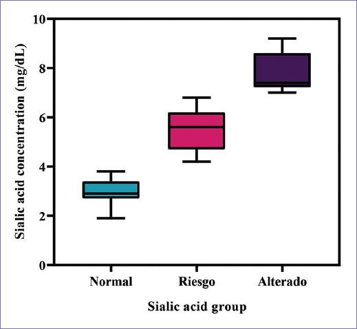
In a survey previously done in San Luis Potosí, S.L.P., by Dr Aida Catalina Hernandez Arteaga et al. "Determination of sialic acid concentration as a biomarker for breast cancer diagnosis on female population," 206 saliva samples were evaluated, 106 from healthy people, and 100 from patients with breast cancer diagnosis. The concentration of sialic acid was determined through SERS method produced by silver nanoparticles reduced with citrate, it was concluded that the SA concentration of those patients in the control group (healthy women) shows a normal distribution with an estimated average measurement of 3.5 mg/dL compared to the patients with breast cancer, where there is a wide dispersion of the concentration that varies from 5 to 40 mg/dL, with an average of 18.3 mg/dL. It is established that salivary sialic acid concentrations higher than 7 mg/dL are an indicator of the presence of a possible cancerous pathology.
Sialic acid
The term sialic acid comes etymologically from the Greek "sialos" which means saliva10. From the diverse natural sialic acids, we can identify as base nucleus N- acetylneuraminic acid11.
N-acetylneuraminic acids are the predominant form of sialic acid and it is almost the only form found in body fluids and human tissue. The chemical formula of sialic acid is C11H19NO9 and it is made by a pyranose ring consisting of five carbon atoms and one oxygen atom, an N- acetyl (N-CH3CO), a carboxyl group (CO-OH), and a glycerol tail (-C3H7O3)12.
In men, they are present in the cerebrospinal fluid, saliva, serum, and urine, but it is more frequently found forming part of the glucidic macromolecular structures such as mucins, glycoproteins and glycolipids. The concentrations of sialic acid in body fluids can reflect the metabolic status and have been recognized as biologic markers for a variety of pathophysiology processes12.
The biologic functions, in which they participate are as follows: synthesis of fundamentally amorphous substance in connective tissue, epithelial secretions, and protecting functions against certain components (antigens), they are part of the cellular membrane, constituting what is known as "cellular recognition mechanism" with connections in the growth processes, senescence, and structural modifications intimately related to pathologic processes such as cancer13.
Sialic acids can be linked in α-2,3 or α-2,6 bond to galactose (Gal) residues, in α-2,6 bond to N-acetyl galactosamine (GalNAc) residues and in α-2,8 bond to another sialic acid residue. The different types of bonds that they form result in a great diversity of structures derived from sialic acid. The transfer of sialic acid to oligosaccharides is catalyzed by a family of enzymes called sialyltransferases (STs), these enzymes are anchored to the membrane of the trans network of the Golgi apparatus. There are 20 known sialyltransferases which show differences in the affinity for the substrate that they recognize and are classified according to the type of bond they form14. In well-differentiated mature cell membranes, sialic acid is normally located at the α-2,3 position. As cells differentiate, the sialyltransferase responsible for achieving this position stops working since the operons of the genes that transcribe for this enzyme are affected, and the glycoprotein begins to express sialic acid at position α-2,6. In certain pathological processes, such as oncogenesis and metastasis, glycosylation alterations occur that are associated with an increase or decrease in its activity. Sialyltransferase α-2,6 is overexpressed in several types of cancer, including breast, colorectal, hepatocarcinoma, and cervical15. Increased sialic acid in the tumor cell membrane has been proposed to promote cell mobility, which could facilitate the separation of cells from the primary tumor14. The determination of sialic acid by SERS is a novel approach technology that requires fewer reagents and could be useful in clinical diagnosis as it is highly sensitive, fast, and inexpensive, the equipment can be portable and the results can be obtained in real time12.
Raman spectroscopy
Raman spectroscopy is a photonic technique used to obtain chemical and structural information on various substances in a few seconds. It is based on the measurement of the light scattered by a material, on which a monochromatic beam is impinged16,17.
Physical principle
The analysis consists of striking a beam of monochromatic light of frequency n on a sample whose molecular characteristics are to be determined. The photons of the incident beam can experience elastic collisions with the atoms of the sample without loss of energy and, therefore, do not provide any information, and inelastic collisions, in which energy transfer occurs that modify the frequency of the scattered photons that form the called Raman scattering, which contains information about the composition and structure of the sample, because the molecules are excited to a different vibrational state than they had before the shock. The energy difference between the incident and dissipated photons is analyzed with an optical spectrometer to generate the vibrational "Raman" spectrum which is unique to each composite type and serves as a "fingerprint" to identify it. The difference in wavelengths between incident and scattered radiation corresponds to the wavelengths of the mid-infrared region, which is between 780 and 3000 nm. Furthermore, since it is only irradiation with light, the technique is not aggressive, which is why it is said to be non-destructive and can be carried out in the environment17.
Amplified Raman spectroscopy on "SERS" surfaces
The SERS effect consists of an increase in the inelastic scattering (Raman signal) of light from certain molecules in the presence of a smooth or rough metallic nanostructure. The Raman scattering of a molecule can be enhanced when placed close to a metallic nanoparticle, leading to the so-called SERS effect. SERS measurements of small molecular systems lead to signal enhancements ranging from 106 to 1012 due to the strong local field arising from plasmon excitation, called the electromagnetic (EM) mechanism, combined with the direct chemical interaction between the molecule and the surface metallic. The SERS effect is determined by three main components: the molecule, the metallic nanostructure, and EM radiation. The absorption and scattering of light by metallic nanoparticles are considered their most important property. The intensification factor due to the SERS effect depends on: the shape and size of the nanoparticles and the distance at which the molecule is on the metal surface (1-10 nm away) since it is a short-range technique12.
Methodology
Study subjects
Sialic acid concentrations were evaluated in saliva samples from 120 women divided into four groups. The positive control consists of 30 patients diagnosed with breast cancer in its different stages, for which the concentration of AS was previously determined in the gynecology division of the Central Hospital "Ignacio Morones Prieto." The control group consists of 30 women who did not use hormonal contraception. The group of women who used hormonal contraceptives was divided into two groups according to the time of hormone intake as follows: intake < 4 years (n = 30) and intake > 4 years (n = 10). The inclusion criteria were: age over 18 years, presence of menstruation, signing of the informed consent, and prior completion of the survey. The exclusion criteria are: diagnosis of diabetes mellitus, arterial hypertension, autoimmune diseases, inflammatory processes, pregnancy or lactation, consumption of anti-inflammatory drugs, and corticosteroids in the past 3 months. The elimination criterion was voluntary withdrawal from the study.
Sample collection
To collect the saliva sample, each volunteer was instructed to perform an oral cleaning that consisted of vigorous tooth brushing and then wait 3 min before depositing 1.0-1.5 mL of saliva in a sterile container. The samples were stored at a temperature of 4°C until use.
Raman-SERS spectroscopy
Raman-SERS measurements were performed using a HORIBA Xplora Plus Raman spectrometer, with the solid-state green laser, 532 nm wavelength, at an estimated power of 5 mW on the sample. The spectrometer is attached to an Olympus BX41 light microscope. The signal is recorded by a CCD (charge coupled device) cooled to −48°C. The laser beam is focused on the sample using an Olympus 10 × objective to give a spot size of approximately 3 μm on the sample. The data acquisition time is 20 s/scan, collecting four coaggregated scans. Light scattering is done using an 1800 slots/mm monochromatic grating.
Synthesis and characterization of metallic nanoparticles (NPs)
The synthesis used is based on the Turkevich method, it involves the reduction of silver ions present in an aqueous solution with sodium citrate. Preparation by chemical reduction consists of two fundamental parts: reduction and stabilization. In relation to stabilization, it is important to maintain the monodisperse state of the NPs. Due to the hydrophobic nature, metallic NPs tend to aggregate into larger complexes by Van der Waals forces. The stabilizing agent contributes negative charges that induce repulsive forces between the particles.
Synthesis of nanoparticles
The reduced citrate NPs-Ag silver nanoparticles were obtained using the Turkevich method, a solution of silver nitrate (2.5 mM) in deionized water (DI) which is heated to 95°C, and a solution of trisodium citrate (2.5 mM). After 15 min, the color of the solution turned grayish-yellow, which indicated the formation of nanoparticles, as seen in the image. After cooling the solution to room temperature, the NPs-Ag obtained were washed by centrifugation at redispersion cycles with DI and stored in the dark until use.
Characterization of nanoparticles
Scanning electron microscopy (SEM) is a technique that allows the structural and morphological characterization of solid materials. For the analysis of NPs-Ag by electron microscopy, a single drop of the sample solution was deposited on a commercial copper grid (lacey carbon formvar). To characterize the morphology and chemical composition of the nanostructures by SEM, a Hitachi STEM-5500 microscope equipped with bright field, STEM mode, and dark field detectors was used. The SEM/STEM microscope has a field emission gun with a spatial resolution of 0.4 nm operated at 30 kV.
Dynamic light scattering (DLS)
DLS is a physicochemical technique used to determine the size distribution of suspended particles. When laser light reaches the many particles in a suspension, it is scattered in all possible directions. If the particles are moving rapidly (small particles), the variation of the scattering intensity is also accelerated. In contrast, slow (large) particles lead to slower variations18.
The size and size distribution of the silver nanoparticles was determined by DLS with the Nano Zetasizer Malvern, model ZEN 3600.
Zeta potential
The zeta potential is a property of materials that measure the electrokinetic potential in colloidal systems; it is usually denoted by the Greek letter ζ19. It indicates the degree of repulsion between adjacent particles, charged in a dispersion20. The importance of the zeta potential is: that its value can be related to the stability of colloidal dispersions. For molecules and particles that are small enough, a high zeta potential confers stability, that is, the solution or dispersion will resist aggregation19.
The Z potential of the silver nanoparticles was determined by DLS with the Nano Zetasizer Malvern, model ZEN 3600.
Sialic acid calibration
For reference, the RAMAN spectra of N-acetylneuraminic acid (Neu5Ac) in solid state, AgNPs colloid, Neu5Ac solution in DI at 200 mg/dL, and solution prepared from AS with NPs-Ag were taken (Fig. 1).

Figure 1 Reference spectra, the contrast of Raman signals (Sialic acid [SA]-Solid) and SERS (AS 200 mg/dL-AgNPS) of the SA is observed. Origin 2018.
The reference Raman spectra were analyzed using AS in solid state, a solution in DI of AS at 200 mg/dL, the colloid of NPs-Ag, and finally the combination of our substrate SERS (NPs-Ag) with the Solution of AS at 200 mg/dL previously measured. Analyzing the spectra obtained, an increase in the intensity of the signal was obtained by combining our substrate together with the AS solution.
In calibrations of the AS at different concentrations with NPs-Ag, commercial sialic acid concentrations were measured at 1, 5, 10, 20, 50, 100, 150, and 200 mg/dL.
All samples were deposited on an aluminum slide with 12 wells, each with a capacity of 75-80 μL, of which a 2:1 mixture of NPs-Ag with AS was used, respectively. The SERS spectra were analyzed obtaining an increase in the intensity of the signal according to the concentration of AS. SERS spectra were collected in the 400-1800 spectral range as an average of four consecutive 20 s laser exposures. The fluorescence background was removed using the Vancouver algorithm.
Processing saliva samples
The collected saliva was centrifuged at 6000 rpm. The supernatants were extracted and mixed with NPs-Ag to later bring them to Raman and thus determine the concentration of AS. Unused sample portions were stored refrigerated at 4°C. To obtain SA concentrations in saliva, SA was re-calibrated in concentrations to be able to compare SA level estimates in saliva samples under the same instrumental conditions.
Statistical analysis of results
The means, frequencies, percentiles, and maximum and minimum, respectively, were determined. Subsequently, the data analysis was carried out to evaluate the type of distribution, finding that they met the parameters to adjust to normal behavior, for which an analysis was carried out with tests for parametric data. The t-student test was used to evaluate the relationship between sialic acid concentrations and contraceptive use, as well as type, timing, and risk factors. Statistical analysis was performed with the SPSS version 1.8 package.
Results
For the characterization of silver nanoparticles by SEM, their morphology, arrangement, and size difference were observed. The size and distribution of the Ag-NPs were determined by DSL, for the first peak a size of 95.45 nm and an intensity of 86.1% was obtained, in the second peak a size of 7.133 nm and an intensity of 13.9, the average size was 40.45 nm.
The zeta potential was measured, which refers to the degree of attraction or repulsion between the particles. A graph was obtained, in which 3 peaks stand out, the mean zeta potential of peak 1 was −36.3 mV and area of 68.1%, in peak 2, it was 65.3 mV, and area of 24.6%, in peak 3, it was −9.49 mV and area of 6.7%; the general average obtained was −42.2 mV with a standard deviation of 18.8 mV.
When analyzing the SERS spectra, an increase in the intensity of the signal was obtained according to the concentration of AS. In figure 2, we can observe a stack, or separate spectra of each of the SA solutions (a) and the enhancement contrast in the SA concentrations (b).

Figure 2 Stack of the proposed solutions of Sialic acid (A) and comparative in intensities (B). Origin 2018.
For this study, 30 saliva samples from women who did not use hormonal contraceptive methods and 40 from women who did use them were analyzed using the Raman-SERS technique (30 with a period of < 4 years and ten with a period of more than 4 years). The age range of the volunteers was from 18 to 40 years with a mean of 23 years, their body mass index (BMI) ranged from 17.7 to 45 Kg/m2 with an average of 27.4 Kg/m2, the age of menarche was from 9 to 19 years with an average of 12.27 years; finally, the concentrations of SA obtained were from 1.9 to 9.2 mg/dL, with a mean of 5.17 mg/dL.
For a better visualization of the sialic acid concentration data, they were divided into three groups, concentration values < 4 mg/dL were considered normal, 4-7 mg/dL at risk and > 7 mg/dL altered (Fig. 3). The aforementioned groups are called "Sialic Acid Group."
Discussion
In the characterization of silver nanoparticles, there were differences in size. Hernandez et al. (2018) carried out AS concentration measurements using nanoparticles of different sizes and distributions, with this, they affirm that the reproduction of the methodology is good even when the synthesis of NPs-Ag are not identical, given that the difference in results in the concentration of AS between one measurement and another, it presents reliable results when the mean values overlap within the experimental uncertainties of the method. According to the zeta potential obtained, Mayoral et al. (2014) describe that the low zeta potential favors the attraction between the particles, thus overcoming the repulsion and forming flocs, instead of dispersion, on the other hand, the high zeta potential colloids are electrically stabilized. Furthermore, the stability of the dispersions depends on the balance between the repulsive electrostatic charges on the colloidal particle and the Van der Waals attractive forces.
Chamorro et al. (2012) mention that, for every 100,000 women, the use of hormonal contraceptives is the cause of 13 more cases of breast cancer annually. In other words, for every 100,000 women who use them, there are 68 cases of breast cancer per year, compared to 55 cases/year among those who do not use them. Thus, corroborating that hormonal contraceptives are a risk factor for breast cancer. As is the case in this study where the concentrations of sialic acid for the volunteers who do use these contraceptives, the range its between normal values (< 4 mg/dL) and altered values (> 7 mg/dL). This may be due to the fact that hormones can become promoters in the carcinogenesis process, in which they activate the altered expression of proto-oncogenes to oncogenes, which lead to the inactivation of tumor suppressor genes, which generate an increase in the division of cancer cells which is a known common denominator in the pathogenesis of various types of cancer in humans21,22.
In figures 4 and 5, it can be deduced that the type of contraceptive has a greater influence compared to the temporality, due to the hormone concentrations handled by the various presentations found on the market, thus resulting in higher concentrations of AS, the Volunteers using the subdermal implant, contraceptive pills, and hormone-impregnated intrauterine devices (IUS), which have concentrations of: 36 mg of levonogestrel and 68 mg of etonogestrel for subdermal implant presentations, 30 μg of levonogestrel for pills of according to NOM-005-SSA2-1993, and 52 mg of levonogestrel for the intrauterine device (López-Olmos, 2014); all of these releasing a certain amount of hormones into the body daily23,24.

Figure 4 Box plot of Sialic acid concentration as a function of time of use of contraceptive methods. GraphPad Prism 8.
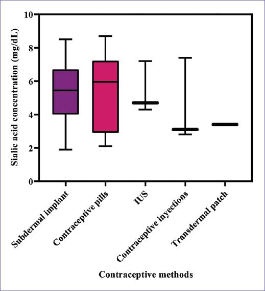
Figure 5 Box plot of Sialic acid concentration as a function of contraceptive methods. GraphPad Prism 8.
Narod et al. (2011) describe that hereditary breast cancer accounts for approximately 5-10% of all breast cancer cases. Hereditary cancer syndromes have been described where there are germline mutations, including mutations in the BRCA1 and BRCA2 genes. The determination of these genes is not performed frequently in most Latin American countries, in contrast to other countries such as the United States, Canada, Poland, Israel, and some of Western Europe, in which they are part of the study battery for evaluate patients who may potentially have hereditary breast cancer. BRCA1 and BRCA2 are tumor suppressor genes that encode proteins that function in the DNA repair process. Therefore, a mutation or a deletion of a tumor suppressor gene would cause a loss of its function and consequently increase the probability that a tumor will develop. Having a family history of some type of cancer increases sialic acid concentrations above 4 mg/dL (risk group and impaired), which corroborates that it is a risk factor for developing cancer, in this case, breast cancer25.
The genetic factor is very important and women who did not use any hormonal contraceptive method had sialic acid concentrations above normal values (< 4 mg/dL), as well as a history of family members with cancer, so it is very important to remember that, the more risk factors, we have, there is a greater predisposition to suffer from some type of cancer. Similarly, emphasis is placed on the volunteers who use a subdermal implant or pills, because they have a greater number of family histories with some type of cancer and according to their concentrations of AS that they are in the risk group and altered, as described above. With this, we emphasize that the type of hormonal contraceptive used is important.
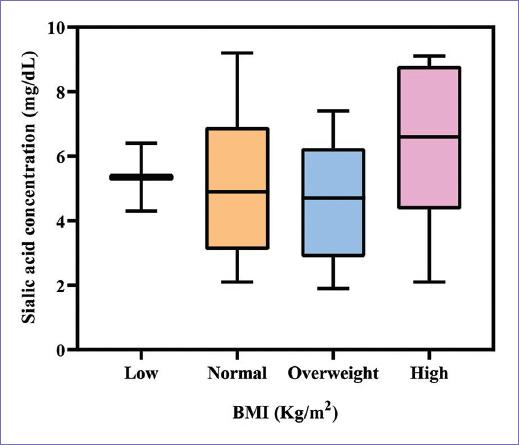
Figure 6 shows the graph of the sialic acid group against the BMI in Kg/m2, those women who presented a high BMI (obesity) are in the risk group or altered. Therefore, we corroborate that a high BMI is a risk factor for presenting high concentrations of AS. It is worth mentioning that, in this study, we used the BMI as a method to estimate the amount of body fat; however, the World Health Organization (WHO) recommends directly using the percentage of body fat, since previous studies have shown its direct relationship with inflammatory processes. Izaola et al. (2015) report that adipose tissue is a metabolically active complex endocrine tissue, which plays a fundamental role on the inflammatory, procoagulant, antifibrinolytic, and vasoactive cascades, which suggests a direct influence on the inflammatory process. In obesity conditions, it has been shown that in approximately 70-80% of individuals there is a remodeling of adipose tissue ("adipose tissue remodeling") both at a structural and functional level that causes an inflammatory reaction. When the resolution of acute inflammation is not resolved correctly, a chronic low-grade inflammatory state is triggered at the local level with local and systemic repercussions known as lipoinflammation.
Conclusions
According to the results obtained, most of the study subjects who presented altered sialic acid concentrations had a family history of some type of cancer and/or a high BMI that classified them as the degree of obesity, which is why it is found that these risk factors have an important role related to breast cancer.
Regarding the time of use, no significant differences were determined between the time of use of hormonal contraceptives; however, regardless of the temporality, the fact of using a hormonal contraceptive method causes an increase in sialic acid. When contrasting the types of contraceptive method used, those who use the IUS already have sialic acid levels higher than 7 mg/dL compared to those who use contraceptive pills or subdermal implants; this is due to the fact that they release a higher concentration of hormones to the body daily. Hence, it is not the time of use, but the type of method according to the amount of hormones released into the body.
For the collection of the samples, emphasis should be placed on their control due to the possible interferences that could arise due to inflammatory processes that the person could have. For this study, we focus on gingivitis, since it is one of the most common inflammatory processes, for which it is proposed to perform an oral examination before sampling and a standardization of the brushing technique performed before sampling, as well as, if possible, ruling out any other inflammatory process so that the result obtained from this biomarker is the real one. It is recommended for later studies to measure the percentage of body fat instead of the BMI, because a person with a normal BMI may have a high percentage of fat, which leads to a possible alteration in the levels of fat sialic acid.
Based on the references and data provided in this study, the importance of identifying modifiable risk factors such as healthy eating habits, physical activity, and monitoring the consumption of steroid hormones is emphasized, all with the aim of to decrease the likelihood of breast cancer.
Finally, the association of sialic acid levels with the use of hormonal contraceptive methods is verified, as well as its relationship with other risk factors for developing breast cancer, such as a history of relatives with some type of cancer and a high BMI.

