


 nueva página del texto (beta)
 Inglés (pdf)
 Artículo en XML
 Referencias del artículo
 Enviar artículo por email
 Citado por SciELO
  Similares en
SciELO
Similares en
SciELO 
 Permalink
Introduction
Corneal diseases are the second cause of blindness in the world. There are about 4.9 million people with bilateral corneal blindness worldwide. Thanks to the improvement of microsurgical techniques and the development of eye banks, a large number of patients can have their eyesight restored through corneal transplantation. There are currently two ways of storing corneal tissue: by short-term hypothermic storage of 2oC to 8oC degrees (on average 4°C) that preserves the corneal tissue for up to 14 days; and by organ culture of 31oC to 37°C that preserves the corneal tissue for up to 35 days. Hypothermic storage is the most commonly used method in the US and Asia, because the cost of acquisition is low compared to organ culture which is the most commonly used method in Europe. Hypothermic storage has the additional advantage of being easy to use and not requiring expensive storage equipment1-3.
The main purpose of corneal preservation media is to maintain the viability of the endothelial cells from the time of collection until the corneal tissue is transplanted. For a cornea to be transplanted for optical purposes, it must have an endothelial cell count of at least 2,000 to 2,200 cells per square millimetre, in accordance with the European Eye Bank Association (EEBA) guidelines4-6.
There is a natural loss of endothelial cells in hypothermic corneal storage media, ranging from 3-9% at 7 days and 15-20% at 14 days according to various studies7-9. These chondroitin sulphate and dextran-based corneal preservation media extend the tissue preservation period to a maximum of 14 days (intermediate-term preservation)10.
However, most surgeons use corneal tissue preserved during the first week, a practice that prevails despite the approval by the FDA (Food and Drug Administration) of a maximum preservation period of 14 days since 199011,12.
The following preservation media are currently available: McCarey-Kaufman medium (Aurora, Buffalo, New York), Cornisol medium (Aurolab, Madurai, India), Life 4°C medium (Numendis, Isanti, Minnesota), Chen medium (Chen Laboratories, Phoenix), Optisol-GS medium (Bausch & Lomb, Inc., Rochester, NY, USA), and Eusol-C medium (ALCHIMIA, Padua, Italy)11,13-15.
The most widely used intermediate-term corneal storage medium in the world is Optisol-GS (Bausch & Lomb, Inc., Rochester, NY, USA), introduced in 1990. This preservation medium contains chondroitin sulphate, non-essential amino acids, ascorbic acid, vitamins, purines, lipids, gentamicin and streptomycin16.
Eusol-C is an intermediate-term hypothermic corneal storage medium, enriched with vitamins, antioxidants and energy precursors, containing gentamicin sulphate (143 μg/ml) as an antibiotic and a red phenol marker to detect pH changes15,17.
Eusol-C was suggested in 2005 by the Federated Scientific Session, Chicago, Illinois, at the Cornea Society and Eye Bank Association of America conference, as an alternative as effective as Optisol-GS in terms of maintaining donor tissue viability, but there are few studies comparing its efficacy to that of Optisol-GS15.
These two corneal preservation media differ in their chemical composition. Optisol-GS contains chondroitin sulphate, non-essential amino acids, ascorbic acid, vitamins, purines, lipids and streptomycin, which are not found in Eusol-C. On the other hand, glutamine and recombinant human insulin are substances found in Eusol-C but not in Optisol-GS16.
Optisol, created by Kaufmann in 1991 and Optisol-GS to which gentamicin sulphate 0.1 mg/ml and streptomycin sulphate 0.2 mg/ml are added, also includes the phenol red marker, but is devoid of antifungal additives. This medium preserves the corneal endothelium for two weeks by combining chondroitin sulphate and dextran17,18.
There is limited evidence comparing Eusol-C to Optisol-GS as an intermediate-term corneal preservation medium. Two published studies compared Eusol-C to Optisol-GS as a corneal preservation medium with transplant-suitable corneas, and found no significant changes in endothelial viability between those two16,19.
The efficacy of Eusol-C as a corneal preservation medium is somewhat controversial, since in the two studies mentioned above there was no significant difference in endothelial viability. However, in the study by Yüksel17 where the efficacy of Eusol-C was evaluated in terms of endothelial viability, a reduction in endothelial cell numbers of 3.1% per day was found during the first week, 24.5% at 8 days and a 50% loss of endothelial cells at 14 +/- 3 days. It is worth mentioning that this study used corneas that were not suitable for transplantation, mainly because they were positive for hepatitis B, HIV or sepsis, which could have been the cause of the greater endothelial loss in these tissues.
Currently, there are no studies comparing these two methods of corneal preservation in Mexican donors. For this reason, as well as the reasons mentioned above, it is important to conduct a study that evaluates endothelial cell viability comparing these two methods that are widely used worldwide.
The main objective of this study was to evaluate the comparative efficacy of two intermediate-term corneal preservation media on the survival of endothelial cells from donated tissues.
Materials and methods
A prospective, longitudinal and comparative study was carried out in the eye bank of Hospital General de México "Dr. Eduardo Liceaga".
Inclusion criteria
Corneoscleral tissue collected from the Hospital General de México was included in the study, where the tissue was obtained from both the right eye (stored in Optisol-GS) and the left eye (stored in Eusol-C) of the same donor suitable for transplantation (Fig. 1). With informed consent by the family, specular microscopy was performed on the same day of extraction and five days later with a cell count greater than 2000 cells/mm² for corneal transplantation for optical purposes.

Exclusion criteria
Cases of ineligible donors due to systemic or ocular pathology and high-risk groups were excluded, as was obtaining a single cornea from the donor.
Corneoscleral disc removal technique
Asepsis and antisepsis of the eyelids and periocular region were performed with 10% iodine solution for three minutes. The donor's face and head were covered with surgical drapes, leaving the eye region uncovered. Sampling from the lower conjunctival fornix was obtained, a topical antibiotic (fourth-generation fluoroquinolone) was applied to the lower conjunctival fornix and a further lower conjunctival fornix sample was collected. A blepharostat was placed, a 360-degree limbal peritomy was performed, and the sclera was scarified and incised 3 mm from the limbus. Then, a cut with 360-degree handle corneal scissors and the corneoscleral disc was carefully separated from the iris and choroidal tissue with two forceps. A conformer was then placed and the eyelids were covered with cyanoacrylate. The corneoscleral tissue was stored in the corresponding preservation medium with the endothelium facing upwards. The right corneal scleral tissue was placed in Optisol-GS preservation medium and the left corneal scleral tissue from the same donor was placed in Eusol-C preservation medium. It was then stored in a cooler with a coolant gel pack at a temperature of 2 to 8oC and refrigerated at 4oC for transport to the eye bank.
The member of staff carrying out the procedure (only highly-trained eye bank staff) wore a surgical cap and mask, as well as a sterile gown and gloves. The procedure took place in the autopsy room at Hospital General de México.
Specular microscopy
Each cornea was examined under a specular microscope (Konan Eye Bank Keratoanalyzer EKA-10, Hyogo, Japan) on the same day as tissue was collected from the right and left corneas and all were assessed under the slit lamp for Descemet's membrane folds, haematic debris and epithelial defects (Fig. 2). Both storage media were kept refrigerated at 4°C in the eye bank, and on day five, both corneas were again examined under a specular microscope. Then, if the corneas were suitable, they were used for optical or tectonic corneal transplantation. Otherwise, they were excluded and sent to the pathology department for final disposal.

Donor's sex, age and co-existing ocular pathologies were recorded. Endothelial cell count (number of cells per square millimetre) was the primary assessment variable. Changes in the coefficient of cell variation (standard deviation divided by mean cell area) were recorded as an index of the extent of variation in cell area and hexagonality (variation in cell shape, determined by the percentage of hexagonal cells) (Figs. 3 and 4). All data was recorded in an Excel spreadsheet.

Figure 3 Initial microscopy and on the fifth day after taking the right corneoscleral tissue, preserved in Optisol-GS medium.
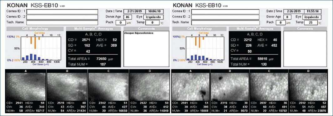
Figure 4 Initial microscopy and on the fifth day after taking the left corneoscleral tissue, preserved in Eusol-C medium.
The data was analysed using descriptive statistics (mean, median and standard deviation). Non-parametric statistics and the Kolmogórov-Smirnov test were performed as well as comparison of the means of related groups using the Wilcoxon test.
Results
In total, 40 corneas were analysed. Twenty corneas were stored in Optisol-GS and the other 20 in Eusol-C. The average age of the donors was 43.85 years, with a range of 17 to 67 years, 5 women and 15 men.
Optisol-GS
The mean endothelial cell count in the group of corneas stored in Optisol-GS medium on the day of tissue harvest was 2598.118, SD 358.976 cells/mm². The coefficient of cell variation was 44.167, SD 5.512 and cell hexagonality was 46.111, SD 7.177 on repeat specular microscopy on day 5. The reported means for endothelial cell count were 2386.389, SD 390.554 cells/mm², the coefficient of variation was 44.889, SD 11.182 and cell hexagonality was 42.889, SD 10.532. The decrease in endothelial cell count at day 5 was 8.149%, the increase in cell variation was 1.634% and decrease in cell hexagonality was 6.988%.
Eusol-C
In the group of corneas stored in Eusol-C, the mean endothelial cell count on the day of tissue collection was 2819.588 SD 491.221 cells/mm², the coefficient of cell variation was 41.444 SD 7.445 and cell hexagonality was 48 SD 5.351. When a new specular microscopy was performed on day 5 after tissue removal, the mean endothelial cell count was 2604.882 SD 529.858 cells/mm², the coefficient of variation was 45.150 SD 7.428 and cell hexagonality was 43.450 SD 7.007. The decrease in endothelial cell count five days after extraction was 7.614%, increase in cell variation was 8.942% and decrease in cell hexagonality was 9.480%.
The Kolmogorov-Smirnov test was performed on the Optisol-GS group and the Eusol-C group. It showed that the distribution of the means of both groups behaved normally at the first and fifth day assessments in both groups (Table 1).
Table 1 Kolmogorov-Smirnov test performed on the Optisol-GS group and the Eusol-C group at baseline and on day 5 after corneal tissue collection
| Optisol-GS | Day 0 | Day 5 |
|---|---|---|
| Endothelial cell count | 2618.15 + 349.22 | 2423.05 + 395.31 |
| Coefficient of variation | 44.6 + 5.38 | 44.7 + 10.77 |
| Hexagonality | 45.65 + 6.98 | 43.65 + 10.23 |
| Eusol-C | Day 0 | Day 5 |
| Endothelial cell count | 2820.35 + 482.73 | 2676.05 + 555.45 |
| Coefficient of variation | 41.05 + 7.14 | 45.15 + 7.42 |
| Hexagonality | 48 + 5.35 | 43.45 + 7.00 |
Once the normality of the distribution had been corroborated, the Wilcoxon test was used to compare the results of the means obtained between Optisol-GS and Eusol-C on the first and fifth days of tissue collection. The endothelial cell count, coefficient of variation and cell hexagonality between the two media showed no statistically significant differences (Table 2).
Discussion
Hypothermic corneal storage media are widely used in the world, as they are easy to obtain and apply, making them an attractive method for corneal tissue preservation15. Donated corneas must remain in the preservation medium during their study, until the time of transplantation. In Mexico, this takes an average of five days. It may take longer for imported corneas. With each day that a cornea is stored in corneal preservation medium, the endothelial cell count decreases. This is important because the endothelial cell count is the most important parameter for deciding whether a cornea is suitable for transplantation20.
In our study, we observed that the reduction in the number of endothelial cells was similar between the two storage media, although Eusol-C medium had a lower percentage of endothelial cell loss, 7.614% at 5 days, compared with Optisol-GS medium which had a loss of 8.149% p=0.006.
Regarding the coefficient of variation on day five, Optisol-GS and Eusol-C had an increase of 1.634% and 8.942% p= 0.948. Finally, cell hexagonality on day five in Optisol-GS medium had a decrease of 6.988% and in Eusol-C medium, it had a decrease of 9.480% p= 0.936.
There have been several reports of endothelial cell loss in Optisol-GS medium, with one of the earliest dating from 1995. Means et al21 reported endothelial cell loss of 0.57 ± 0.30 cells per day, with average cell damage of 9.5% in the first week, 11% in the second week and 16% in the third week.
Nelson et al22 in a comparative study between Optisol-GS medium and Chen medium, reported a loss at 7 days of 5 ± 5% of basal endothelial cells in Optisol-GS medium and 11 ± 10% in Chen medium, as well as an increase in the coefficient of cell variation.
In 2003, in a comparative study between Optisol-GS medium and Eusol-C medium, Camposampiero et al19 reported a decrease in average cell density during the first week in Optisol-GS medium of 6.2 ± 7.4% and in Eusol medium of 6.9 ± 10.4%. However, the number of elapsed days between the two media is not comparable, as for Optisol-GS, the number of elapsed days is 6.1 ± 1.9 days, and for Eusol-C, the number of elapsed days is 1.6 ± 1.1 days.
In 2015, Kanavi et al16 compared Optisol-GS with Eusol-C medium, with two determinations at day 1 and day 7, without finding significant changes in mean endothelial cell count, coefficient of variation or cell hexagonality. They considered Eusol-C medium to be a suitable substitute for eye banks working with a short time interval between preservation and transplantation.
Yüksel et al17 in a study of non-transplant corneas preserved in Eusol-C medium reported an average endothelial loss of 3.1% per day in corneas preserved from day 9 to day 24, with a 24.5% loss of endothelial cell viability after day 8 of preservation and a 50% loss of viability on day 14 of preservation.
In 2022, Gimenes et al23 compared Optisol-GS medium to seven different corneal preservation media in a review article: Chen, Cornea Cold, Cornisol, Dexsol, Kerasave, Sinasol and Eusol-C media. They found that most of the corneal storage media had similar performance in preserving corneal tissue after seven days. However, when the preservation time is longer than 10 days, Cornisol is superior to Optisol-GS (p=0.049).
In our study, we reported the number of endothelial cells lost in the first five days in both media at comparable times and with corneas suitable for transplantation and found that the number of viable endothelial cells in Eusol-C medium is slightly higher than those preserved in Optisol-GS medium. However, a sample with a larger number of corneas is needed to confirm these results and statistically evaluate the differences between the two preservation media.
If these results are confirmed, Eusol-C could be used more frequently as a storage medium. It has the added advantage of being a cheaper storage medium compared to Optisol-GS16.

