


 nueva página del texto (beta)
 Inglés (pdf)
 Artículo en XML
 Referencias del artículo
 Enviar artículo por email
 Citado por SciELO
  Similares en
SciELO
Similares en
SciELO 
 Permalink
Introduction
Reconstruction of the lower lip in total defects or > 90% implies extensive knowledge of facial anatomy and surgical techniques in the reconstructive algorithm to preserve the sphincter function of the mouth.
The main cause of total defects of the lower lip is oncological resections, mainly caused secondary to the appearance of squamous cell cancer, which represents more than 90% of the types of lip cancer. Squamous cell cancer presents as ulcerated and exophytic lesions, slow-growing, with potential lymph node spread in the submandibular and submental regions. Its diagnosis is clinical and histopathological and should be complemented to the diagnostic approach with computed tomography images for lesions larger than 2 cm. The treatment is surgical resection with negative margins, and depending on the clinical stage, a lymphatic resection will be performed toward the corresponding lymph node replacement, and adjuvant chemotherapy and radiotherapy1.
The classic reconstructive algorithm will depend on the size of the defect and location, with oral competence and plane reconstruction as its main objective2.
The reconstructive algorithm in lower lip defects is summarized in: small defects (< 30% or 1/3 part), intermediate (30-60% or up to 2/3 parts), and total defects (> 90%), and its management is with direct closure options, local flaps, and free flaps, respectively.
For intermediate defects (2/3 parts), the option is local flaps, the most used are those defects that involve the commissure: the Eastlander flap (interpolation flap, full thickness, triangular in shape, based on the labial arteries, and donor site the oral commissure), and in central lesions the Abbe flap (same characteristics as the Eastlander with a central donor site), including modifications thereof. In major central defects, the Karapandzic, Gillies, or Webster-Bernard flaps (Bernard1) are used.
The gold standard for total lower lip defects is the radial free flap with graft harvesting from the palmaris longus covered by it, providing internal and external coverage, and continuity of the orbicularis oculi muscle3.
To present an alternative for total lower lip defects comparable to the gold standard, a review of reconstructive techniques is carried out. The technique described by Fujimori is optimized, in which internal and external coverage is achieved with preservation of oral continence and restoration of the continuity of the orbicularis oris muscle, providing less morbidity, and adequate functional and esthetic results.
Material and methods
Three elderly male patients recruited by the Head and Neck Surgical Oncology service of the General Hospital of Mexico “Dr. Eduardo Liceaga” with a clinical Stage III diagnosis of epidermoid cancer in the entire lower lip (It is the only inclusion criterion of the study). The patients were candidates for management by means of surgical resection and lymph node dissection and were recruited by the Plastic and Reconstructive Surgery service of the same Hospital, a reconstructive surgical plan was given.
Reconstruction is performed using a Gate flap or Fujimori flap with a modification to the original technique described by Ryosuke Fujimori (Fujimori4). An unilateral musculocutaneous flap based on the facial artery, with restoration of the oral sphincter, was performed. The same work team performs surgical procedure.
Results
Three elderly male patients are presented: 62, 60, and 70 years old, diagnosed with clinical Stage II epidermoid cancer, candidates for oncological resection, and immediate reconstruction of the entire lower lip (Fig. 1).

Figure 1 Patients diagnosed with clinical stage III epidermoid cancer, candidates for oncological resection and immediate reconstruction of the entire lower lip.
Surgical technique
Anatomical references are taken; the path of the facial artery is identified and marked, checking it with a portable 8 mHz Doppler. Subsequently, including the artery, the dimensions of the flap are drawn, taking into account the size of the defect resulting from the oncological resection in both axes, its upper limit will be the piriformis fossa and the lower one or pivot point even up to the mandibular ridge (Fig. 2). The oncosurgical team performs the tumor resection, in the first stage (Fig. 3). Subsequently, the second stage is performed, which consists of dissection in the cephalocaudal direction, performing a musculocutaneous flap, partially including the levator labii muscle and zygomaticus minor and major. The facial artery is ligated at the upper limit of the flap and included, preserving the buccinator muscle and oral mucosa. Along its course, the superior labial artery will be ligated and the flap is rotated 90° (Fig. 4).

Figure 2 The surgical marking of the gate flap described by Fujimori in 1980 is shown. Bilateral marking is performed and intrasurgical, the decision is made as to which side to use at the expense of oncosurgical margins and vascularity of the donor site.
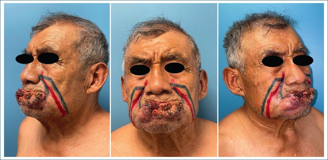
Figure 3 Patients in the immediate postoperative period of oncological resection showing defects involving the entire lower lip.
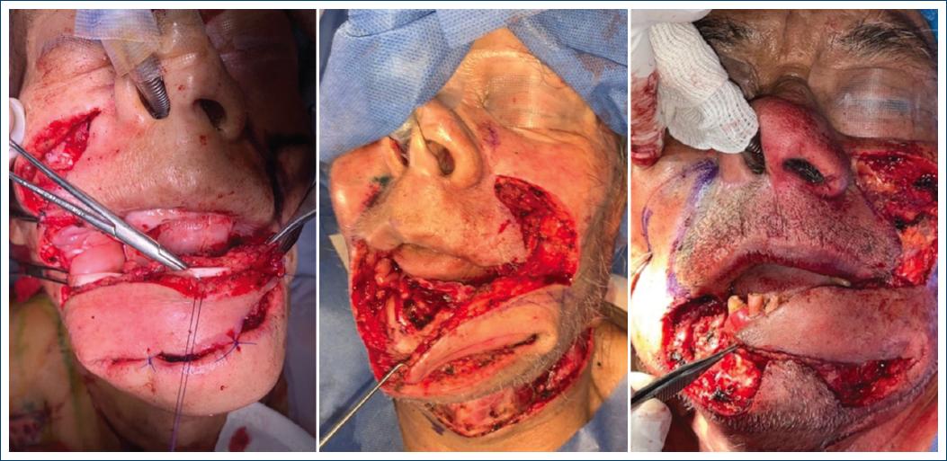
Figure 4 Trans-surgical images when performing a 90° rotation of the flap. Left image: the palmaris longus tendon can be seen passing through a bridge made subcutaneously in the flap.
Next, the Palmaris longus tendon is taken and sutured with two simple stitches with 3-0 nylon to the upper portion of the ipsilateral orbicularis muscle and a pulley system is made in the modiolus, checking the traction of the orbicularis. Continuing, a bridge is made through the flap between the subcutaneous cellular tissue and its muscular portion until a new pulley system is created in the contralateral modiolus, and it is concluded by fixing it with 3-0 nylon to the contralateral orbicularis oculi muscle, with two points simple. Traction of the tendon graft is checked to ensure that the sphincter function of the mouth is adequate. The bloody area of muscle tissue in the oral cavity is covered with a full-thickness graft, taken in our case from the inguinal region, fenestrations are made to avoid seroma and hematomas, and it is fixed on the periphery with simple 4-0 polyglactin sutures. It is concluded by placing a nasogastric tube to protect the integrity of the graft in the oral mucosa (Fig. 5).

Surgical time for surgical resection and supraomohyoid neck dissection for oncosurgery was 90-120 min. Second surgical time modified gate flap is performed in 100 min with two surgical teams. Post-surgical management: a nasogastric tube is placed and oral fasting and enteral feeding are indicated for 7 days, then oral administration is restored with a soft consistency diet.
Follow-up
The first two patients progressed adequately and did not present complications associated with the flap or the graft and adequate oral competence (Fig. 6). The third patient (70 years old) died at the 4th month due to complications associated with oncological pathology.

Discussion
The gold standard for coverage of defects > 90% is free flaps, with the Chinese flap using the palmaris longus tendon being the main one3.
Traditional local flaps for coverage of total or subtotal defects such as the Karapandzic flap5 (bilateral lip advancement myocutaneous flap, with a design of continuation of the lip defect toward the nasolabial folds) Gillies flap6 (or Fan-flap, which consists of advancing a full-thickness unilateral flap from the lip where the resection was performed, continuing the incision towards the labiomental sulcus and continuing over nasolabial fold) and Webster-Bernard7 (modification of the Bernard flap, being a full-thickness flap, advanced, with excision of skin triangles based on the nasolabial fold, extending to the cheek, and moving them medially until they meet the contralateral flap, together with Webster’s contribution with the excision of four triangles of cheek skin to allow advancement) provide adequate coverage of the defect, however, without preservation or partial preservation of the function, and obtaining as a result the main adverse effect: the microstoma.
The flaps described by Alic et al.8 (modification of the Fujimori flap as it is unilateral full-thickness) and Gürel et al.9 (carrying it out unilaterally or bilaterally as full-thickness Fujimori, with marking on the oral mucosa respecting papilla of the Stenon duct, in order not to injure it) with modifications of the original Fujimori technique, provide skin coverage without achieving oral continence or restoration of the sphincter of the mouth and represent a high risk of injury to the Stenon duct by including the mucosa oral, just as they do not give continuity to the oral sphincter.
There are reconstructive options with regional flaps such as the pectoral, deltopectoral, and mental flap for coverage of large defects; however, the esthetic and satisfaction result is affected from our point of view, as well as the impossibility of restoring oral competence.
This surgical proposal provides less morbidity of the donor site and less risk of complications associated with reconstruction, under the following premises: (1) it is unilateral, (2) the oral mucosa and buccinator muscle are respected, abolishing the risk of injury to the Stenon duct, (3) the length of the orbicularis muscle is restored by replacing it with a palmaris longus graft to preserve sphincter function, and (4) microstomia is avoided by not involving subunits of the lip for reconstruction.
Our technique is reproducible and requires an adequate knowledge of the anatomy of the face and neck, in addition to a close relationship with the oncosurgical team to preserve the facial artery.
It can be considered in results even superior to the current gold standard since it creates less donor site morbidity and provides the same oral competence with the use of the tendon graft, adding the pulley system.
Conclusion
The reconstructive gate flap option modified by the authors implies less morbidity as it is performed unilaterally, without presenting risk of injury to the Stenon duct; in addition, to recreating the oral sphincter through the pulley system with the palmaris tendon graft compared to the other techniques, where the main benefit is the coverage of the defect and not the oral competence that is achieved through this flap. In addition, we managed to avoid the most notorious adverse effect of the total reconstructions of the entire lip: the microstoma, by respecting the integrity of our upper lip and the mucosa of our cheeks. The degree of satisfaction of the patients and the surgical team was favorable.
We consider the need to include a larger population to carry out an adequate statistical analysis of morbidities, complications, and evolution to have convincing evidence to support our surgical proposal as the gold standard for sequelae of this nature.

