


 nueva página del texto (beta)
 Inglés (pdf)
 Artículo en XML
 Referencias del artículo
 Enviar artículo por email
 Citado por SciELO
  Similares en
SciELO
Similares en
SciELO 
 Permalink
Introduction
Herpesviruses that predominantly infect psittacines (e.g., parakeets, parrots, macaws, and cockatoos) belong to the subfamily Alphaherpesviridae and the genus Iltovirus. These viruses are double-stranded DNA with a viral tegument and envelope.1 Through their replication cycle, herpesviruses can establish latent infections and reactivate spontaneously; as a result, infected individuals may serve as lifelong reservoirs of the virus.2,3 The initial herpesvirus infection is acute and usually occurs in cells or epithelial tissues where virus replication is efficient.3,4 Four genotypes of herpesvirus have been identified so far, including virulent and non-virulent strains, all of which have the potential to cause disease.5 Most disturbingly, infected birds can act as asymptomatic carriers of herpesvirus. For example, the Patagonian Conure (Cyanoliseus patagonus), Nanday Conure (Nandayus nenday), and golden parrot (Aratinga solstitialis) have been implicated in outbreaks of herpesvirus infection without showing signs of the disease. New world Psittaciformes such as macaws and Amazona spp. are considered highly susceptible, whereas old world Psittaciformes appear to be more resistant to herpesvirus.6-9 Birds that survive infection become latent carriers, and stressors, such as concomitant infections, malnutrition, reproductive periods, thermal variations, and environmental changes, can cause carriers to release the virus in their faeces without showing any symptoms.10
The clinical presentation of herpesvirus may be acute and lack clinical signs. However, when clinical signs are present, they include depression, anorexia, diarrhoea, regurgitation, bristling plumage, elimination of yellow-greenish urates, tremors and, less frequently, conjunctivitis and sinusitis.6 Additionally, lesions may be observed in various organs, such as the liver, spleen, kidneys, and intestines. The liver may be enlarged and may exhibit multiple foci of yellowish discolouration with haemorrhagic centres. The kidneys may be enlarged, and the intestinal mucosa may be hyperaemic or haemorrhagic. Herpesviruses can induce the formation of intranuclear inclusion bodies, which are frequently found in the liver but have also been identified in other organs, such as the kidneys, spleen, pancreas, and large intestines.11
The illegal trafficking of exotic birds in Mexico is concerning because it may promote the entry of birds with latent infections and thereby endanger the health of endemic psittacines.12 The objective of this study was to identify the presence of herpesvirus in endemic and exotic psittacines in Mexico, based on the results of polymerase chain reaction (PCR) assays and pathological studies.
Materials and methods
Study population
Fifty psittacines in captivity were involved in the study. Forty-five were clinically healthy, and five presented with clinical features suggestive of herpesvirus infection. The study sample included 13 native birds representing six species (Amazona autumnalis, Amazona oratrix, Amazona albifrons, Amazona farinosa, Ara militaris, and Cacatua galerita) and 37 exotic birds representing nine species (Agapornis spp., Nymphicus hallandicus, Myiopsitta monachus, Cyanoliseus patagonus, Ara ararauna, Eclectus roratus, Melopsittacus undulatus, Eupsittula canicularis, and Aratinga mitrata). The study was approved by the FESC Internal Committee on Animal Used, Care, and Experimentation code C14_11.
Sample collection
Three methods for obtaining proctodaeal cells-(1) swabs, (2) interdental brushes, and (3) enemas-were evaluated prior to performing the study. These methods were initially performed in one bird with clinical signs of being infected with herpesvirus, and based on the results, the enema procedure was selected for the collection of cells from the 45 clinically healthy psittacines. After immobilizing each bird, 0.6 ml of Hank’s 1× solution (In Vitro S.A. Mexico city, Mexico) was introduced through the cloaca using an insulin syringe without a needle. Then, the area was lightly massaged, and the solution was recovered in a microtube. The microtubes were centrifuged at 6000 rpm, and the pellets were resuspended in phosphate-buffered saline (PBS). The resulting suspensions were used for DNA extraction and for Wright’s stained smears that were observed under a microscope.
Polymerase Chain Reaction (PCR)
The primers used for PCR were designed based on sequences available in GenBank from psittacidae herpesvirus genotypes 1-4 (Accession numbers: genotype 1 [NC_005264, AF261756, JF313327, JF338866, JF338872, JF33886, AY282622, AB510906, AY421992, AY421979, and AY326284 ], genotype 2 [AY282623 and AY282630 ], genotype 3 [AY282641 and AY282635 ], and genotype 4 [AY282660 and AY282669 ]). BioEdit and Primer3 Input V4.0 were used to design the primers. Two pairs of primers located in a portion of the UL16 gene, which is related to the expression of the viral tegument protein and, possibly, virion morphogenesis, were generated: PROFw 5´ATGCGCGTATCCGTATCTAA3´, PRORv 5´ACAAAAGACAAAACGCATGG3´ and RAVFw 5´CGAGCGACTTCTCAACGA3´, RAVRv 5´CACAGCTGGGACTTAGATACG3´. These primers produced 301bp and 336bp amplification products, respectively.
The PCR reaction mixture included Buffer 1X (Kapa taq, Kapa Biosystems, Cape Town, South Africa), 0.08-U/µl Taq polymerase (Kapa taq, Kapa Biosystems, Cape Town, South Africa), 224 µM DNTPs (Kapa taq, Kapa Biosystems, Cape Town, South Africa), 2 mM MgCl2 (Kapa taq, Kapa Biosystems, Cape Town, South Africa), 600 nM forward and reverse primers (Eurofins), and 500 ng of DNA in a 30-µl mixture. The amplification conditions were as follows: initial denaturation at 95 °C for 5 min; 40 cycles at 95 °C for 40 s (denaturation), 54 °C for 40 s (hybridization), and 72 °C for 30 s (elongation); and one final step of extension at 72 °C for 7 min. DNA from a psittacine without apparent clinical signs of herpesvirus infection was used as a negative control, and DNA from a red-lored parrot (Amazona autumnalis) with lesions suggestive of herpesvirus infection (necrotic hepatitis with intranuclear inclusion bodies, nephromegaly, and cerebral congestion) was used as a positive control. As expected, amplification products were corroborated in the positive control but not in the negative control. The amplification products were separated by electrophoresis, stained with ethidium bromide, and visualized using a transilluminator.
Birds with clinical signs suggestive of herpesvirus infection that died were submitted for necropsy. The following organs were collected, fixed in 10% buffered formalin, and embedded in paraffin for histopathological studies: kidneys, liver, intestines, brain, and lungs.
Results and discussion
Herpesvirus can be identified via PCR by detecting extracellular viral particles during phases of active infection as well as during periods of latency. Herpesviruses generate latency in nervous system, lymphoid, and epithelial cells.13 In the present study, three methods for obtaining proctodaeal epithelial cells from psittacines were evaluated, and an enema was found to be the most efficient. Smear loops5 and swabs7 have also been used to obtain intestinal epithelial cells. In this study, the swab method collected a small population of nucleated cells (fewer than three cells per observed field/100×), similar to that obtained using an interdental brush. In contrast, the enema method proved to be the most useful for obtaining nucleated proctodaeal cells from psittacines (Figure 1). This method collected a satisfactory number of cells (seven cells per observed field/100×) and resulted in less trauma and stress for the birds than the other two methods tested.
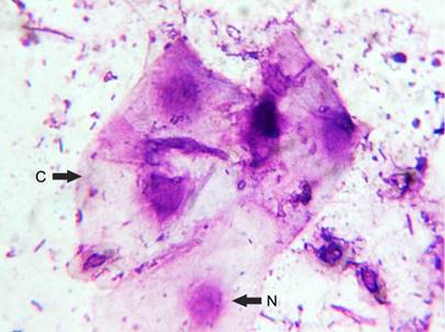
Figure 1 Epithelial cells collected from the proctodeum, which is the last portion of the colon and rectum. The cytoplasm (C) and nucleus (N) (Wright’s stain, 100×) are observed.
PCR was performed to detect herpesvirus in the collected cells. Primers that hybridized to a portion of the UL16 gene, a region that is frequently used for the identification of herpesviruses in psittacines, were used.5,11,14 The expected amplification was achieved in two M. monachus birds (Table 1 and Figure 2) using PRO primers (301 bp) and in 7 psittacines (Agapornis spp: A. ararauna, A. autumnalis [n = 2 ], M. monachus [n = 2 ], and A. mitrata [Table 1 ]) with the RAV primers (336 bp). However, in paraffin-embedded tissue specimens, the herpesvirus genome could not be amplified by PCR, possibly because of fragmentation, base modification, or oxidative damage in the formalin-fixed tissues. These changes degrade the sample material and negatively affect the DNA yield, as demonstrated in a study of a previously described DNA extraction method.15,16
Table 1 The common and scientific names of the psittacines included in the study population, their status in Mexico, and the PCR results.
| Common name | Scientific name | Status in country | PCR amplification product (bp) |
|---|---|---|---|
| Agaporni | Agapornis spp. | Exotic | Negative |
| Agaporni | Agapornis spp. | Exotic | Negative |
| Agaporni | Agapornis spp. | Exotic | Negative |
| Agaporni | Agapornis spp. | Exotic | Negative |
| Agaporni | Agapornis spp. | Exotic | Negative |
| Agaporni | Agapornis spp. | Exotic | Negative |
| Agaporni | Agapornis spp. | Exotic | Negative |
| Agaporni | Agapornis spp. | Exotic | Positive (336) |
| Agaporni | Agapornis spp. | Exotic | Negative |
| Agaporni | Agapornis spp. | Exotic | Negative |
| Cockatiel | Nymphicus hollandicus | Exotic | Negative |
| Cockatiel | Nymphicus hollandicus | Exotic | Negative |
| Cockatiel | Nymphicus hollandicus | Exotic | Negative |
| Cockatiel | Nymphicus hollandicus | Exotic | Negative |
| Cockatiel | Nymphicus hollandicus | Exotic | Negative |
| Monk parakeet | Myiopsitta manachus | Exotic | Negative |
| Monk parakeet | Myiopsitta manachus | Exotic | Negative |
| Monk parakeet | Myiopsitta manachus | Exotic | Negative |
| Monk parakeet | Myiopsitta manachus | Exotic | Positive (336) |
| Burrowing parrot | Cyanoliseus patagonus | Exotic | Negative |
| Blue-and-yellow macaw | Ara ararauna | Exotic | Positive (336) |
| Eclectus parrot | Eclectus roratus | Exotic | Negative |
| Red-lored parrot | Amazona autumnalis | Endemic | Negative |
| Budgerigar | Melopsittacus undulates | Exotic | Negative |
| Sulphur-crested cockatoo | Cacatúa galerita | Endemic | Negative |
| Agaporni | Agapornis spp. | Exotic | Negative |
| Military macaw | Ara militaris | Endemic | Negative |
| Military macaw | Ara militaris | Endemic | Negative |
| Mealy parrot | Amazon farinose | Endemic | Negative |
| Yellow-headed amazon | Amazona oratrix | Endemic | Negative |
| Red-lored parrot | Amazona autumnalis | Endemic | Positive (336) |
| Monk parakeet | Myiopsitta manachus | Exotic | Negative |
| Monk parakeet | Myiopsitta manachus | Exotic | Positive (301) |
| Monk parakeet | Myiopsitta manachus | Exotic | Negative |
| Red-lored parrot | Amazona autumnalis | Endemic | Positive (336) |
| Agaporni | Agapornis spp. | Exotic | Negative |
| Monk parakeet | Myiopsitta manachus | Exotic | Positive (301) |
| Monk parakeet | Myiopsitta manachus | Exotic | Positive (336) |
| Mitred parakeet | Aratinga mitrata | Exotic | Positive (336) |
| Monk parakeet | Myiopsitta manachus | Exotic | Negative |
| Monk parakeet | Myiopsitta manachus | Exotic | Negative |
| Orange-fronted parakeet | Eupsittula canicularis | Exotic | Negative |
| Monk parakeet | Myiopsitta manachus | Exotic | Negative |
| White-fronted parrot | Amazona albifrons | Endemic | Negative |
| Red-lored parrot | Amazona autumnalis | Endemic | Negative |
| Yellow-headed amazon | Amazon oratrix | Endemic | Negative |
| Red-lored parrot | Amazona autumnalis | Endemic | Negative |
| Monk parakeet | Myiopsitta manachus | Exotic | Negative |
| Monk parakeet | Myiopsitta manachus | Exotic | Negative |
| Red-lored parrot | Amazona autumnalis | Endemic | Negative |
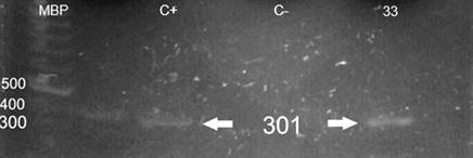
Figure 2 The 301-bp amplification product (PRO primers) of the herpesvirus UL16 gene from DNA obtained from the proctodeum cells of an M. monachus (33). Mbp = base pair marker; C + = positive control; C- negative control.
Only five psittacines exhibited clinical signs suggestive of herpesvirus infection (Amazona autumnalis [n = 2 ] and Myiopsitta monachus [n = 3 ]), such as diarrhoea, dyspnoea, ataxia, and posterior limb paralysis. On necropsy, congestion in the blood vessels of the brain (Figure 3), enlarged livers with multiple zones of pale-yellow parenchyma (Figure 4), dark-brown kidneys with asymmetric discolouration, and haemorrhagic lungs were observed. Microscopically, mononuclear infiltrates were observed in various organs and predominantly consisted of lymphocytes and plasma cells (Figure 5a). In the liver, kidneys, and intestines, multiple foci of coagulative necrosis surrounded by mononuclear infiltrates were identified (Figures 5b, c, d). Additionally, neurophagia and satellites were identified in the grey matter of the brain (Figure 6a), and intranuclear inclusion bodies exhibiting basophilic colouring were identified in the liver (Figure 6b) and kidneys (Figure 6c). Lesions similar to those found in the liver and kidneys in this study have been previously reported. Although only 10% of the psittacine samples studied here exhibited clinical signs suggestive of herpesvirus infection, intranuclear inclusion bodies in the liver and kidneys were observed and were associated with severe mononuclear infiltrates in two psittacines. These findings suggest a strong tissue reaction to virus replication and infection in the tissues.17,18 As described elsewhere and confirmed here, PCR is a useful technique for the identification of herpesvirus in apparently sick birds, although previous PCR-based detections were achieved using cell cultures for viral isolation.5,11,18,19

Figure 3 Congestion in the blood vessels of the brain. Highly congested and prominent blood vessels are indicated by arrows.

Figure 4 Liver with hepatomegaly. Rounded edges and blackish-red coloration interspersed with areas of apparently normal color were observed. Additionally, pale-yellow areas were distributed in the parenchyma.
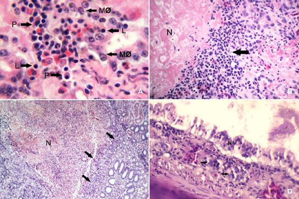
Figure 5 a) Liver with mononuclear infiltrate (MNI) (100× magnification). The image shows the parenchyma at the interstitial level with a mononuclear inflammatory infiltrate characterized by the presence of lymphocytes (L), plasma cells (P), and macrophages (MØ). b) Liver with necrosis and mononuclear infiltrate (40× magnification). Zones of coagulative necrosis (N) delimited by the inflammatory mononuclear infiltrate (arrow) are evident. c) Kidney with necrosis and mononuclear infiltrate (10× magnification). Renal parenchyma with extensive areas of coagulative necrosis (N) between the cortex and the medulla were observed. These areas were characterized by a marked loss of architecture and cellular detail and were delimited by mononuclear inflammatory cells (arrows). d) Necrotic intestine (100× magnification). The intestine developed severe coagulative necrosis (N) throughout the mucosa. Additionally, inflammatory infiltrates of mononuclear cells were observed near the basal membrane of the mucosa (arrows).

Figure 6 a) Brain with neuronophagia and satellites (40× magnification). Neurophagia (NF) is evident in the gray matter and is characterized by glial cells phagocytizing neurons and invading the perineural space surrounded by satellites (S). b) Liver with intranuclear inclusion bodies (INIB) (100× magnification). Multiple inclusion bodies (arrows) were observed in the liver parenchyma. c) Kidney with mononuclear infiltrate (MNI) and intranuclear inclusion bodies (100× magnification). In the renal parenchyma of the cortex at the tubular epithelium level, severe cellular swelling leading to coagulative necrosis was observed. As a result, the occurrence of pyknotic nuclei and karyolysis and presence of intranuclear inclusion bodies (INIB) were exacerbated. MNI took place in parallel.
In the present study, 90 % (45/50) of the birds were clinically healthy, and PCR results revealed that of these 45 birds, eight were positive for herpesvirus. These results demonstrate that PCR is a good technique for diagnosing latent herpesvirus infections in psittacines. In contrast, herpesvirus infection was confirmed by PCR in only one psittacine with suggestive clinical signs and lesions of herpesvirus infection (data not shown) because an enema could only be performed in one of the birds that exhibited clinical signs. In the other four cases, only organ collection was conducted. These results suggest that the PCR primers used in this study were adequate for the detection of latent infections in proctodaeal cells. Given that all herpesviruses remain in their natural hosts, generating latent infections and stress-induced asymptomatic reactivation that favour virus excretion and transmission to new susceptible individuals,3,4) it is important to emphasize that among the psittacines identified as positive by PCR were several Monk parakeets (M. monachus) (5/9), an Agaporni, and a Mitred parakeet, which are exotic birds. Two domestic birds (a blue-and-yellow macaw and red-lored parrot) were also identified.
Conclusions
The method of obtaining proctodaeal epithelial cells by enema proved to be efficient and allowed us to identify herpesvirus DNA in clinically healthy birds, thereby demonstrating latent infections. The results of PCR, along with the histopathological findings, revealed herpesvirus infection in clinically healthy endemic and exotic psittacines in Mexico, highlighting the need for broader studies on psittacine populations and the establishment of regulations by the country’s health authorities to regulate the entry of imported birds, thereby preserving the health of Mexico’s psittacines.

