


Servicios Personalizados
Revista
Articulo
 texto en
  Inglés (pdf)
 Artículo en XML
 Referencias del artículo
 Enviar artículo por email
Indicadores
-
 Citado por SciELO
 -
 Accesos
Links relacionados
-
 Similares en
SciELO
Similares en
SciELO
Compartir

 Permalink
Abanico veterinario
versión On-line ISSN 2448-6132versión impresa ISSN 2007-428X
Abanico vet vol.11 Tepic ene./dic. 2021 Epub 21-Mayo-2021
https://doi.org/10.21929/abavet2021.5
Original Article
Mycobacterium avium ssp. paratuberculosis identification and seroprevalence in sheep flocks in Aguascalientes
1
 http://orcid.org/0000-0002-3627-0534
1
http://orcid.org/0000-0002-3793-4582
2
http://orcid.org/0000-0003-1123-1878
2
http://orcid.org/0000-0002-3793-4646
1Centro de Ciencias Agropecuarias, Universidad Autónoma de Aguascalientes. Av Universidad 940, col. Ciudad Universitaria, CP 20131, Aguascalientes, Aguascalientes. México.
2Facultad de Medicina Veterinaria y Zootecnia (CEIEPAA), UNAM. Carretera Federal Tequisquiapan a, Ezequiel Montes Km 8.5, CP 76790, Tequisquiapan, Querétaro, México.
3*Autor para la correspondencia: García-Munguía Carlos, Departamento de Veterinaria y Zootecnia, Universidad de Guanajuato. Carretera Irapuato-Silao km 9, CP 36500 Irapuato, Guanajuato, México. cagamu@hotmail.com, gepallas@correo.uaa.mx, tequezada@correo.uaa.mx, gris@unam.mx, edithmc@unam.mx, lchavezglz@hotmail.com
With the objective of identifying the presence of Paratuberculosis (PTB), an infectious disease caused by Mycobacterium avium ssp paratuberculosis (MAP), in sheep, through pathological studies, bacterial culture and IS900 PCR, as well as estimating seroprevalence to MAP. The present cross-sectional study, was conducted in 16 different flocks, with the serum of 2415 adult sheeps, and analyzed by Enzyme-Linked ImmunoSorbent Assay (ELISA); nine sheep were used with clinical signs suggestive of PTB, from which samples were obtained for the identification studies; obtaining 51.3% of animals seropositive to MAP (1239/2415), in 100% of the herds (16/16); Bacterial isolation and its identification by PCR IS900 were founded in five of the nine cases (5/9) corresponding to 31.25% of the herds (5/16). Confirming the presence of Mycobacterium avium ssp paratuberculosis, and a high frequency of seropositive animals to MAP in flocks of Aguascalientes.
Keywords: Paratuberculosis; sheep; seroprevalence; Mycobacterium
Con el objetivo de identificar la presencia de Paratuberculosis (PTB), enfermedad infecciosa causada por el Mycobacterium avium subsp paratuberculosis (MAP), en ovinos, a través de estudios anatomopatológicos, cultivo bacteriano y PCR IS900, así como estimar la seroprevalencia a MAP. El presente estudio, de tipo transversal, se realizó en 16 diferentes rebaños con el suero de 2415 animales adultos y analizados por Ensayo por Inmunoabsorción Ligado a Enzimas (ELISA); se emplearon nueve ovinos con signos clínicos sugerentes a PTB, de los cuales se obtuvieron muestras para la realización de los estudios de identificación; obteniendo un 51.3 % de animales seropositivos a MAP (1239/2415), en el 100% de los rebaños (16/16); el aislamiento bacteriano y su identificación por PCR IS900 en cinco de los nueve casos hallados (5/9) correspondiendo al 31.25% de los rebaños (5/16). Conformando la presencia del Mycobacterium avium subsp paratuberculosis, así como una elevada frecuencia de animales seropositivos a MAP en rebaños de Aguascalientes.
Palabras clave: Paratuberculosis; ovinos; seroprevalencia; Mycobacterium
INTRODUCTION
Paratuberculosis (PTB) or Johne's disease (JD), is a chronic infectious disease that causes granulomatous enteritis in ruminants and eventually other species, with a wide worldwide distribution; caused by Mycobacterium avium ssp. paratuberculosis (MAP) (Coelho et al., 2010). Classified by the World Organization for Animal Health (OIE) as a communicable disease, with socio-economic and public health impact, suggesting the participation of MAP in Crohn's disease in humans (OIE, 2017; Balseiro et al., 2019;Gioffré et al., 2015). Three different strains of MAP have been described: type I (Sheep), type II (Cattle) and type III (Bison or intermediate), according to their preference for infecting different species of ruminants and their different requirements for their bacterial isolation (Begg and Whittington, 2010; Windsor, 2015). The disease is characterized by signs of progressive emaciation, submandibular edema, and eventually the death of the animals. The main route of infection is fecal-oral, with an incubation period that ranges from one to four years, which is why signs of the disease can be observed from one year of life (Chávez et al., 2004), once MAP enters the intestine, it lodges in macrophages, constituting a granulomatous infection where variable amounts of bacteria can be observed (Begg and Whittington, 2010; Windsor, 2015). The diagnosis, prevalence and impact of the disease are scarce and inconclusive both in Latin America and in Mexico (Chávez et al., 2004; Begg and Whittington, 2010; Fernández et al., 2014).
In Mexico, the presence of PTB in sheep has been detected in various states of the country, such as San Luis Potosí, Guanajuato, Querétaro, Mexico City, the State of Mexico and Veracruz (Estévez et al., 2006). Particularly, in Aguascalientes State, the activity of sheep farming, as in other states of the country, has grown since 1990 by more than 100%, with a meat production of approximately 612 tons in 2018, being the Valle area, made up of the municipalities of Aguascalientes, San Francisco de los Romo, Rincón de Romos, Jesús María, Pabellón de Arteaga and Asientos, where 85% of the State's sheep meat production and livestock activity in general are concentrated (SIAP, 2018).
It is very important to highlight that in this region there are no records of the diagnosis and identification of the diseases that are capable of affecting sheep herds and the adverse effect that these can cause on productivity. In Aguascalientes State there are unquantified cases of adult animals with progressive emaciation and mortality, however, a diagnosis is not made and it is impossible to determine if there is PTB.
Therefore, the objective of this work is to identify the presence of MAP through pathological studies, culture and PCR in sheep with a clinical picture suggestive of PTB, as well as to determine the seroprevalence against MAP by means of ELISA in sheep herds from the Aguascalientes Valley.
MATERIAL AND METHODS
Study design
This descriptive-cross-sectional study was carried out in Aguascalientes State, Mexico, with the herds and study animals selected according to the non-probabilistic method "for convenience" (Trusfield, 2018). In this way, 16 sheep herds were selected with populations ranging between 60 and 340 individuals, making up a total study population of 2,415 animals (Figure 1 and Table 1), where the herd was required to have clinical signs of chronic progressive emaciation suggestive to PTB in at least one specimen older than one year; considering each herd as an experimental unit.
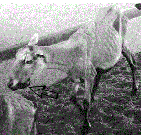
Table 1 Paratuberculosis seroprevalence results (ELISA), autopsies performed and bacterial isolation results, by herd in the Aguascalientes Valley
| Herd number | Seroprevalence % | Positive results ELISA-PPA-3 | Animals older tan 1 year | Specimens with clinical sings suggestive of PTB /Necropsy performed | Baterial isolation and positive PCR | Location by municipality |
|---|---|---|---|---|---|---|
| 14 | 91.3 | 168 | 184 | +/1 | - | Rincón de Romos |
| 6 | 84.4 | 287 | 340 | +/1 | + | Aguascalientes (sur) |
| 3 | 76.3 | 122 | 160 | +/0 | Nr | Rincón de Romos |
| 11 | 73.1 | 117 | 160 | +/1 | + | Asientos |
| 10 | 64.4 | 125 | 194 | +/1 | - | Jesús María |
| 9 | 61.4 | 72 | 140 | +/1 | + | Asientos |
| 4 | 58.6 | 99 | 169 | +/1 | + | Aguascalientes (sur) |
| 15 | 56.8 | 50 | 88 | +/1 | - | San Francisco de los Romo C |
| 13 | 54.5 | 36 | 66 | +/0 | Nr | Pabellón de Arteaga |
| 16 | 46.7 | 56 | 120 | +/0 | Nr | Aguascalientes (Nte) |
| 1 | 43.8 | 35 | 80 | +/1 | - | Jesús María |
| 8 | 28.3 | 17 | 60 | +/0 | Nr | Pabellón de Arteaga |
| 5 | 12.9 | 8 | 62 | +/0 | Nr | Aguascalientes (Nte) |
| 7 | 9.5 | 31 | 326 | +/0 | Nr | Aguascalientes (sur) |
| 12 | 8.1 | 8 | 99 | +/0 | Nr | San Francisco de los Romo |
| 2 | 4.8 | 8 | 167 | +/1 | + | San Francisco de Los Romo |
| 16 | 51.3 | 1239 | 2415 | 16/9 |
Nr: Not perfomed
Sampling
Blood serum. 5 mL of blood were obtained by venipuncture of the jugular, which was collected in tubes without additive for obtaining serum; later the samples were stored at - 20 °C until their use in the ELISA test.
ELISA. The previously obtained sera were analyzed by means of the indirect enzyme immunoassay (ELISA) using the protoplasmic antigen PPA3 (Allied, Mo.), which has a specificity of 75% and a sensitivity of 64% according to the protocol used by García (1992).
Obtaining samples for histopathological study and isolation of MAP. Taking Timoney (1988) as a reference and observing the clinical course of the animals and the epidemiological characteristics of the 9 herds, a presumptive diagnosis was established in 9 animals; who presented signs of progressive emaciation, hair or hairy wool, submandibular edema, lesions and loose stools. Thus, understanding that 56.25% of the study herds presented at least one suggestive case to PTB.
Subsequently, the 9 animals were sacrificed with an overdose of sodium pentobarbital (NOM-033-SAG/ZOO-2014) according to the guidelines established in the “Ethics Committee for the use of animals in teaching and research of the Autonomous University of Aguascalientes”.
The confirmation of these cases was carried out through necropsy and identification of lesions, thus ruling out the presence of possible lesions suggestive of differential diagnoses of pseudotuberculosis (Corynebacterium pseudotuberculosis), digestive alterations due to the presence of foreign bodies, neoplasms, chronic pneumonias and severe parasitosis, (Straub, 2004; Windsor, 2014).
After that, samples of intestinal sections of the ileum and jejunum were taken, which showed thickening of the intestinal mucosa that suggested lesions associated with paratuberculosis. These sections were fixed in buffered formalin (pH 7.6) for subsequent histological processing, using Hematoxylin-Eosin (HE) and Ziehl-Neelsen (ZN) stains, the latter in order to demonstrate the presence of acid-fast bacilli (BAAR) (Estévez, 2006). Similarly, samples for bacterial isolation were obtained from 30-cm sections of the ileum and jejunum that showed lesions suggestive of PTB (Chávez et al., 2004; Dennis et al., 2011), being deposited in sterile plastic containers for its conservation at -20 ºC until its processing in the laboratory.
Bacterial culture from intestine. In turn, samples taken from the intestine were scraped from the intestinal mucosa with a sterile scalpel, processing them for decontamination and concentration of mycobacteria, according to the methodology described by Sánchez and Guerrero, 2006. Later, the material from the interface obtained was inoculated in the Löwentein-Jensen (L-Jm) culture media with mycobactin, placing three to eight drops of each of the samples, distributing it over the entire surface of the medium. From each sample, two different tubes were inoculated with L-Jm medium, one containing mycobactin and the second without mycobactin. Later they were incubated at 37 ° C, keeping them in an inclined position and without humidity. Bacterial growth was verified every four weeks until week twelve, the media where no growth was observed, continued in incubation until week 40 (De Juan et al., 2006; Whittington, 2010).
PCR IS900. Molecular identification was carried out from the growth of the MAP colonies, by means of endpoint PCR, using the primers IS900P3N (5'-GGG TGT GGC GTT TTC CTT CG-3 ') and IS900P5N (5'-ATTTCGCCGCCACCGCCACG-3 ') (Chávez et al., 2004).
Estimation of seroprevalence. The prevalence was estimated based on the proportion of animals that were positive in the ELISA test, in relation to the total population of adult animals in each of the herds. The information generated in the study was evaluated and contrasted using Descriptive Statistics tests, using “Analysis for one variable”, with the support of the “Statgraphics Centurion XV” TM package (Fernandez et al., 2002).
RESULTS
Seroprevalence. The mean seroprevalence percentage of the 16 herds was 51.3%, with a minimum value of 4.8% and a maximum value of 91.33% with a 95.0% confidence interval for the mean of: [32.7173, 62.3415]. The individual seroprevalence results for each herd are described in Table 1. In the sixteen herds in which the ELISA tests (PPA- 3) were performed, positive cases were identified, obtaining a seroprevalence calculated at “herd level” 100% (Table 1).
Anatomopathological study. In the 9 animals that were necropsied, they presented emaciation, hairy wool or hair, submandibular edema (Figure 1), presence of ascites, fat degeneration, as well as apparent thickening of the intestinal mucosa at the ileum and jejunum (Figure 2), in addition to an increase in the size of the mesenteric lymph nodes and their fusion, not finding evidence of alterations or lesions suggestive of another differential etiology to cases of PTB. Through the histopathological study, it was possible to observe in 8 of the 9 animals studied (88.8%), the presence of enteritis, granulomatous lymphadenitis with the presence of macrophages, giant cells and the presence of countless Acid-Alcohol Resistant Bacteria (ABB) by staining from Ziehl-Neelsen (ZN). Determined as multibacillary lesions (Figure 3). While in 1 of the 9 animals studied (11.2%), the presence of multifocal granulomatous lesions with a low number of bacilli (BAAR) inside macrophages was observed, coinciding with a paucibacillary lesion.

Figure 2 Macroscopic lesions corresponding to intestinal thickening characteristic of granulomatous enteritis

Figure 3 Histopathological lesions of multibacillary ID, Ziehl-Neelsen stain 400X where multiple BAARs are observed in intestinal mucosa
Bacterial isolation. After 12 weeks of incubation, in 5 cases (55.5%), development of yellow, creamy, convex and circular colonies characteristic of MAP were observed in L- Jm media added with mycobactin. While in 4 of the cases (44.5%), no colony development was observed until week 40. All the media that were not added with mycobactin did not show growth.
PCR IS900. From the five bacterial isolates obtained, amplification products of the IS900 sequence corresponding to 310 base pairs (bp) were obtained, thus confirming the presence of MAP from these isolates obtained (Figure 4).

Figure 4 Photograph of a 2% agarose gel stained with ethidium bromide. Where amplification products with a size of 310 bp are observed, from DNA of bacterial colonies obtained from intestinal tissue, where the molecular weight marker 100 bp, C + positive control, negative control lane 1 (water ), lanes 3 to 7 amplification product obtained from isolates 03, 04, 05, 06 and 09, which correspond to an IS900 insert fragment of MAP (*).
Thus, in 100% of the sample (16 study herds) MAP seropositivity was obtained; in turn, in 31.25% of the sample (5 herds) the presence of MAP was confirmed by bacterial isolation and identification by PCR IS900 from the animals that presented clinical symptoms at the time of the study (Table 2).
DISCUSSION
The mean seroprevalence obtained in the present study (51.3%) is reported to be high, compared to other studies conducted to measure prevalence both in the country and worldwide, considering that the disease internationally has a high prevalence in different species of ruminants, with 20% and up to 40% (Whittington et al., 2019). In Mexico, various studies indicate serological evidence for MAP such as those reported by Jaimes et al., (2008) in San Luis Potosí, Mexico; where the seroprevalence results in sheep by the Agar-gel immunodiffusion method was 44%. While in Nayarit state, 28.94% was reported in sheep, through ELISA tests (Mejía et al., 2015). In Latin America, few studies carried out in sheep show a wide variability in the prevalence of MAP, where prevalences from an average 4.3% in sheep are reported (Fernández et al., 2014), to 54.08% of seroprevalence to MAP in sheep in Brazil, (Medeiros et al., 2012).
Given the few studies, and a wide variation in the methodology and diagnostic test used to assess the prevalence of PTB, Espechit et al. (2017) and Whittington et al., (2019) point out that this results in a great variability of the results, and consequently, an underestimation of the presence and true impact of paratuberculosis.
Despite the high frequency results obtained, at the time of the study, only clinical cases (9/16) were found in 56.25% of the herds studied, where all of them show multibacillary or paucibacillary histopathological lesions, characteristic of the disease where if it is considered that these occur in individuals older than one year of age, with a prolonged time of infection coupled with the presentation of a characteristic clinical picture, highly suggestive of PTB, they can be considered a definitive diagnosis of MAP individually, hardly ruled out by other tests such as culture and PCR as mentioned by several authors (Pérez et al., 1999; Dennis et al., 2011; Windsor, 2015; Whittingtong et al., 2019).
Considering that the nine animals that were necropsied presented lesions characteristic of PTB, only in 55.5% (5/9) was bacterial isolation achieved despite presenting multibacillary lesions in eight of the nine. The difficulty of isolating MAP, the culture medium used considering the possible variation of the type of MAP present, "S" or "C", which present a difference in nutritional requirements for growth (Elguezabal et al., 2011; Whittington, 2011; Stevenson, 2015; Whittington et al., 2019), or at the time of the bacterial decontamination process, where the number of viable bacteria for cultivation and isolation can be reduced. The isolation and cultivation of the microorganism in sheep is extremely difficult, being in sheep herds that fecal culture detects fewer than clinical cases and requires more than 12 months for incubation, as reported by Windsor (2015), as well as Whittington, 2010.
The subsequent confirmation and identification of the isolates obtained from MAP by means of IS900 PCR, carried out from the five cultures obtained, confirmed the presence of MAP. Some authors currently suggest that the identification of the bacteria by PCR be carried out from the samples obtained from the tissues, speeding up the time of the results, without directly depending on the cultures obtained, since the diagnostic times are prolonged by the characteristics of MAP isolation, indicating that this test has a similar efficacy with cultures (Kawaji et al., 2007; Nilsen et al., 2008; Whittington et al., 2019).
According to authors such as Kumthekar, et al., (2013)) and Konbon (2018), determining the degree of infection of the disease in herds allows establishing adequate control programs that significantly impact the cost-benefit; primarily considering test-scrap. Well, given the infeasibility of discarding and considering the limited availability of some producers for the vaccination of herds, the control measures must be framed towards a strengthening of biosecurity.
Considering thus the early separation of the lambs and the elimination of animals with initial clinical signs of the disease, it may be a measure that positively impacts the reduction of the prevalence of the disease (Robee, 2011; Windsor 2015). Emphasizing that professionals in the animal health sector and leaders of the productive sector have a clear knowledge of the degree of presence and impact of the disease, both clinically, economically and public health; thus achieving control proposals (Roche, 2015; Windsor, 2015; Wittington et al., 2019)
CONCLUSION
The presence of Mycobacterium avium ssp paratuberculosis is confirmed as an etiological agent of the clinical disease, in addition to observing the high frequency presence of animals seropositive to MAP, in herds of the Central Valley of Aguascalientes state, Mexico which makes it a high-impact pathology in sheep production in the state.
LITERATURA CITADA
Balseiro A, Perez V, Juste R. 2019. Chronic regional intestinal inflammatory disease: A trans-species slow infection. Comparative Immunology, Microbiology and Infectious Diseases. 62:68-100. https://doi.org/10.1016/j.cimid.2018.12.001 [ Links ]
Begg D, Whinttington R. 2010. Paratuberculosis in Sheep. In: Paratuberculosis, Organism, Disease, Control. United Kingdom. CAB International. Pp.157-168. http://doi.org/10.1079/9781845936136.0157 [ Links ]
Chávez G, Trigo TF, Svastova P, Pavlik P. 2004. Identificación del polimorfismo genético de aislamientos de Mycobacterium avium subsp. Paratuberculosis de caprinos del centro de México. Veterinaria México. 35:75-84. https://www.redalyc.org/pdf/423/42335107.pdf [ Links ]
Coelho AC, Pinto ML, Coelho AM, Aires A, Rodríguez J. 2010. A seroepidemiological survey of Mycobacterium avium subsp. paratuberculosis in sheep from North of Portugal. Pesquisa Veterinária Brasileira. 30(11):903-908. http://dx.doi.org/10.1590/S0100-736X2010001100001 [ Links ]
Dennis M, Reddacliff LA, Whittington RJ. 2011. Longitudinal study of clinicopathologycal features of Johne`s diseases in sheep naturally exposed to Mycobacterium avium subs paratuberculosis. Veterinary. Pathology. 48:565-575. http://dx.doi.org/10.1177/0300985810375049 [ Links ]
De Juan L, Álvarez J, Romero B, Bezos J, Castellanos E, Aranaz A, Mateos A, Domínguez L. 2006. Comparison of four different culture media for isolation and growth of type II and type I/III Mycobacterium avium subsp. paratuberculosis strains isolated from cattle and goats. Applied and Environmental Microbiology. 72:5927-5932. http://dx.doi.org/10.1128/AEM.00451-06 [ Links ]
Elguezabal N, Bastida F, Sevilla IA, González N, Molina E, Garrido JM, Juste RA. 2011. Estimation of Mycobacterium avium subsp. paratuberculosis Growth Parameters: Strain Characterization and Comparison of Methods. Applied and Environmental Microbiology. 77: 8615-8624. http://dx.doi.org/10.1128/AEM.05818-11 [ Links ]
Espeschit IF, Schwarz GG, Faria CS, Souza CC, Paolicchi FA, Juste RA, Carvalho IA, Moreira AS. 2017. Paratuberculosis in Latin America: a systematic review. Tropical Animal Health and Production. 49:1557-1576. http://dx.doi.org/10.1007/s11250-017-1385-6 [ Links ]
Estévez DI, Hernández CR, Trujillo AM, Chávez GG. 2006. Detection of Mycobacterium avium subsp. Paratuberculosis in goat and sheep flocks in México. Small Ruminant Research. 72:209-213. http://dx.doi.org/10.1016/j.smallrumres.2006.10.017 [ Links ]
Fernandez SJ, Correa NM, Fernando RN. 2014. Systematic review of the prevalence of Paratuberculosis in cattle, sheep, and goats in Latin America and the Caribbean. Tropical Animal Health and Production. 46:1321-1340. http://dx.doi.org/10.1007/s11250-014-0656-8 [ Links ]
Fernandez SF, Sánchez JM, Córdoba A, Largo AC. 2002. Estadística descriptiva. 2da ed. Madrid, España: Editorial Esic. Pp. 566. ISBN: 978-847-35630-62 [ Links ]
García MJ, Chávez G, Adúriz JJ, Pérez V, Juste RA, Badiola JJ. 1992. Prevalence of paratuberculosis in infected goat flocks and comparison of different methods of diagnosis. Proc. III Int. Coll. PTBC. 157-163. http://www.paratuberculosis.net/publications.php [ Links ]
Gioffré A, Correa MM, Alvarado PM, Vaca R, Morsella C, Fiorentino MA, Romano MI. 2015. Tipificación molecular de Mycobacterium avium subsp. paratuberculosis mediante análisis de repetición en tándem de número variable de múltiples locus. Revista Brasileña de Microbiología. 46:2: 557-564. https://doi.org/10.1590/S1517-838246220140283 [ Links ]
Jaimes NG, Santillán FM, Hernández CO, Córdova LD, Guzmán RC, Arellano RB, Díaz AE, Tenorio GV, Cuéllar OA. 2008. Detección de Mycobacterium avium subespecie paratuberculosis, por medio de PCR-anidada a partir de muestras de heces de ovinos. Revista Veterinaria México. 39(4): 377-386. https://www.redalyc.org/articulo.oa?id=42339402 [ Links ]
Kawaji S, Taylor DL, Mori Y, Whittington RJ. 2007. Detection of Mycobacterium Avium Subsp. paratuberculosis in ovine faeces by direct quantitative PCR has similar or greater sensitivity compared to radiometric culture. Veterinary Microbiology. 125:36-48. http://dx.doi.org/10.1016/j.vetmic.2007.05.002 [ Links ]
Konboon M, Bani YM, Pithua PO, Rhee N, Aly SS. 2018. A nested compartmental model to assess the efficacy of paratuberculosis control measures on US dairy farms. PLoS One. 13:10. https://doi.org/10.1371/journal.pone.0203190 [ Links ]
Kumthekar S, Manning EJ, Ghosh P, Tiwari K., Sharma RN, Hariharan H. 2013. Mycobacterium avium subespecie paratuberculosis confirmada tras la vigilancia serológica de pequeños rumiantes en Granada, West Indies. Revista de investigación de diagnóstico veterinario. 25(4): 527-530. https://journals.sagepub.com/doi/10.1177/1040638713490688 [ Links ]
Medeiros JM, Garino JF, Torres MR, de Medeiros CV, Riet CF. 2012. Frequência de anticorpos para paratuberculose em bovinos no semiarido paraíbano. Pesquisa Veterinária Brasileira. 32:697-700. https://doi.org/10.1590/S0100-736X2012000800003 [ Links ]
Mejía MK, Lemus FC, González MC, Palomares RG, Díaz AE, Gutierréz HJ. 2015. Factores de riesgo asociados a Mycobacterium avium subsp. Paratuberculosis en rebaños ovinos de Nayarit, México. Revista Científica FCV-LUZ. 5:294-302. https://www.redalyc.org/pdf/959/95953315005.pdf [ Links ]
Nielsen SS, Toft N. 2008. Ante mortem diagnosis of paratuberculosis: A review of accuracies of ELISA, interferon-gamma assay and fecal culture techniques. Veterinary Microbiology. 129:217-235. http://dx.doi.org/10.1016/j.vetmic.2007.12.011 [ Links ]
NOM-033-SAG/ZOO-2014. Norma Oficial Mexicana: Métodos para dar muerte a los animales domésticos y silvestres. México. Diario Oficial de la Federación, 26/08/2014. https://www.dof.gob.mx/nota_detalle.php?codigo=5405210&fecha=26/08/2015 [ Links ]
OIE (World Organisation for Animal Healt). 2017. Manual of diagnostic test and vaccines for terrestrial animals Vol I. Recuperado en noviembre de 2017. http://www.oie.int/es/normas-internacionales/manual-terrestre/acceso-en-linea/ [ Links ]
Pérez V, Tellechea J, Corpa JM, Gutiérrez M, García MJ. 1999. Relation between pathologic findings and cellular immune responses in sheep with naturally acquired Paratuberculosis. American Journal of Veterinary Research. 60:123-127. https://pubmed.ncbi.nlm.nih.gov/9918160/ [ Links ]
Roche S, Jones BA, Meehan M, Massow M, Kelton D. 2015. Evaluating the effect of focus farms on Ontario dairy producers’ knowledge, attitudes, and behaviour toward control of Johne’s disease. J Dairy Sci. 98:5222-040. https://doi:10.3168/jds.2014-8765 [ Links ]
Robbe AS. 2011. Control of Paratuberculosisin Small Ruminants. Vet Clin Food Anim. 27:609-620. https://doi:10.1016/j.cvfa.2011.07.007 [ Links ]
Sánchez CC, Guerrero GC. 2006. Procedimientos en microbiología clínica. Recomendaciones de la Sociedad Española de Enfermedades Infecciosas y Microbiología Clínica. ISBN: 84-609-2287-1. https://seimc.org/contenidos/documentoscientificos/procedimientosmicrobiologia/seimc-procedimientomicrobiologia1.pdf [ Links ]
SIAP (Servicio De Información Agroalimentaria Del Gobierno De México). 2018. Estadísticas de la producción pecuaria 2018.Recuperado en septiembre de 2018. http://infosiap.siap.gob.mx/gobmx/datosAbiertos.php [ Links ]
Stevenson K. 2015. Genetic diversity of Mycobacterium avium subspecies paratuberculosis and the influence of strain type on infection and pathogenesis: a review. Veterinary Research. 46:64. http://dx.doi.org/10.1186/s13567-015-0203-2 [ Links ]
Straub OC. 2004. Maedi-Visna virus infection in sheep. History and present knowledge. Comparative Immunology. Microbiology and Infectious Diseases. 27:1-5. https://doi.org/10.1016/S0147-9571(02)00078-4 [ Links ]
Timoney JF, Gillespie JH, Scott FW, Barlough JE. 1988. Hagan and Bruner’s Microbiology and Infectious Diseases of Domestic Animals. Ithaca, United States: Cornell University Press. Pp. 912. ISBN: 9780-801-41896-9. [ Links ]
Trusfield M. 2018. Veterinary Epidemiology. 4ta ed. UK: Blackwell publishining. Pp. 888. ISBN: 978-1-118-28028-7. [ Links ]
Whittington RJ. 2010. Cultivation of Mycobacterium avium subsp. paratuberculosis. 2da ed. Wallingford, UK. Editorial Cabi. Pp. 266-304. ISBN: 978-1-789-24341-3. [ Links ]
Whittington RJ, Marsh IB, Saunders V, Grant IR, Juste R, Sevilla IA, Manning EJ, Whitlock RH. 2011. Culture phenotypes of genomically and geographically diverse Mycobacterium avium subsp. paratuberculosis isolates from different hosts. Journal of Clinic Microbiology. 49:1822-1830. http://dx.doi.org/10.1128/JCM.00210-11 [ Links ]
Whittington RJ, Donat K, Weber MF. 2019. Control of paratuberculosis: who, why and how. A review of 48 countries. BMC Veterinary Research. 15:198. http://dx.doi.org/10.1186/s12917-019-1943-4 [ Links ]
Windsor PA. 2014. Managing control programs for ovine caseous lymphadenitis and paratuberculosis in Australia, and the need for persistent vaccination. Veterinary Medicine: Research and Reports. 5:11-22. https://doi.org/10.2147/VMRR.S44814 [ Links ]
Windsor PA. 2015. Paratuberculosis in sheep and goats. Veterinary Microbiology. 181:161-9. http://dx.doi.org/10.1016/j.vetmic.2015.07.019 [ Links ]
Received: August 28, 2020; Accepted: January 15, 2021; Published: January 30, 2021
 Este es un artículo publicado en acceso abierto bajo una licencia
Creative Commons
Este es un artículo publicado en acceso abierto bajo una licencia
Creative Commons
