Services on Demand
Journal
Article
Indicators
-
 Cited by SciELO
 -
 Access statistics
Related links
-
 Similars in
SciELO
Share
Abanico veterinario
On-line version ISSN 2448-6132Print version ISSN 2007-428X
Abanico vet vol.10 Tepic Jan./Dec. 2020 Epub May 07, 2021
https://doi.org/10.21929/abavet2020.25
Clinical Caso
Report of rabies in Tepezcuintles Cuniculus paca (syn. Agouti paca) in captivity in Yucatan
1Facultad de Medicina Veterinaria y Zootecnia, Universidad Autónoma de Yucatán. Mérida, Yucatán, México.
2Práctica privada. Tuxtla Gutiérrez, Chiapas, México.
3Práctica privada. Mérida, Yucatán, México.
4Práctica privada. Mérida, Yucatán México.
The objective of this case study is to report the presence of rabies in three Cuniculus paca specimens, of kept in captivity, in Yucatan, Mexico. The specimens’ clinical signs were recorded before the death of each one, the brain was collected and sent to a certified animal pathology laboratory, to detect the Lyssavirus´ nucleoprotein by the immunofluorescence technique. The clinical picture of each of the affected specimens was described. The results showed positivity to the immune reaction.
Keywords: Lyssavirus; rabies; zoonosis; wildlife
El objetivo del presente estudio de caso es informar la presencia de rabia en tres ejemplares de Cuniculus paca, mantenidos en cautiverio en Yucatán, México. Se registraron los signos clínicos de los ejemplares previos al deceso de cada uno, se colectó el cerebro para ser enviado a laboratorio certificado de patología animal, donde se aplicó la técnica de inmunofluorescencia para detectar la nucleoproteína del Lyssavirus. Se describió el cuadro clínico de cada uno de los ejemplares afectados. Los resultados mostraron positividad a la reacción inmunológica.
Palabras clave: Lyssavirus; rabia; zoonosis; fauna silvestre
INTRODUCTION
Rabies is a zoonosis with a wide geographic distribution, the etiological agent is a neurotropic virus of the genus Lyssavirus, of the family Rhabdoviridae, of the order Mononegavirales, which causes irreversible lesions in the central nervous system (CNS); same that lead to death from paralysis (Torres et al., 2019). The presence of the rabies virus spreads throughout the planet, except Antarctica, the prevalence is especially high in regions of Africa and Asia, mainly in children under 15 years of age (Frantchez and Medina, 2018). There are susceptible hosts in Yucatán that are skunks of the genera Spilogale and Conepatus, hematophagous bat of the genera Desmodus and Diphilia, raccoons (Procyon lotor) and badger (Nasua narica) (Ortega-Pacheco and Jiménez, 2017).
The clinical picture of classical human rabies is recognized as having three stages: incubation, prodrome, and acute neurological. The first phase occurs from the entry of the virus by laceration or bite, generally lasts from one to three months; but it can last a few days when virus penetration is near the neck or head, and even directly to the nervous tissue; but it can also remain for several years before continuing to the next stage, for this reason it can go unnoticed. The second stage generally lasts 2 to 10 days, the virus has migrated through the central nervous system, and the patient exhibits signs of fever, headache, malaise, irritability, nausea, and vomiting. The third phase lasts from hours to a week, manifests as encephalitic or paralytic rabies. Mainly there is fever, hypersalivation, excessive sweating, piloerection, pupillary abnormalities, cardiac arrhythmias, pulmonary edema, diaphragm and bulbar paralysis (Frantchez and Medina, 2018).
There are two types of rabies, encephalitic or furious and paralytic; the furious is the most frequent, the signs are: hyperexcitability due to stimuli, such as noise or light, hallucinations, excessive salivation, hydrophobia and aerophobia. The paralytic shows paralysis of the hind limbs and urinary incontinence (Frantchez and Medina, 2018).
The diagnosis of rabies is made mainly by immunofluorescence (IF) in brain tissue, by cell culture, inoculation in mice or genotyping by PCR (OMS, 2013); because clinical observation only indicates suspicion of rabies in the early stages of the disease course, it manifests when CB-ir neurons´ functions (GABA-ergic) are altered, in the terminal phase of rabies clinical picture, when the virus has spread over most areas of the brain (Lamprea and Torres-Fernández, 2008). In C. paca the presence of rabies has only been reported in a previous case in Guatemala (Díaz et al., 1962). These authors also report the presence of rabies in the following species of wildlife: tacuatzin (Philandel opossum fuscogriseus and Didelphys marsupiales), bats (Molossus sinaloae, Phyllostomidae sp, and Artibeus palmorum), fox (Urocyon cinereargenteus), raccoons) (Procyon lotus), coyotes (Canis latrans) and skunks (Conepatus mapuritus).
The objective of this report is to communicate the presence of rabies in three Cuniculus paca specimens kept in captivity in Yucatán, México.
CLINICAL CASE PRESENTATION
The clinical cases appeared in the Xmatkuil Wildlife Conservation Management Unit, located in Mérida city, Yucatán, Mexico. The first clinical case was an adult female (number 03), who on March 24, 1999 presented irritability, anorexia and emaciation (decreased body condition). Six days after the beginning of the clinical picture, she manifested intolerance to the male presence without an identification number, with whom she shared a corral, preventing her access to the burrow; causing superficial skin bite wounds, which were treated with antiseptics. Three days after being attacked, the male (animal without number) showed slight irritability; however, he continued eating food, albeit irregularly; but throughout the days she showed a decrease in his body condition. On the 12th day of observation of the first case, the female presented moderate hind limb ataxia and photophobia. In the male, new bite lesions were observed on body sides and on the penis, which led to the separation of animals in different pens.
On day 14, female 03 prostrated and died, proceeding to perform a necropsy and extraction of the brain. Her brain tissue was kept refrigerated at 10 °C for immunofluorescence examination at the Central Regional Laboratory of Mérida, Yucatán (LCRM). On the same day, severe depression and anorexia were detected in the male, dying the following day (9 days post-bite); carrying out the collection of encephalon to carry out the same laboratory examination.
In the first week of April, juvenile female number 02, showed irritability, anorexia and loss of body condition; starting day one of observation of the new event. The following day the female prostrated, showing severe hind limb ataxia. On the third day she showed marked dyspnea and anorexia continued. During days four and five after the onset of the clinical picture, greater aggressiveness and photophobia were noted.
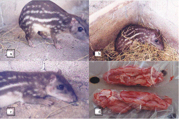
On day 13, hirsute hair, hypothermia and hind limb paralysis were observed; on the 14th day she woke up severely depressed and died. The aforementioned study procedures were carried out on her corpse. The brains of the three investigated animals were positive for rabies by immunofluorescence (Figures 1 and 2). Table 1 shows the summarized data of the three cases. Figure 3 shows tepezcuintles affected by rabies virus and one of the brains that was sent to the laboratory for immunofluorescence examination.

Figure 1 Evidence of positivity to rabies by immunofluorescence test in brain tissue of Cuniculus paca, female 03 and male without number

Figure 2 Evidence of positivity for rabies by immunofluorescence test in brain tissue of Cuniculus paca, juvenile female 2
Table 1 Summary information on animals that died from rabies
| Animal number and sex | Duration of the process | Date of death | Diagnosis by immunofluorescence in brain tissue | Observations in the animal |
| 03 Female | Subacute Started 24/03/1999 Duration 14 days |
6/04/1999 | Positive for rabies. Study with folio number 34520 |
-marked aggressiveness. -irritability. -Hind limb ataxia. -hypersalivation. -photophobia. -anorexia. emaciation. |
| S/N Male | Subacute, bitten on 30/03/1999 Duration 9 days |
7/04/1999 | Positive for rabies. Study with folio number 34520 | -irritability. -decreased body condition. -severe depression. -photophobia. |
| 02 Juvenile female l | Subacute Started 6/04/1999 Duration 14 days |
19/04/1999 | Positive for rabies.Study with folio number 34680 | -irritability. -aggressiveness. -decreased body condition. -hypothermia. -oaralysis of hind limbs (ataxia). -photophobia. -apnea. -anorexy. bristly hair |

Figure 3 Tepezcuintles Cuniculus paca male with signs of rabies and brain. a) un-numbered male Cuniculus paca, bitten by female 03, b) juvenile female 02 tepezcuintle prostrated on the 11th day of the clinical picture, c) female 02 juvenile on the 13th day of the clinical picture and d) the two halves of the brain of juvenile female 02.
DISCUSSION
In general, the clinical signs presented in the three cases showed similar signs. It was observed that in females the furious phase was more evident than in the male; however, unlike what is described in the medical literature for other animal species, tepezcuintles (C. paca) that suffered attacks in the pens did not attempt to bite their wounds. The male of the second case did not present photophobia or hypersalivation, the paralytic phase duration was nine days and not hours, as mentioned by Frantchez and Medina (2018). Although the primary aggressor who introduced the virus corresponding to the first and third cases could not be identified, it is important to consider that there are clinical behavioral differences in diseased animals, according to the species of the aggressor that caused the infection. When it is a carnivore like the dog, it becomes predominantly furious, and on the other hand, when it is a chiroptera (especially the hematophagous bat, Desmodus rotundus), the paralytic phase predominates, as manifested in cattle (Ibáñez and Chang, 2019; Sánchez et al., 2019). Because the behavioral habits of parsnips are nocturnal, it is assumed that the virus is unlikely to have been introduced by bats. In the cases of tepezcuintles, only in one male (case No. 2), bitten by his sick partner, could the original source of infection be determined; finding that only photophobia and hypersalivation differentiated the clinical pictures between the couple.
It is possible to suppose that, in pictures´ severity observed between females and the male, the wounds that constituted the gateway to the rabies virus had some relationship, since no lacerations or solutions could be found in females during the clinical inspection of continuity in the skin. On the other hand, the male showed a frank wound caused by teeth, which had saliva residues in the hair. Frantchez and Medina, (2018), mention that the body location and the severity of the wounds largely determine the incubation period duration, the disease course and the transition between the aggressive and paralytic phases. This may explain the reason why the male tepezcuintle showed a shorter duration of its pathology, by virtue of the fact that it was bitten on the penis, which is abundantly vascularized and innervated by the sympathetic and parasympathetic nervous system (Vozmediano and Bonilla, 2010) and in the middle region of the body.
The LCRM is certified by the National Institute of Epidemiological Reference of Mexico, to make the diagnosis of rabies, by means of the IF technique in brain tissue, therefore the diagnosis is highly reliable; in addition, this technique has a sensitivity of up to 99% (Secretaría de Salud, 2012).
LITERATURA CITADA
DIAZ LH, Santamaría JG, Otal AU. 1962. Datos sobre la situación de la rabia en Guatemala. Salud Pública de México. Epoca V. 4(2): 247-251. ISSN: 1606-7916. http://saludpublica.mx/index.php/spm/article/viewFile/4177/4058 [ Links ]
FRANTCHEZ V, Medina J. 2018. Rabia: 99.9% mortal, 100% prevenible. Revista Médica del Uruguay. 34(3):164-171. ISSN 1688-0390. http://www.scielo.edu.uy/pdf/rmu/v34n3/1688-0390-rmu-34-03-86.pdf [ Links ]
IBAÑEZ MM, Chang RE. 2019. La Rabia en la Patagonia. Desde La Patagonia Difundiendo Saberes. 16(28): 24-28. ISSN 2618-5385. https://repositorio.inta.gob.ar/xmlui/bitstream/handle/20.500.12123/6555/INTA_CRPatagoniaNorte_EEABariloche_ChangReissig_E_La_Rabia_En_La_Patagonia.pdf?sequence=1&isAllowed=y [ Links ]
LAMPREA N, Torres-Fernández O. 2008. Evaluación inmunohistoquímica de la expresión de calbindina en el cerebro de ratones en diferentes tiempos después de la inoculación con el virus de la rabia. Colombia Médica. 39 (3): 7-13. ISSN: 0120-8322. https://www.redalyc.org/articulo.oa?id=28309602 [ Links ]
OMS ORGANIZACION MUNDIAL DE LA SALUD. 2013. Reunión Consultativa de expertos de la OMS sobre la rabia: segundo informe. Serie de informes técnicos de la OMS. No. 982. ISBN 978 92 4 069094 3. https://www.paho.org/panaftosa/index.php?option=com_docman&view=download&slug= consulta-expertos-oms-sobre-rabia-espanol-0&Itemid=518 [ Links ]
ORTEGA-PACHECO A, Jiménez M. 2017. La rabia canina, una zoonosis latente en Yucatán. Revista Biomédica. 28 (2):61-63. ISSN 2007-8447. https://www.researchgate.net/publication/328397960_La_rabia_canina_una_zoonosis_latente_en_Yucatan [ Links ]
SANCHEZ MP, Díaz SOA, Sanmiguel RA, Ramirez AA, Escobar L. 2019. Rabia en las Américas, varios desafíos y “Una sola salud”: artículo de revisión. Revista de Investigación Veterinaria. 30(4): 1361-1381. http://www.scielo.org.pe/scielo.php?pid=S1609-91172019000400001&script=sci_abstract [ Links ]
SECRETARIA DE SALUD. Dirección General de Epidemiología. Grupo Técnico Interinstitucional del Comité Nacional para la Vigilancia Epidemiológica (CoNaVE). 2012 Manual de Procedimientos Estandarizados para la Vigilancia Epidemiológica de la Rabia en Humano. ISBN sin número. http://187.191.75.115/gobmx/salud/documentos/manuales/27_Manual_RabiaenHumano.pdf [ Links ]
TORRES MBB, Domínguez MY, Rodríguez NJA. 2019. La rabia como enfermedad re- emergente. Medicentro Electrónica. 23(3): 238-248. ISSN-e 1029 3043. https://www.medigraphic.com/pdfs/medicentro/cmc-2019/cmc193g.pdf [ Links ]
VOZMEDIANO RCh, Bonilla PR. 2010. Recuerdo y actualización de las bases anatómicas del pene. Archivos Españoles de Urología. 63(8): 575-580. ISSN 0004-0614. http://scielo.isciii.es/scielo.php?script=sci_arttext&pid=S0004-06142010000800002 [ Links ]
Received: May 01, 2020; Accepted: August 19, 2020; Published: September 10, 2020


 text in


