


 texto en
  Inglés (pdf)
 Artículo en XML
 Referencias del artículo
 Enviar artículo por email
 Citado por SciELO
  Similares en
SciELO
Similares en
SciELO 
 Permalink
INTRODUCTION
Selenium (Se) is an essential trace element in most living organisms, which has mainly antioxidant properties (Kruzhel et al., 2014, Marek and Stanislaw, 2013). The content of Se in soil below 0.010 mg/kg of dry matter can cause signs of deficiency in animals and amounts less than 0.5 mg/kg in soils are considered insufficient for animals (Hefnawy and Tórtora-Pérez, 2010; Kruzhel et al., 2014). In Mexico there is a deficiency of Se in soils, mainly in the highland zone (Ramírez-Bribiesca et al., 2001), this affects animal production, mainly in ruminants, where selenium absorption is known to vary from 11 to 35%, while in non-ruminants it is 77 to 85% (Hefnawy and Tórtora-Pérez, 2010; Kruzhel et al., 2014; Lescure et al., 2009; Silva et al., 2000).
The deficiency of Se can bring both productive and reproductive consequences (Haenlein and Anke, 2011, Hefnawy and Tórtora-Pérez, 2010). In addition, this deficiency has resulted in immunosuppression with consequences for animals. Intraruminal boluses are an effective form of dosage, can transport drugs and/or minerals in a small space and release it gradually. They are relatively easy to administer manually through the oral cavity, where they are retained in the reticulum-rumen (Edwards et al., 2011; Vandamme and Ellis, 2004).
The objective was to evaluate intraruminal boluses loaded with Se on the consumption of food, weight gain and IgG against Mannheimia haemolytica in goats; as well as comparing mineral management schemes and evaluating the balance between selenium administered and that quantified in erythrocytes.
MATERIAL AND METHODS
Standard of ethics and experimental well-being
The animals were managed under the rules of the Internal Committee for the Care and Use of Experimental Animals (CICUAE) approved by the Cuautitlán School of Higher Studies of the National Autonomous University of Mexico (FESC-UNAM), with registration key CICUAE- FESC C15_03.
Characteristics, management and division of the group of study animals
20 goats of Alpina race, of 7 months of age, with an average weight of 22,914 kg ( 4,146 kg, of the caprine production module of the Cuautitlán School of Higher Studies were used. The animals had a period of adaptation in pens with concrete floor, the food was offered once a day and water ad libitum; they were dewormed with Closantel at a dose of 10 mg / kg PV. Subsequently, the animals were housed in individual metabolic cages with 1.50 m in length, 0.5 m in width and 1.2 m in height to show the possible regurgitation of the boluses and to measure in a more precise way the individual food consumption of the study animals. The animals were divided into three study groups as follows: a group supplemented with Beef-Se parenteral Selenium, considering a dose of 0.25 mg Se per kilogram PV, which was administered twice during the experiment subcutaneously with intervals 15 days (GSeP) (7 animals); a group supplemented with bolus with sodium selenite (0.050 mg) (GBSe) (7 animals) and a control group (GC) (6 animals) without supplementation. A bacterin-toxoid (Toxo Bac Pneumonias) elaborated in the National Institute of Forestry, Agriculture and Livestock Research (INIFAP) was used to stimulate the immune response in the animals used for this study.
The animals were fed ground milled alfalfa, which contained 94.13% dry matter, 17.12% crude protein and 1.11 (gSe/g of food sample. Consumption was calculated by subtracting the weight of the food at the beginning of the week, minus the remaining amount; at this value the rejection of food was subtracted to calculate the real consumption per animal and per group. The weight gain of each of the animals involved in the study was evaluated. The animals were weighed weekly in the morning before receiving feed, using a 100 kilogram Torey® capacity scale. Blood samples were taken at intervals of 7 days for 56 days, by venipuncture of the external jugular vein; vacutainer needles and tubes (vacuum system) Vacutainer® caliber 20G- X 38mm and tubes with vacuum with EDTA and for obtaining Vacutainer® BD serum were used.
Intraruminal skittles and laboratory tests
The elaboration of Se boluses was carried out by means of the technique of fusion granulation. For the quantification of total Se in erythrocytes, the atomic absorption spectrophotometry technique with hydride generator was used (Ghany-Hefnawy et al., 2007). In the evaluation of absorbances for IgG against Mannheimia haemolytica, an ELISA test was performed (Morales-Alvarez et al., 1993).
Experimental design
A completely randomized design was used, with three treatments; treatment 1 corresponded to the group supplemented with parenteral selenium (GseP), treatment 2 corresponded to the group supplemented with selenium through an intraruminal bolus (GBSe) and 3 to the control group (GC). In treatments 1 and 2, 7 repetitions were used and in 3, 6 repetitions. The dependent variables were food consumption, weight gain and Se concentration in erythrocytes and serum IgG against Mannheimia haemolytica.
RESULTS
The results are presented in figures and tables, showing the mean, standard error and p-value of the three experimental groups. The intraruminal boluses had an average weight of 7,994 g ( 0.020 g; the average density was 2.665 g / ml, ( 0.007 g / ml; average hardness of 25,017 Kp ( 0.929 kp, length of 19,030 mm ( 0.043 mm and width of 15,010 ( 0.003 mm respectively.
Table 1 evaluates the consumption of food in each of the experimental groups for every 7 days of study. For days 0 and 7 of the experiment, no significant differences were observed in the three study groups; on day 14, a difference in feed intake was observed between GSeP and C, where the former had a higher feed intake; this difference is not observed between GBSe and GSeP or between GBSe and C for this day; on day 28 GSeP and C present differences in consumption, where C has higher feed intake; there are no differences between C and GBSe and between GBSe and GSeP; on day 35 GSeP has the highest feed intake unlike GBSe and C; on day 42 there is a difference in consumption between GSeP and GBSe, where the latter has the highest feed intake, this difference is not observed between GBSe and C or between GSeP and C. Finally, on days 49 and 56 there is no difference in food consumption for the three study groups.
Table 1 Average and standard errors of food consumption of the study groups
| Day | Bolus Selenium Group (GBSe) | Parenteral Selenium Group (GSeP) | Control (C) | Significance |
|---|---|---|---|---|
| 0 | 6.433±0.326 | 6.124±0.302 | 6.407±0.401 | NS |
| 7 | 7.214±0.654 | 6.904±1.085 | 7.873±0.289 | NS |
| 14 | 8.487±0.663 | 8.494±0.417* | 7.598±0.298* | * |
| 21 | 7.091±0.2992 | 6.973±0.352 | 7.426±0.460 | NS |
| 28 | 5.154±0.474 | 4.631±0.553* | 5.673±0.442* | * |
| 35 | 5.546±0.257 | 7.806±1.577* | 5.303±0.608 | * |
| 42 | 8.661±0.490* | 7.652±0.337* | 7.726±1.218 | * |
| 49 | 6.073±0.708 | 6.386±0.606 | 6.531±0.599 | NS |
| 56 | 6.180±0.584 | 6.189±0.542 | 6.567±0.714 | NS |
GBSe, selenium applied in intraruminal bolus, GSeP, parenteral selenium, NS, not significant (p> 0.05), * significant difference (p <0.05) parental)
Table 2 shows the weight gain in the study animals. On day 0 there are no significant differences; on day 7 C it presents the highest weight gain unlike GBSe and GSeP; by day 14 C has the lowest weight gains unlike the groups supplemented with selenium; on day 21 GSeP has the highest weight gains unlike GBSe and C; On day 28 GBSe has the highest weight gains unlike GSeP and C; on day 35 there are no significant differences between the three groups; on day 42 again GBSe has the highest weight gains unlike GSeP and C; on day 49 there is a significant difference between GSeP and C where the first has greater weight gain, this is not observed between GBSe and GSeP, as well as between GBSe and C. On day 56 there is a significant difference between GBSe and GSeP, where the two have greater weight gains unlike C, but without significant difference between them.
Table 2 Average and standard errors for weight gain of the study groups
| Día | Bolus Selenium Group (GBSe) | Parenteral Selenium Group (GSeP) | Control (C) | Significance |
|---|---|---|---|---|
| 0 | 0 | 0 | 0 | NS |
| 7 | 0.060±0.101 | 0.307±0.084 | 1.713±0.994* | * |
| 14 | 0.300±0.200 | -0.100±0.310 | -0.700±0.300* | * |
| 21 | 0.167±0.167 | 0.833±0.167* | 0.500±0.000 | * |
| 28 | 2.167±0.088* | 1.400±0.305 | 1.500±0.000 | * |
| 35 | 0.833±0.328 | 0.700±0.305 | 0.633±0.067 | NS |
| 42 | 0.733±0.067* | 0.233±0.120 | 0.300±0.000 | * |
| 49 | 0.467±0.145 | 0.500±0.115* | 0.233±0.145* | * |
| 56 | 3.567±0.348* | 3.933±0.133* | 1.667±0.437 | * |
GBSe, selenium applied in intraruminal bolus, GSeP, parenteral selenium, NS, not significant (p> 0.05), * significant difference (p <0.05) parental)
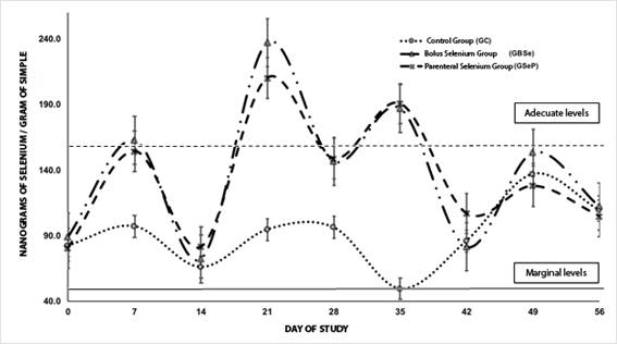
In figure 1 on the concentration of Se in the study groups, it is observed that for day 0 there is no difference between groups (p> 0.05) averaging 84,115 ng of Se per g of sample. By day 7, the supplemented groups have, on average, higher levels of Se, with a significant difference with GC (p <0.05) (158,617 ng vs 97,256 ng); on day 14, no differences were observed in Se concentrations in erythrocytes in the study groups with an average of 73,359 ng; on day 21 it is observed that the supplemented groups have, on average, higher Se concentrations without significant difference between them (p> 0.05), but significantly different from GC (P <0.05) (223.668 ng vs 94.720 ng); the same happens for day 28 (147,836 ng vs 96,310 ng) and on day 35 (188,890 ng vs 49,707 ng). From day 49 to 56 no significant differences were observed (p> 0.05) between groups, averaging 91,614 ng, 139,457 ng and 108,869 ng respectively.
In Figure 2 the absorbances obtained for IgG in serum of the study groups are observed. From day 0 to day 28, no significant differences were observed in the study groups (p> 0.05); from day 35 until day 49, significant differences were observed (p <0.05) between the groups supplemented with Se and GC; on day 35 the groups supplemented with Se average 1,096 absorbance unlike GC which has 0.839 for IgG; on day 42 the supplemented groups presented greater absorbances than GC (p <0.05), averaging 1.318 against GC of 0.996; On day 49 GBSe and GSeP have an average absorbance of 1,251 and the GC has an absorbance of 0.934; on day 56 no significant differences were observed for the study groups (p> 0.05), averaging an absorbance of 0.960.
DISCUSSION
Intraruminal boluses have the advantage of releasing the active ingredient that they are containing for longer periods of time than when they are supplemented in a parenteral form, so they can be safer; for example, in the prevention of selenium deficiency without having to periodically administer the mineral to the animals. However, there is the possibility that the animals regurgitate the bolus, split it and swallow it again, releasing the active principle as selenium immediately, and there may be a risk of intoxication. To avoid this, two types of qualities are used; one has to do with the use of density, which ensures that the object will stay close to the bottom of the reticle-rumen and the other with the geometric shape, which reduces the possibilities of regurgitation (Vandamme and Ellis, 2004).
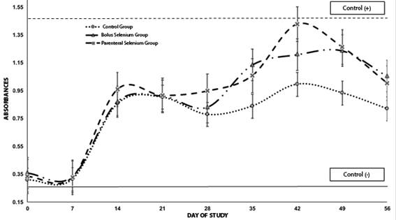
Figure 2 Absorbances for IgG in serum of the study groups, in the experiment the lower continuous line, shows the absorbance of the negative control and the upper dashed line shows the absorbance for the positive control
The boluses manufactured for this work, fulfilled the density characteristics required to avoid regurgitation, Cardinal, 2000, mentions that densities between 2-3.5g / ml prevent the regurgitation of boluses. In this work the boluses presented a density of 2.665g / ml, so they fall within the range required to prevent bolus expulsion, which was not observed in this work. Regarding the geometric shape, this proved to be suitable for administration in kids, the bolus was easily administered manually without the need of a specialized instrument; the oral mucosa of the animals was not injured, nor was the esophagus of the animals injured. Finally, the hardness of the boluses ensures that they do not fracture during administration; the hardness obtained for these boluses was 25,017, which proved to be effective, since there were no fractures due to the handling of the bolus or the administration thereof.
With the application of intraruminal boluses, it is sought to reduce the frequency of treatments, the labor for the handling of the animals and the administration of the medication; it is also sought that the treatment is efficient, that reduce the traumatic and stressful effects for the animal, that it offers availability for prolonged periods and that diminish the collateral effects to the minimum, in particular the possible poisoning. It is worth mentioning that with regard to the productive parameters, it was observed in this work that the food consumption did not present significant differences in the study groups (p> 0.05),
In the work carried out by Simoes Cortinhas et al., 2012, in dairy cows supplemented with selenium, they do not find significant differences in food consumption, as happened in the present experiment. Therefore, it is considered that although this parameter was not positively influenced in this study, the measurement of feed intake may be useful in this experiment as an indicator that the animals did not receive toxic doses of Se, since when this occurs, the consumption of food is affected negatively as mentioned by Lopez-Arellano et al., 2015. Regarding the evaluation of weight gain, on day 14 GC loses weight significantly (p < 0.05), unlike the supplemented groups; it is from day 21 that the animals supplemented with Se have better weight gains than GC, until the end of the experiment on day 56 (p <0.05).
In work done on the weight and weight gain of animals with respect to selenium supplementation Chauhan et al., 2016, observed that lambs supplemented with better weight gain than the control group. On the other hand, Cristaldi et al., 2005, found no differences in the average weights of castrated sheep at the end of their experiment; however, weight gains were not measured as in the present experiment, so although there are reports of positive effects in terms of Se supplementation on weight gain, there are also unfavorable results on this parameter. Qin et al., 2007 explains that in many studies no effect of Se supplementation on weight gain has been observed when there are normal or marginal Se concentrations in the diet.
In this work the animals did not have marginal concentrations of Se in erythrocytes, due to the amount of Se in the diet (1.11 (gSe/g of sample), which could explain the lack of differences in the productive parameters, such as the gain of weight or food consumption. On the other hand, regarding the estimation of selenium in biological samples, Stefanowicz et al., 2013 mention that although plasma is commonly used as an indicator in studies on the status of Se in the organism, it can decay rapidly, regardless of the actual status of the mineral in the organism during acute phases of response; while trials were erythrocyte levels are more robust and not the concentrations are affected by such factors as the systemic inflammatory response.
Qin et al., 2007, mention that the analysis could be done in whole blood, since the hemolysis of the erythrocytes during the handling of the sample could cause false high values in plasma. Therefore, studies of Se levels in erythrocytes are a good option to avoid false levels, as a result of mishandling the sample. With regard to supplementation, it was observed that there was a significant increase in selenium levels in the groups supplemented by the two routes from day 14 (p <0.05). In the study by Van Ryssen et al., 2013, they mention that small ruminants below 50 ng of Se per g of fluid are considered marginally deficient and below this value there could be repercussions on the health of ruminants.
The animals of the present study began the experiment with an average of 84 ng of Se per gram of sample, which are levels close to the marginal ones. By day 28 the animals supplemented with Se reach their maximum level, with an average of 147 ng of Se, being able to be considered adequate levels, taking into account those reported in serum and whole blood; so it is considered then that, in the present work, the supplementation of Se was achieved to increase the levels of the mineral in the animals up to adequate levels in erythrocytes, although without differences between the two forms of supplementation. For this work it was proposed that the supplementation of Se through two administration routes would have differences in the concentrations of Se erythrocytes; however, there were no differences associated with the supplementation route, although it should be mentioned that the parenteral route required two administrations with 15-day intervals to maintain levels in erythrocytes; this could be a disadvantage with respect to the bolus, since the bolus only required one administration and there was no regurgitation in this study.
In the work done by Gutiérrez Olvera et al., 2005 in sheep, Se boluses of 5 g with 50 mg were used for intraruminal release, which maintained the release of Se until day 120, with Se concentrations in the animals of 148 ng/g sample. In our work on day 28 an average of 147 ng of Se/g of sample was obtained for the two supplementation routes, so for this work both routes seem to be effective for the maintenance of adequate levels of Se in kids; however, only these levels were measured for 56 days, which would require a longer study to know the behavior of the bolus. On the other hand, the parenteral dose recommended by Ramírez-Bribiesca et al., 2004 of 0.25 mg/kg of live weight managed to increase Se levels in erythrocytes until reaching adequate levels, as shown by the results obtained. The author mentions that the safety of the dose of Se for the animals depends on the concentration of the mineral in blood before administering the treatments.
In the work done by Davy et al., 2016, three different methods were evaluated for the supplementation of Se in ruminants: through premixes, subcutaneous injection and intraruminal boluses, the experiment lasted 90 days and found that the best form of supplementation of It is through the intraruminal boluses, which maintained adequate levels of Se in the blood for a longer time than the subcutaneous injection, the author mentions that the injection can be an easier method in what refers to the resistance of the cattle to the bolus administration, manipulator skill and sometimes time; however, the bolus is more effective due to less handling of the animals, less stress and labor costs.
Regarding the humoral immune response, no significant differences in IgG absorbances were observed in the study groups during the first 28 days of the experiment (p> 0.05); however, groups supplemented with Se show significantly higher absorbances for IgG from day 28 after administration of the bacterin-toxoid and until the end of the experiment. In the work done by Rodinova et al., 2008 sheep were supplemented with Se and IgG concentrations were measured in the mothers and their offspring, finding that the supplemented animals had higher IgG concentrations than the non-supplemented animals. Alhidary et al., 2016, evaluated the antioxidant status and the immune response in camels supplemented with long-acting boluses, finding data similar to ours in regard to the behavior of the antibodies after the antigenic challenge.
Alhidary et al., 2016, found that in the first immunization no differences were observed in the supplemented groups and the controls, after the second immunization, greater differences were observed between the supplemented and non-supplemented groups. The authors mention that trace minerals are able to regulate the immune response, so they are often used as immunostimulants, having importance in the immune response both innate and adaptive; which could be observed through the humoral immune response.
CONCLUSIONS
In the present work, the consumption of food was not significantly influenced by selenium supplementation. With regard to weight gain, no significant difference was observed between the study groups; however, it was possible to increase selenium concentrations in erythrocytes in the supplemented animals; although no differences were observed between the forms of supplementation. Finally, the humoral immune response increased with Se supplementation.

