SciELO - Scientific Electronic Library Online

 
vol.34 número4Técnica de Rendezvous y prótesis metálica autoexpandible como tratamiento para síndrome de asa aferenteTerapia combinada con coil y cianoacrilato guiada por ultrasonido endoscópico en una várice gástrica fúndica índice de autoresíndice de materiabúsqueda de artículos
Home Pagelista alfabética de revistas  

Servicios Personalizados

Revista

Articulo

Indicadores

Links relacionados

  • No hay artículos similaresSimilares en SciELO

Compartir


Endoscopia

versión On-line ISSN 2444-6483versión impresa ISSN 0188-9893

Endoscopia vol.34 no.4 Ciudad de México oct./dic. 2022  Epub 30-Ene-2024

https://doi.org/10.24875/end.23000014 

Clinical cases

Successful re-intervention with antegrade insertion of the metal stent into the hepaticojejunostomy through the hepaticogastrostomy route

Re-intervención exitosa con inserción anterógrada de prótesis metálica en la hepático-yeyuno-anastomosis a través de la hepático-gastro-anastomosis

Juan L. Romero-Flores1 

Eva Juárez-Hernández2 

Iván López-Mendez1  3  * 

1Endoscopy Unit

2Translational Research Unit

3Transplant and Hepatology Unit. Medica Sur Clinic and Foundation, Mexico City, Mexico


Abstract

Endoscopic ultrasound-guided hepaticogastrostomy (EUS-HGS) is a therapeutic tool that allows for biliary drainage when conventional endoscopic retrograde cholangiopancreatography is technically or clinically unsuccessful. For those cases where re-intervention of HGS stent is necessary to treat obstruction, this kind of approach could be an option. We describe a novel technique to treat EUS-HGS obstruction.

Keywords Endoscopic ultrasound; Hepaticogastrostomy; Stent; Obstruction

Resumen

La hepático-gastro-anastomosis guiada por ultrasonido endoscópico es una herramienta terapéutica que permite el drenaje biliar, cuando la colangiopancreatografía retrógrada endoscópica convencional no tiene éxito técnico o clínico. Existen escenarios en donde se presenta la disfunción de esta anastomosis por lo que es necesaria la reintervención. Han sido descritos distintos abordajes, sin embargo, hasta el momento no existe una técnica estándar. En este caso describimos una técnica endoscópica diferente para la disfunción de la hepático-gastro-anastomosis.

Palabras clave Ultrasonido endoscópico; Hepático-gastro-anastomosis; Prótesis; Stent; Obstrucción

Introduction

Endoscopic ultrasound-guided hepaticogastrostomy (EUS-HGS) is a therapeutic tool that allows for biliary drainage when conventional endoscopic retrograde cholangiopancreatography (ERCP) is technically or clinically unsuccessful; around 5.6% of patients present stent obstruction at long-term follow-up1. Here, we describe a novel technique to treat EUS-HGS obstruction.

Case report

A 63-year-old man, diagnosed with a Klatskin tumor in 2019, was initially treated with ERCP, with the placement of two uncovered self-expandable metal stents (UC-SEMS) to the left and right hepatic ducts. After a positive response to oncologic treatment, a biliodigestive derivation (Roux-en-Y surgery) was carried out. In March 2021, the patient presented tumor progression with acute cholangitis; hence, an EUS-HGS was performed with a SEMS (Giobor Stent Taewoong Medical, Seoul, Korea). In April 2022, the patient presented acute cholangitis; the treatment of choice was UC-SEMS biliary stent (70 mm × 10 mm Wall Flex Biliary RX Stent; Boston Scientific Corp) placement in the HGS. In December 2022, the patient reported a new episode of acute cholangitis2.

A therapeutic endoscopic re-intervention was done. An indirect canulation through the HGS stent approach was decided, and with a diagnostic gastroscope (190 Olympus Medical Systems, Tokyo, Japan), the HGS SEMS was identified. Lumen obstruction from abundant cellular debris was observed.

With a sphincterotome (TRUEtome 44; Boston Scientific Corp), the HGS stent was cannulated, and a 0.035 mm guidewire (Jagwire Revolution High-Performance Guidewire, Boston Scientific Corp) was advanced up to the left hepatic duct. (Fig. 1) The cholangiography showed stent dysfunction and dilatation of intrahepatic ducts.

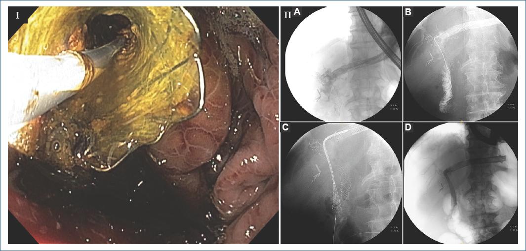
Figura 1 I. Selective cannulation of the hepaticogastrostomy with conventional sphincterotome. II. Cholangiogram. A: intrahepatic biliary dilatation with diffuse infiltration of cholangiocarcinoma. B: selective cannulation of the main left hepatic duct with the guidewire; contrast shows the hepaticojejunostomy. C: placement of the UC-SEMS from the hepaticogastrostomy to the hepaticojejunostomy. D: complete tract from the hepaticogastrostomy to the hepaticojejunostomy. 

The guidewire was repositioned until it reached the hepaticojejunostomy (HJ). Then, an 8.5 × 10 Fr biliary dilatation catheter (Soehendra, Cook Medical, Winston Salem, NC, USA) was advanced until it reached the HJ. Later, an antegrade UC-SEMS biliary stent (80 mm × 10 mm WallFlex Biliary Stent, Boston Scientific Corp) was advanced from the HGS stent to the HJ. The patient improved clinically, with cholangitis resolution. No procedure-related adverse events were encountered.

Discussion

Patients with hepatobiliary malignancy have benefited from increased survival rates in recent years, due to new drug chemotherapies and indicated surgery in borderline resectable and locally advanced hepatobiliary cancer3.

With an increased survival rate, the probability of EUS-HGS obstruction and dysfunction increases over time because of an increased mucosal hyperplasia and biliary sludge that accumulates inside the stent4. Some studies have shown that stent length >3 cm has been associated with an increased patency time5, but very few studies have reported a concise technique to recover patency through the original stent.

A recent case report by Atalla et al.6 showed a similar technique to treat biliary malignant stenosis in a patient with modified anatomy, by introducing an anterograde stent through the puncturing of the left hepatic duct and placing a metal stent in the common bile duct.

The "through the mesh" technique, described by Minaga et al.7, is another example of unconventional intervention of an already obstructed stent. They reported that puncturing the mesh of a metal stent and then placing a guidewire in which they would later install another metal stent could be an alternative to treat this type of patients.

Argon plasma trimming is another recently described technique, in which patency is recovered through the coagulation of mucosal hyperplasia and sludge8.

Another described technique is the EUS-HGS dysfunction approach through direct visualization with a cholangioscope; this device allows a direct visualization of the stenosis zone and a more accurate maneuverability with the guidewire and sphincterotome9.

Conclusion

Although there is no gold standard for re-intervention of HGS stents, these techniques are promising to offer alternatives in the growing scenario of patients that with the new surgical and oncological treatments have been surpassed the survival rates and, in consequence, increased the probability of EUS-HGS dysfunction and the need of re-intervention.

The approach for patients with HGS dysfunction with an anterograde insertion of UC-SEMS is a technique that could be considered an additional treatment option.

References

1. Jagielski M, Zieliński M, Piątkowski J, Jackowski M. Outcomes and limitations of endoscopic ultrasound-guided hepaticogastrostomy in malignant biliary obstruction. BMC Gastroenterol. 2021;21:202. [ Links ]

2. Yokoe M, Hata J, Takada T, Strasberg SM, Asbun HJ, Wakabayashi G, et al. Tokyo Guidelines 2018:diagnostic criteria and severity grading of acute cholecystitis (with videos). J Hepatobiliary Pancreat Sci. 2018;25:41-54. [ Links ]

3. Eshmuminov D, Aminjonov B, Palm RF, Malleo G, Schmocker RK, Abdallah R, et al. FOLFIRINOX or gemcitabine-based chemotherapy for borderline resectable and locally advanced pancreatic cancer:a multi-institutional, patient-level, meta-analysis and systematic review. Ann Surg Oncol. 2023;30:4417-28. [ Links ]

4. Ogura T, Higuchi K. Endoscopic ultrasound-guided hepaticogastrostomy:technical review and tips to prevent adverse events. Gut Liver. 2021;15:196-205. [ Links ]

5. Ogura T, Yamamoto K, Sano T, Onda S, Imoto A, Masuda D, et al. Stent length is impact factor associated with stent patency in endoscopic ultrasound-guided hepaticogastrostomy. J Gastroenterol Hepatol. 2015;30:1748-52. [ Links ]

6. Atalla H, Shiomi H, Sakai A, Masuda A, Kodama Y. Combined bridging and antegrade stent placement during transmural treatment for malignant hilar biliary obstruction in a patient with surgically altered anatomy. VideoGIE. 2020;6:87-9. [ Links ]

7. Minaga K, Takenaka M, Miyata T, Ueda Y, Kitano M, Kudo M. Through-the-mesh technique after endoscopic ultrasonography-guided hepaticogastrostomy:a novel re-intervention method. Endoscopy. 2016;48:E369-70. [ Links ]

8. Yane K, Katanuma A, Maguchi H, Takahashi K, Osanai M, Kin T, et al. Successful re-intervention with metal stent trimming using argon plasma coagulation after endoscopic ultrasound-guided hepaticogastrostomy. Endoscopy. 2014;46 Suppl 1 UCTN:E391-2. [ Links ]

9. Yonamine K, Koshita S, Kanno Y, Ogawa T, Kusunose H, Sakai T, et al. Endoscopic ultrasound (EUS)-guided antegrade intervention for a hepaticojejunostomy anastomosis obstruction under peroral cholangioscopy via an EUS-guided hepaticogastrostomy route. Endoscopy. 2022;54:E788-9. [ Links ]

FundingNone.

Ethical disclosures

Protection of human and animal subjects. The authors declare that no experiments were performed on humans or animals for this study.

Confidentiality of data. The authors declare that no patient data appear in this article. Furthermore, they have acknowledged and followed the recommendations as per the SAGER guidelines depending on the type and nature of the study.

Right to privacy and informed consent. The authors declare that no patient data appear in this article.

Use of artificial intelligence for generating text. The authors declare that they have not used any type of generative artificial intelligence for the writing of this manuscript, nor for the creation of images, graphics, tables, or their corresponding captions.

Received: March 29, 2023; Accepted: August 10, 2023

* Correspondence: Ivan López-Méndez E-mail: yahvelopezmendez@gmail.com

Conflicts of interest

None.

Creative Commons License Instituto Nacional de Cardiología Ignacio Chávez. Published by Permanyer. This is an open ccess article under the CC BY-NC-ND license