SciELO - Scientific Electronic Library Online

 
vol.31 número4Relevancia del reprocesamiento de los endoscopios en el control de infeccionesNeurofibromatosis y tumor estromal gastrointestinal diagnosticado por enteroscopia anterógrada. Reporte de caso índice de autoresíndice de assuntospesquisa de artigos
Home Pagelista alfabética de periódicos  

Serviços Personalizados

Journal

Artigo

Indicadores

Links relacionados

  • Não possue artigos similaresSimilares em SciELO

Compartilhar


Endoscopia

versão On-line ISSN 2444-6483versão impressa ISSN 0188-9893

Endoscopia vol.31 no.4 Ciudad de México Out./Dez. 2019  Epub 01-Nov-2021

https://doi.org/10.24875/end.m19000169 

Casos clínicos

Endoscopic ultrasound fine needle biopsy of the liver with the new 20G procore needle in a patient with abnormal liver function tests and without solid liver lesion

Toma de biopsia hepática con un nuevo tipo de aguja, ProCore 20G, en un paciente con alteración de las pruebas de funcionamiento hepático pero sin lesiones focales

Félix I. Téllez-Ávila1  * 

Gilberto Duarte-Medrano1 

Fredy Chable-Montero2 

1Departamento de Endoscopia Gastrointestinal, Instituto Nacional de Ciencias Médicas y Nutrición Salvador Zubirán;. Ciudad de México, México

2Departamento de Patología, Hospital San Ángel Inn Universidad. Ciudad de México, México


Abstract

Conventionally, liver biopsies are obtained by surgery, through percutaneous or transjugular. All that are good options but all have disadvantages as costs, complications risk, or invasiveness grade. Recently, endoscopic ultrasound-guided liver biopsies have showed good diagnostic yield in patients with solid liver lesions; however, in patients without solid lesions have been unsatisfactory. This problem has related to the kind of needles used. Recently, needles with a new design for liver biopsies have been created.

Key words Endoscopic ultrasound; Biopsy; Liver

Resumen

Tradicionalmente las biopsias hepaticas se obtienen a través de cirugía, vía percutánea o transyugular. Todas son buenas opciones pero tienen desventajas como costos, riesgos o el grado de invasión. Recientemente la toma de biopsias hepáticas guiadas por ultrasonido han mostrado buenos resultados en pacientes con lesiones focales, sin embargo en pacientes sin lesiones los resultados no han sido satisfactorios. Esta falta de resultados adecuados se ha asociado al tipo de agujas utilizadas. Recientemente se han diseñado agujas para su uso exclusivo para toma de biopsias hepáticas.

Palabras clave Hígado; Ultrasonido endoscópico; Biopsias

Introduction

Conventionally, the options for obtaining liver samples have included percutaneous biopsy (PCB) and the vascular approach. PCB is the most common and has a diagnostic yield between 67% and 94%, with potential complications described in 0.09-3.1% of the cases1. The transjugular approach, which is used as an alternative is not without complications (0.5-6.5%)2. There is evidence that endoscopic ultrasound (EUS) is a useful method for liver pathology25. However, there are some reports with conflicting results mainly in patients with abnormalities in liver function tests (LFTs) without solid liver lesions6.

Clinical case

A 64-year-old woman who went to medical assessment because during general evaluation abnormalities in the LFTs was documented with elevated alanine aminotransferase (ALT) and aspartate aminotransferase (AST) (ALT 220 IU/L and AST 280 IU/L) with elevated alkaline phosphatase in 350 IU/L. Rest of the LFT were normal. Patient was asymptomatic. In abdominal ultrasound only hepatic steatosis was reported without any other abnormalities. A computed tomography scan of the abdomen reported as normal. Viral hepatitis tests were reported as negatives as well as antibodies antinuclear. LFTs were repeated on two occasions and were founded with similar results. For this reason and due to the desire of the patient, we decided to perform a liver biopsy guided by EUS (EUS fine-needle biopsy [FNB]).

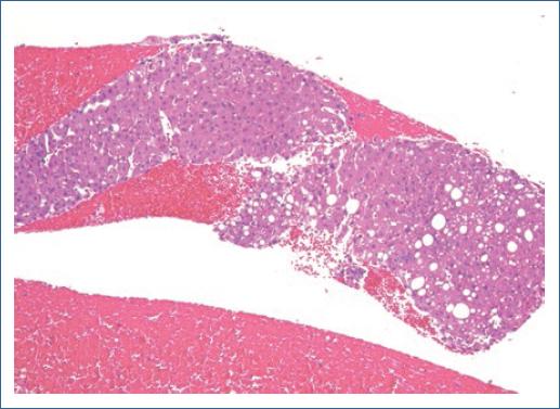
The above procedure was carried out with the new needle EchoTip ProCore 3-20-c (Cook Medical, Limerick, Ireland) which is specifically designed to take liver biopsies. EUS FNB was performed by the conventional manner with capillarity technique (Video 1) and with one single pass. Histopathological result was of “non-alcoholic steatohepatitis” with the presence of evaluable liver tissue sample including 10 portal tracts (Fig. 1).

Figura 1 Evaluable liver tissue sample 

Discussion

Actually, there are some controversies about if tissue samples of the liver obtained by EUS guidance are a good method for diagnostic approach mainly because the tissue sample obtained with EUS FNA is mainly useful for “cytologic evaluation” and does not allow to see the “architecture” of the parenchyma. EUS FNB is considered as a good option for obtaining better tissue samples compared to EUS FNA7. The previous reports of EUS-guided tissue samples that have included patients with non-specific alterations in LFTs and biopsy were taken randomly from parenchyma and have been showed discrepancy in the results25.

In the present case, the EUS FNB was made with a needle with a new design specifically for achieve a “tissue sample that allows to see architecture in the histologic evaluation.” This specific point could be the difference in many patients with liver diseases traditionally of “difficult diagnosis.” Of course, this is only one case report and we need studies with good simple size that allows a real evaluation. For now, we can say that the new device could represent, in the future, a good option for EUS FNB in patients with abnormal LFTs without a solid liver lesion.

References

1. Elsayes KM, Ellis JH, Elkhouly T, Ream JM, Bowerson M, Khan A, et al. Diagnostic yield of percutaneous image-guided tissue biopsy of focal hepatic lesions in cancer patients:ten percent are not metastases from the primary malignancy. Cancer. 2011;117:4041-8. [ Links ]

2. Pineda JJ, Diehl DL, Miao CL, Johal AS, Khara HS, Bhanushali A, et al. EUS-guided liver biopsy provides diagnostic samples comparable with those via the percutaneous or transjugular route. Gastrointest Endosc. 2016;83:360-5. [ Links ]

3. Diehl DL, Johal AS, Khara HS, Stavropoulos SN, Al-Haddad M, Ramesh J, et al. Endoscopic ultrasound-guided liver biopsy:a multicenter experience. Endosc Int Open. 2015;3:E210-5. [ Links ]

4. Gor N, Salem SB, Jakate S, Patel R, Shah N, Patil A, et al. Histological adequacy of EUS-guided liver biopsy when using a 19-gauge non-tru-cut FNA needle. Gastrointest Endosc. 2014;79:170-2. [ Links ]

5. Stavropoulos SN, Im GY, Jlayer Z, Harris MD, Pitea TC, Turi GK, et al. High yield of same-session EUS-guided liver biopsy by 19-gauge FNA needle in patients undergoing EUS to exclude biliary obstruction. Gastrointest Endosc. 2012;75:310-8. [ Links ]

6. Dewitt J, McGreevy K, Cummings O, Sherman S, Leblanc JK, McHenry L, et al. Initial experience with EUS-guided tru-cut biopsy of benign liver disease. Gastrointest Endosc. 2009;69:535-42. [ Links ]

7. Duarte-Medrano G, Téllez-Ávila FI. Diagnostic yield ProCore™needle biopsy guided by endoscopic ultrasound in patients with a first non diagnostic EUS-FNA and lesions difficult to diagnose by EUS-FNA. Endoscopia. 2013;25:100-4. [ Links ]

Recibido: 01 de Septiembre de 2019; Aprobado: 14 de Septiembre de 2019

* Correspondencia: Félix Ignacio Téllez Ávila E-mail: felixtelleza@gmail.com

Creative Commons License Instituto Nacional de Cardiología Ignacio Chávez. Published by Permanyer. This is an open ccess article under the CC BY-NC-ND license