


 nueva página del texto (beta)
 Inglés (pdf)
 Artículo en XML
 Referencias del artículo
 Enviar artículo por email
 Citado por SciELO
  Similares en
SciELO
Similares en
SciELO 
 Permalink
Introduction
Kidney stone incidence depends on geographical, climatic, ethnic, dietary and genetic factors, and so accordingly, the prevalence rates for urinary stones vary from 1% to 20%1,2. Many studies have shown there to be a strong family history in patients with kidney stones, especially in those with recurrence. In a comprehensive genetic study in Iceland of 5954 patients with kidney stones significantly more kidney stones were determined to be seen in family members compared to the general population3,4. Genetic polymorphism also causes nephrolithiasis. The most known polymorphic genes are the calcium-sensing receptor (CASR), vitamin D receptor (VDR), matrix Gla protein, and plasminogen activator, urokinase5,6. Furthermore, the concordance rate of stone disease in monozygotic twins is substantially higher than in dizygotic ones (32.4% vs. 17.3%) demonstrating that genetic factors play a vital role in the formation of nephrolithiasis7.
Homeodomain interacting protein kinase 2 (HIPK2) has been shown to be a new androgen receptor regulator. HIPK2 and androgen have been shown to mediate kidney tubular epithelial cell injury and apoptosis8,9. In a recent study, Lin et al. found that the HIPK2 gene polymorphism also increased kidney stone risk in Chinese males10. Some studies have stated that renal tubular epithelial cell (RTEC) damage is closely associated with early basic lesions of kidney stones11,12. The shedding and death of these cells as a result of damage expose the basal membrane. Stone formation is facilitated by crystals adhering to these parts. Information related to HIPK2 polymorphism and renal stone formation is new-found and inconclusive. To the best of our knowledge, there is only one study in literature which has investigated the effect of HIPK2 polymorphism on nephrolithiasis10. This study aimed to investigate whether HIPK2 polymorphism is associated with renal stone formation in a Turkish population.
Materials and methods
Approval for the study was granted by the Hospital Local Ethics Committee (E1-20-334). The clinical trials registration ID of this study is NCT04804436. Informed consent was obtained from all the patients. Between August 2018 and October 2019, patients with idiopathic calcium nephrolithiasis were recruited to the study, and patients with other stone compositions were excluded. The age and gender of patients were recorded. The study included a total of 129 patients with calcium nephrolithiasis and 67 age- and gender-matched healthy controls. Blood and urine tests were performed on all patients. Nephrolithiasis was diagnosed on the basis of non-contrast enhanced computed tomography (CT). Stone specimens were obtained after shock-wave lithotripsy and analyzed with X-ray diffraction. Patients with a history of chronic urinary tract infection, renal failure, gastrointestinal diseases, increased levels of vitamin D, sarcoidosis, primary hyperoxaluria, polycystic kidney disease, gout, renal tubular acidosis, primary or secondary hyperparathyroidism, and a history of cancer or anatomic abnormalities were not included in the study.
Blood samples were collected into EDTA tubes. The DNA of patients was extracted with a QIAsymphony® automated DNA isolation system (Qiagen Inc. Mississauga, ON, Canada). The real-time PCR amplification was performed using Human rs2058265, rs6464214, and rs7456421 TaqMan® SNP Genotyping Assays (Thermo Fisher, Waltham, MA, USA) in a final volume of 20 mL reaction mixture, including 10 ng of genomic DNA, 5 mL of TaqMan® Universal PCR Master Mix, and 0.5 mL of 40× TaqMan® assay. Thermal cycling conditions were as follows: initial denaturation at 94°C for 3 min, then 40 cycles of 94°C for 15 secs, and 60°C for 1 min. The Rotor-Gene Q Series Software Version Q 2.3.1 (Rotor-Gene Q Series, Qiagen) was used for allelic discrimination.
Data analysis was performed using SPSS for Windows, version 20 software (SPSS Inc., Chicago, IL, USA). Continuous variables showing normal distribution were reported as mean ± standard deviation values, and when not normally distributed, as median (minimum-maximum) values. Categorical variables were stated as number (n) and percentage (%). The Chi-square test was utilized to compare the differences in the genotype and allele frequencies between the patient and control groups. All statistical tests were two-tailed. A value of p < 0.05 was considered statistically significant.
Results
The evaluation was made of a total of 196 subjects, 129 with calcium nephrolithiasis, aged 52 ± 12 years, and 67 age-matched control subjects with no familial history of urinary stone diseases, abnormal urine analysis findings, and no findings of kidney stones on non-contrast-enhanced CT examinations.
The incidence of single nucleotide polymorphism (SNP) in single allele and double alleles in the rs2058265 region was found to be 25% and 3.1%, respectively, in the nephrolithiasis patient group, and 29.9% and 9% in the control group (p = 0.13). The rates of SNP in the rs6464214 region in single allele and double alleles were found to be 20.3% and 8.6%, respectively, in the nephrolithiasis patient group, and 26.9% and 11.9% in the control group (p = 0.37). No statistically significant difference was determined between the groups in respect of these two results. The SNP incidence in double alleles in nephrolithiasis patients at rs7456421 was statistically significantly lower than in the control group (25.6% and 52.2% respectively, p = 0.001). SNP incidence at rs7456421 in single allele was statistically significantly higher in nephrolithiasis patients than in the control group (66.7% and 44.8% respectively p = 0.013) (Fig. 1).
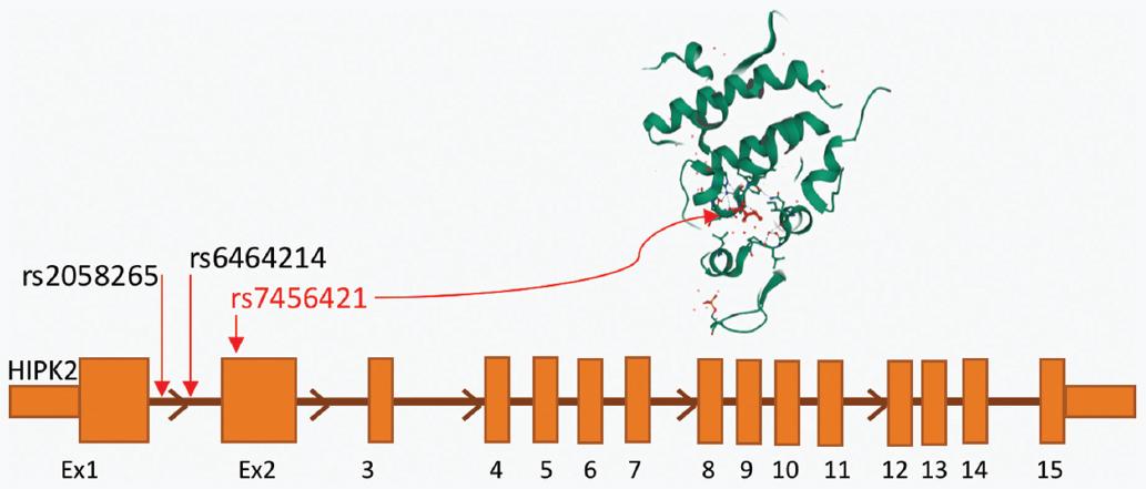
Figure 1 The position of SNPs reported in this publication on the HIPK2 gene is shown at the bottom of the figure. The "kinase domain" of the HIPK2 protein (from uniProt) affected by the rs7456421 change is shown (in the circle) at the top of the figure. The HIPK gene consists of 15 exons and the SNPs included in the study are located in the first parts of the gene. rs 2058265 and rs6464214 are located at intron 1 of HIPK2, rs7456421 is located at exon 2 of HIPK2 and affects the "kinase domain" of the HIPK2 protein.
The genotype and allele frequencies of the rs2058265, rs6464214, and rs7456421 HIPK2 polymorphisms in patients with kidney stones and the control subjects are listed in table 1. The distributions of the genotype and allele of the three polymorphisms (rs2058265, rs6464214, and rs745642 in HIPK2) were not associated with an increased risk of kidney stones in a Turkish population. With the exception of SNP incidence at rs7456421 in a single allele, the three polymorphisms in HIPK2 were higher in the control group in this Turkish population.
Table 1 Comparisons of the allele frequencies of the polymorphisms in patients with nephrolithiasis and healthy controls
| Polymorphism | Patients with nephrolithiasis (n, %) | Control group (n, %) | p-value |
|---|---|---|---|
| rs2058265 | |||
| Single allele | 32 (25%) | 20 (30%) | 0.13 |
| Double alleles | 4 (3.1%) | 6 (9%) | |
| rs6464214 | |||
| Single allele | 26 (20.3%) | 18 (27%) | 0.37 |
| Double alleles | 11 (8.6%) | 8 (12%) | |
| rs7456421 | |||
| Single allele | 86 (66.7%) | 30 (44.8%) | 0.001* |
| Double alleles | 33 (25.6%) | 35 (52.2%) | |
| 129 (100%) | 67 (100%) |
*Statistically significant.
Discussion
Calcium oxalate stones are the most common type of renal stones and occur in approximately 75%-80% of cases13. Genetic analysis can identify susceptible individuals who may develop calcium stones and helps in understanding the stone formation mechanism and predicting the response to drugs and nutrients. Studies of families and twins have proven the importance of genetic predisposition in calcium stones. It has been shown that stone prevalence is higher among relatives of patients with nephrolithiasis when compared to relatives of healthy controls. Family studies have also demonstrated that the genetic transition pattern of the stone is not mendelian and is compatible with the complex polygenic substrate14,15.
Findings of genetic studies demonstrated that Claudin 14, CASR, Osteopontin, and VDR genes may be implicated in human calcium nephrolithiasis. The pathogenetic significance of these genes has not been fully established, although the expression of these genes has been seen to be altered in patients with stones16-18.
Increasing evidence has shown that RTEC damage is closely related to early lesions responsible for the formation of renal stones11,12. These damaged epithelial cells undergo some pathophysiological changes and the adaptive response of these cells plays an important role in kidney stone formation19. An increase in HIPK2 expression in response to DNA damage and oxidative stress stimulates apoptosis through phosphorylation and triggers p53 activation. It has been suggested that these events have a potential role in the apoptosis of RTECs20-22. Lin et al. investigated the relationship between HIPK2 polymorphism in the rs2058265, rs6464214, and rs7456421 regions and kidney stones in a Chinese population, and reported that no difference could be found between nephrolithiasis patients and the control group in terms of these three gene polymorphisms. When only males were evaluated, a significantly increased risk of kidney stones was detected with polymorphism present in these three regions (rs2058265: OR = 2.48, rs6464214: OR = 2.46, rs7456421 OR = 2.84). The authors attributed this situation to the fact that HIPK2 and androgens are involved in RTEC damage and apoptosis. Damaged RTECs play an important role in stone formation by forming the nucleus of kidney stones. HIPK2 may therefore upregulate androgen receptors in male patients. Oxalate synthesis increases in the liver and stone formation is facilitated by increasing androgen receptors8-11,23. In the current study, the role of HIPK2 polymorphism in rs2058265, rs6464214, and rs7456421 regions in nephrolithiasis formation was evaluated in a Turkish population. Interestingly, the study results showed that HIPK2 SNP in these three regions was significantly higher in both alleles of control group subjects than in stone patients. The results were similar when only male patients were evaluated. Only the HIPK2 SNP in the rs7456421 region in a single allele was found to be higher in patients with kidney stones than in the control group. HIPK2 polymorphism, which stimulates RTEC damage and therefore kidney fibrosis, is expected to play an important role in the formation of renal stones when the mechanisms described above are considered logically. However, in this study population, no explanation could be found for why HIPK2 polymorphism was detected at higher rates in both alleles of the control group subjects. This demonstrates that a single gene is not responsible for the stone formation mechanism and that environmental factors play an important role together with many genetic changes. The relationship between HIPK2 polymorphism and stone formation and the underlying pathophysiological mechanisms are more complex than expected. Apart from these two studies, there is no other that has evaluated the association between HIPK2 polymorphism and stone formation.
There were some limitations to this study, primarily that the relatively low number of patients could have caused Type II statistical error. Although secondary diseases which could cause stone formation were excluded, confounding factors cannot be completely excluded as several environmental factors are responsible for stone formation. Moreover, that there was no investigation of other genetic disorders which could cause similar stone formation could also be considered a limitation.
Conclusions
Our study showed that the distributions of the genotype and allele of the three polymorphisms (rs2058265, rs6464214, and rs745642 in HIPK2) were not found to be associated with an increased risk of kidney stone in a Turkish population. Further studies with large patient series are needed to clarify the association of HIPK2 polymorphisms with nephrolithiasis.

