


 nova página do texto(beta)
 Inglês (pdf)
 Artigo em XML
 Referências do artigo
 Enviar este artigo por email
 Citado por SciELO
  Similares em
SciELO
Similares em
SciELO 
 Permalink
Introduction
Measurement of intra-abdominal pressure (IAP) is commonly performed in post-operatory and critically ill patients1,2, as an increase in IAP could derive in intra-abdominal hypertension, abdominal compartment syndrome, and failure in another body organs3-5. Hence, an opportune monitoring of IAP allows a prompt diagnosis and adequate treatment of such patients6. There currently exists a variety of techniques for measuring of IAP, both directly and indirectly through the urinary bladder, inferior vena cava, stomach, or vesicle7-15. A review of the principles, advantages, and disadvantages of the diversity of techniques for the measurement of IAP is beyond the scope of this article and reader is encouraged to read reference16. Although there is no gold standard, the Kron's method, based on measuring the intravesical pressure, is widely used in clinical practice for measuring the IAP; it has the inconveniences of water infusion toward the bladder, long measuring time for each data point as well as long waiting time in between consecutive measurements. Hence, there is still a need for improvements in the techniques for measuring IAP, for example, to minimize or reduce contamination of the instruments with patient's urine which results in high measurement costs.
Nowadays, it is undeniable that healthcare costs are relevant for patients but also for healthcare professionals, administrators and government officials. In developing countries, such as Mexico and some others in Latin America, this topic is of paramount importance because we keep dependence on medical supplies imports, the ever-escalating costs will not end and will keep impacting the availability of supplies in our health-care systems. Accordingly, novel initiatives focused on reducing the costs of substituting imports with products generated by local research and development are welcome. Substitution of imported medical devices through local technological innovation represents a viable and desired alternative because it not only diminishes health-care costs but also generates employment. Accordingly, a reduction in costs associated with the measurement of IAP would be beneficial in undeveloped countries.
The main contribution of this study is to introduce a different medical mechanical pressure transducer based on a disposable membrane which represents a physical barrier between the patient's urine and measurement instruments. The transducer was validated in vitro tests by comparing it with a water column, graded in cmH2O that simulated the IAP, as previously reported17, as well as with direct IAP measurements from patients in the intensive care unit (ICU). Human tests were performed by comparing the pressure transducer device versus the Kron's method taken as reference.
Materials and methods
The novel transducer has a hermetic chamber and two valves to achieve pressure measurements (Fig. 1). When the catheter connection valve (A) is plugged, the liquid or gas enters the transduction chamber (B), where the transducer sends air with a pressure proportional to the gas or liquid entering the measuring valve (C), where an electronic pressure measurement device displays the measured pressure in mmHg or cmH2O. The transducer chamber of the proposed pressure transducer was designed using SolidWorks computer-assisted design software (Waltham, MA, USA) and fabricated using the fused filament deposition printing technique which is a rapid and low-cost three-dimensional (3D) printing technique, based on depositing successive layers of melted printing material to conform a 3D object. The printing material was acrylonitrile butadiene styrene (ABS) plastic and the cost of the ABS plastic employed was < 2 dollars. The transducer chamber was divided into two subchambers separated by a thin Nylon membrane that allows the transduction of IAP to the sensor and avoids contamination of the instrument with the patient's urine.

Figure 1 Schematic of the disposable transducer. A: connecting valve to catheter. B: transduction chamber. C: air pressure measuring valve. Bottom: actual view of the disposable transducer.
In vitro test simulating IAP measurements
Using as a simulator of the bladder, a serum bag filled with distilled water was used, and a uniform pressure was exerted on the bag in random form over a range from 5.3 to 33.4 cmH2O by tightening up the screws located on the four corners of a mechanical press. Simultaneous measurements were performed with the disposable pressure transducer and a standard water column manometer graded in cmH2O, Figure 2.

Figure 2 Schematic of in vitro simulated intra-abdominal pressure experiment. A: press. B: water bag. C: catheter. D: three-way stopcock valve connector. E: mechanical pressure transducer with a proximal and a distal valve. F: pressure measurer/calibrator. G: H2O water column graded manometer.
A total of 159 pairs of measurements were performed at different pressures ranges: one-hundred and two pairs of measurements between 0.0 and 21.0 cmH2O (0-15 mmHg), thirty-one pairs between 21.1 and 27.0 cmH2O (16-20 mmHg), and the remaining twenty-six pairs between 27.1 and 34.0 cmH2O (21-25 mmHg). All measurements were performed at a temperature between 20 and 25° C. Data for the pressure transducer were distributed as shown in the stem and leaf plot below, where the decimal point is one digit(s) to the right of the | symbol:
−5 | 9
−6 | 1 1 2 6
−7 | 1 3 3 8 9
−8 | 0 1 6 8 9 9
−9 | 0 0 1 3 5 6 7 7 8 9 9 9
−10 | 1 1 3 5 7 8 9
−11 | 0 1 1 2 5 6 7 7 8
−12 | 1 2 2 3 5 6 8 8
−13 | 1 2 3 4 6 8 9
−14 | 0 0 8 9
−15 | 1 2 2 8
−16 | 2 3 3 3 4 8
−17 | 0 3 3 5 5 6 6 8 9
−18 | 0 2 2 5 6 7 8 8 8
−19 | 1 4 4 5 8
−20 | 2 2 3 4 5 7
−21 | 2 7
−22 | 0 2 2 2 2 8
−23 | 0 0 2 4 6 8 9 9
−24 | 1 7 8
−25 | 3 6 6 6 8 9
−26 | 3 4 5 6 9
−27 | 0 3 3 8 9
−28 | 1 2 3 4 5 8 9
−29 | 5 6
−30 | 2 7 8 9
−31 | 1 3 4 7
−32 | 2 3 3
−33 | 4 6.
Clinical tests
SUBJECTS
The research protocol was submitted to the research committee of Hospital Juárez de México, the study was approved and assigned the registration number HJM 0609/19-I. The studied sample consisted of 20 measurements performed on patients with routine monitoring of IAP admitted to the intensive care unit; those identified with ≥ 10 mmHg were selected for the study. Informed consent was granted by a family member. Patients age ranged from 19 to 56 years (41 ± 14 years), weight 94 ± 19 kg, and height 1.65 ± 0.05 m.
DATA ACQUISITION
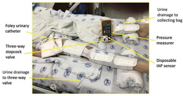
The measurements were made in patients through the indirect method of Kron's, which is the most used method and regarded as the standard to measure the IAP due to due to its low cost and its worldwide acceptance. The Kron's method involves placing a Foley urinary catheter through the urethra until it lodges in the bladder in patients placed in the dorsal decubitus position. Three three-way valves (Luer Lock) are placed in the urine drain line, where a bag of saline solution is connected to the first Luer Lock valve and an H2O column graduated in cmH2O is connected to the second valve. Particularly, in this work, in the third Luer Lock valve was connected to the new pressure transducer device to make sequential measurements of IAP with both methods. Then, the valves are placed in position to allow the urine draining to the bladder and when the urine stops coming out, the urine drainage duct is blocked in such a way that 25 ml of saline can be infused into the bladder to measure with the H2O column. For the data acquisition, an inspiratory pause was placed on the ventilator and the transducers were placed at the level of the median axillary line or the symphysis pubis as "a reference to 0 cmH2O" and the saline was allowed to flow through their respective Luer Lock valves to the connected transducer and the data were acquired. Finally, the Luer Lock valves were placed in the position to drainage of urine. Figure 3 shows an example of the experimental setup during the data acquisition in a patient.
Measurement results were distributed for the pressure transducer device, as shown in the stem and leaf plot below, where the decimal point is one digit(s) to the right of the | symbol:
As can be seen, 11 pairs of measurements were acquired on the pressure range from 0 to 10 mmHg (0-13.595 cmH2O), seven on the range from 10 to 20 mmHg (13.60-27.19 cmH2O), and two on the range from 20 to 30 mmHg (27.19-40.79 cmH2O). These last three patients were sent to the operating room to decompression surgery after measurements were retested. All measurements were performed at a temperature between 20 and 25° C.
Statistical tests
Statistical analysis was performed using the software R (Core Team 2016, R Foundation for Statistical Computing, Vienna, Austria). Statistical tests performed for both experiments were as follows: (a) correlation analysis, (b) linear regression analysis, and (c) Bland-Altman analysis, to determine the existing relationship between the water column manometer graded in H2O and the disposable pressure transducer device.
Results
In vitro measurements simulating IAP
Correlation analysis between the pressure transducer device and the water column manometer graded in H2O showed a Pearson's product-moment correlation r = 0.9980317, with a 95% confidence interval equal to 0.9973070 and 0.9985615, and p-value < 2.2 × 10⁻¹⁶. The fitted line obtained through linear regression analysis was transducer H2O = 0.9863731 * manometer H2O + 0.3816354, with r2 = 0.9961, and p-value < 2.2 × 10⁻¹⁶. Figure 4A presents the linear regression results for the in vitro experiment. From Bland-Altman analysis, we found that the proposed pressure transducer produced a bias of 0.135 cmH2O (p-value = 0.0006132) and a standard deviation of 0.488 cmH2O. Accordingly, the 95% confidence interval was −0.821-1.091 cmH2O. The maximum error was 2.2 cmH2O. Figure 4B shows the corresponding Bland-Altman plot for the in vitro experiment.

Figure 4 In vitro measurements of intra-abdominal pressure using the proposed pressure transducer and the water column manometer of H2O as reference (n = 159). A: regression curve; red line indicates the regression line and the dashed line indicates the identity line. B: Bland-Altman plot: Solid horizontal blue line and solid horizontal red lines indicate the bias and 95% limits of agreement, respectively.
Clinical tests for measuring of IAP
In measurements from patients, we found an absolute error equal to 1.39 ± 1.00 mmHg, which corresponds to a relative error equal to 18.31 ± 16.27% when normalized to reference measurements. The coefficient of variation was computed by dividing the standard deviation of the absolute error by its mean and was found to be equal to 0.72.
We found a Pearson's product-moment correlation r = 0.9735539, with 95% confidence interval equal to 0.9329757 and 0.9896960, and p-value < 5.46 × 10⁻¹³. The corresponding linear model found through linear regression analysis between the proposed pressure transducer (manual) and the reference device (automatic) for IAP was piaAutomaticmmHg = 1.10991* piaManualmmHg - 0.97373, with r2 = 0.9449, and p-value < 5.46 × 10⁻¹³. Figure 5A presents the corresponding regression results for the experiments with patients. When Bland-Altman was applied, we found that the proposed pressure transducer produced a bias of 0.17 mmHg (p-value = 0.6661) and a standard deviation of 1.773549 mmHg resulting in 95% limits of agreement equal to −3.302 and 3.645 mmHg. The maximum error found was equal to 4.411 mmHg. The Bland-Altman analysis corresponding to the highest measurements from patients is shown in Figure 5B.

Figure 5 Measurements of intra-abdominal pressure in patients with abdominal complications using the proposed pressure transducer (manual) and the reference device (automatic) (n = 20). A: regression curve; red line indicates the regression line and the dashed line indicates the identity line. B: Bland-Altman plot: Solid horizontal blue line and solid horizontal red lines indicate the bias and 95% limits of agreement, respectively.
Finally, a summary of the performance of the disposable pressure transducer device when compared to the Kron's protocol for IAP measurements is presented for each patient in table 1 in terms of the absolute and relative errors.
Table 1 Biometrics and performance of the proposed pressure sensor (PIA manual) and the comparison to the reference (PIA automatic) for each of n = 20 patients
| Patient No. | Age (years) | Weight (kg) | Height (m) | Body mass index (kg/m2) | PIA automatic (mmHg) | PIA manual (mmHg) | Absolute error (mmHg) | Relative error (%) |
|---|---|---|---|---|---|---|---|---|
| 1 | 47 | 120 | 1.65 | 44.08 | 14.90 | 13.43 | 1.47 | 10.92 |
| 2 | 19 | 52.4 | 1.56 | 21.53 | 13.10 | 14.18 | 1.08 | 7.61 |
| 3 | 19 | 52.4 | 1.56 | 21.53 | 12.00 | 12.69 | 0.69 | 5.41 |
| 4 | 47 | 120 | 1.65 | 44.08 | 21.20 | 18.66 | 2.54 | 13.63 |
| 5 | 47 | 120 | 1.65 | 44.08 | 5.30 | 6.72 | 1.42 | 21.09 |
| 6 | 47 | 120 | 1.65 | 44.08 | 3.80 | 5.22 | 1.42 | 27.26 |
| 7 | 56 | 66 | 1.62 | 25.15 | 3.70 | 2.24 | 1.46 | 65.27 |
| 8 | 36 | 96.3 | 1.68 | 34.12 | 6.70 | 8.21 | 1.51 | 18.38 |
| 9 | 36 | 96.3 | 1.68 | 34.12 | 3.40 | 6.72 | 3.32 | 49.38 |
| 10 | 36 | 96.3 | 1.68 | 34.12 | 8.90 | 10.45 | 1.55 | 14.81 |
| 11 | 25 | 93.6 | 1.6 | 36.56 | 12.50 | 13.43 | 0.93 | 6.94 |
| 12 | 25 | 93.6 | 1.6 | 36.56 | 10.00 | 9.70 | 0.30 | 3.08 |
| 13 | 25 | 93.6 | 1.6 | 36.56 | 8.20 | 7.46 | 0.74 | 9.88 |
| 14 | 25 | 93.6 | 1.6 | 36.56 | 4.30 | 5.22 | 0.92 | 17.69 |
| 15 | 55 | 93.4 | 1.7 | 32.32 | 3.30 | 2.99 | 0.31 | 10.55 |
| 16 | 55 | 93.4 | 1.7 | 32.32 | 8.10 | 8.96 | 0.86 | 9.55 |
| 17 | 55 | 93.4 | 1.7 | 32.32 | 13.20 | 12.69 | 0.51 | 4.05 |
| 18 | 55 | 93.4 | 1.7 | 32.32 | 25.00 | 20.90 | 4.10 | 19.64 |
| 19 | 55 | 93.4 | 1.7 | 32.32 | 28.00 | 27.61 | 0.39 | 1.41 |
| 20 | 55 | 93.4 | 1.7 | 32.32 | 6.70 | 4.48 | 2.22 | 49.63 |
| Mean | 41.00 | 93.73 | 1.65 | 34.35 | 10.62 | 10.60 | 1.39 | 18.31 |
| SD | 13.79 | 19.15 | 0.05 | 6.63 | 7.15 | 6.37 | 1.00 | 17.27 |
SD: standard deviation.
Discussion
Evaluation of novel measurement instruments requires to quantify their performance through their characteristics that specify how good their measurements are18. In this study, we evaluated such characteristics for a novel disposable pressure transducer device. The relationship between the proposed pressure transducer and the water column was found to be strongly linear through the Pearson correlation coefficient (r = 0.9980317 and r = 0.9735539, for the in vitro and clinical measurements, respectively). Due to the high linear correlation in both experiments, Bland-Altman analysis was employed to verify the data distribution across the measuring range. Bias was found in both experiments, the in vitro experiment was statistically significant (p-value = 0.0006), but the patients experiment was not (p-value = 0.6661).
In the linear regression analysis, in both experiments, the slope of the regression line (m), defining the sensibility of the instrument, was very close to 1 (m = 0.986373 and m = 1.10991). For the in vitro and clinical measurements, the values reflect the values measured with the proposed pressure transducer are almost the same those measured with the water column. While the y-intercept (b) was close to 0 (b = 0.381635 and b = −0.97373, for in vitro and clinical measurements, respectively), this indicates that the value added in the whole range inherent to the proposed disposable transducer is almost 0. Accordingly, the measurements performed with the proposed pressure transducer are almost the same than those made with the water column, which corresponds to a desirable characteristic in the novel measurement instrument. It is worth mentioning that some instruments currently available in the market for pressure measurement have reported in vitro experimental results with a higher zero deviation range than the one found in our pressure transducer device in this study16,19, but we think they do not offer any clinical advantage.
A characteristic that can affect a measurement, or a set of measurements, is the cost of the device. Elevated costs of medical devices in health-care centers around the world, particularly in their critical areas, demand to find more affordable and efficient solutions continuously. Such solutions would allow redirecting economic resources toward innovation in infrastructure, processes, and materials indispensable to offer high-quality medical care at lower costs. As a consequence of high resources consumption, the treatment of critically ill patients in the intensive care units represents a considerable portion the health-care expenditures, which already account for 1% of the gross domestic product in the United States of America20,21. In Mexico, the cost of the device of the Kron's method is between US$ 1.71 and US$ 2.85, while the cost of this new device to clients will be around US$ 1.15 with the advantage of avoiding urine contamination. This result represents approximately half of price the Kron's method. Therefore, a lower cost production of a pressure measurement device, as the one proposed in this study, as well as its portability in all acute care areas of health-care centers, especially those without monitors available, constitute a qualitative and competitive advantage concerning costs.
Conclusions
In the present work, a new novel disposable sensor was tested to measure IAP in humans. Both in-vitro and clinical tests were performed. Based on the provided results, we found that the proposed disposable sensor seems promising for its use in clinical applications. We consider that this and similar efforts contributes to the development of low cost solutions that would be beneficial in countries like Mexico.

