


 nova página do texto(beta)
 Inglês (pdf)
 Artigo em XML
 Referências do artigo
 Enviar este artigo por email
 Citado por SciELO
  Similares em
SciELO
Similares em
SciELO 
 Permalink
Introduction
Ankle fractures are among the most common injuries treated by orthopaedic surgeons. The failure to obtain an anatomic reduction of the ankle mortise leads to altered loading of the tibiotalar joint1,2,3 and subsequent post-traumatic arthritis4,5,6,7 with poor functional outcomes.8 Once malposition is diagnosed, corrective surgery is indicated to avoid long-term disability.
A re-operation rate of 1.6% due to malreduction has been described and the most common cause for re-operation is syndesmotic malreduction.9 Forty-six percent of the re-operated patients have at least two different malreduction sites, and a medial malleolus malreduction is present in 38% of the cases.9
The current literature includes several reports of lateral malleolus poor reduction, rotational malunion, and syndesmosis postoperative incongruence.10,11,12 However, the existing literature overlooks the medial malleolus malunion after internal fixation of ankle fractures and lacks solutions for its treatment. Osteotomy through the original fracture line pattern and internal fixation has been described as an effective solution.13 Based on our experience, this type of osteotomy has high risk of jeopardizing the medial malleolus, and stable fixation is hard to obtain.
Here, we present two case studies of medial malleolus rotation malunion as well as an alternative treatment option.
The «box resection» - Why, How, and When to use it
Rationale: the tibiotalar joint is unstable in lateral direction. To prevent such tendency, the ankle relies on the lateral malleolus, syndesmosis, deltoid ligament, and aligned medial malleolus (where the deltoid ligament is attached). If the medial malleolus is laterally deviated (lateral translation or lateral rotation), all the constructions are doomed to fail because the talus will be laterally displaced, preventing syndesmosis fixation in the correct position.
An osteotomy through the original fracture pattern and re-fixation is the most valid option. The problem arises when the malunited medial malleolus fragment is small, friable, and very difficult to handle, thereby leading to a failed reconstruction. In such cases, we propose a new solution: a bony resection of the articular surface of the medial malleolus that removes the internal block and allows the talus to occupy its original place, creating a congruent mortise. We entitled this technique «box resection».
The indication for this procedure is a malunited medial malleolus causing lateral talar translation, in which a displacement osteotomy and re-fixation planning raises the concern for poor internal fixation stability due to a small or friable fragment.
Surgical tecnhique: An antero-medial approach to the ankle joint is made. The deltoid ligament is first detached at its anterior medial malleolus insertion, and the «box resection» is then applied: the articular (lateral) facet of the medial malleolus is removed with an osteotome in vertical alignment. The amount of bone resection should be the necessary for the talus to correctly realign with the tibial plafond. To complete the «box resection», the most lateral 1 or 2 mm of the distal tibial plafond are removed with a small osteotome introduced in a horizontal orientation. A new «shoulder» is created for the talus to get back to its original position. With the talus repositioned, we proceed with correcting the previous malunited syndesmosis. The syndesmosis is reduced with a clamp and fixed with a flexible system, with the ankle in neutral position. An open syndesmosis debridement is required if the fibrous tissue is compromising anatomical reduction.
Through the medial and lateral approaches, under direct visualization, we can confirm the correct tibiotalar congruency, followed by the evaluation of the ankle sagittal motion (plantar flexion and dorsiflexion). To prevent medial instability, the anterior bundles of the deltoid ligament are re-inserted with anchors.
Case 1
The first case is of a 27-year-old male, referred to our clinic seven months after internal fixation of an ankle fracture. A dorsiflexion deficit caused an abnormal gait due to a hard and sudden block at -30o in the right ankle (Figure 1). Plantar flexion was normal. The patient AOFAS Score was 78 points.

The AP ankle radiograph (Figure 2) showed an apparent congruent joint with a symmetrical clear space between the tibial articular surface and the talar dome, without internal augment. On a second look, an irregularity on the lateral facet of the medial malleolus could be noticed, as well as the incorrect vertical alignment between the tibia and the talus (Figure 2, zoom with circles).

Figure 2: Case 1. Right ankle AP radiograph seven months after the osteosynthesis. Zoom view of the irregularity of the medial malleolus articular (lateral) facet and incorrect tibiotalar vertical alignment (right-superior corner).
The CT Scan confirmed a medial malleolus rotational malunion with a bone fragment that blocked the ankle dorsiflexion. There was also a talar lateral translation causing tibiotalar incongruency and syndesmosis malreduction, with diastasis (Figure 3).

Figure 3: Case 1. CT Scan image showing the malunion of the medial malleolus; the dorsiflexion mechanical block was due to diminished ankle mortise dimensions, which became clinically relevant at 30º due to the increased talar dome size in its anterior half.
The patient was submitted to hardware removal but the dorsiflexion block persisted. As our goal was to remove the dorsiflexion mechanic block and to restore tibiotalar congruency, we decided to perform the «box resection» (Figure 4 and 5). This allowed an immediate medial talus translation. A syndesmosis debridement and open reduction with a clamp was performed and stabilized with a flexible system (TightRope ® Arthrex). We conducted a deltoid ligament re-insertion with anchors (Figure 6). At the end of the procedure, complete dorsiflexion was accomplished with a stable range of motion. Tibiotalar congruency was confirmed in the AP radiograph (Figure 7).

Figure 4: «Box resection» performed in the medial malleolus. The white box shows vertical and horizontal osteotome orientation.
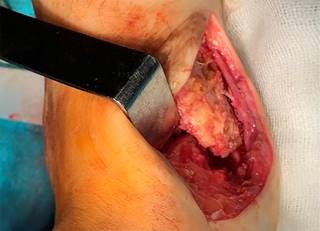
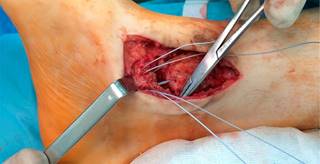

Figure 7: Case 1. One week after reconstructive surgery. AP ankle radiograph showing a congruent joint and syndesmosis reduction.
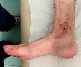
Two weeks after surgery, the patient gained 15o in dorsiflexion, comparing with pre-operative mobility, and started a rehabilitation program. He had a surgical site infection, fully treated with two-week empirical oral antibiotic (amoxicillin/clavulanic acid). Twelve months after surgery, the patient regained almost normal range of motion, with 5o degrees of dorsiflexion (10o less than the contralateral side) (Figure 8) and normal gait without pain. A final AOFAS score of 100 points was obtained.
Case 2
The second case refers to a 79-year-old female submitted to internal fixation of a trimalleolar fracture with one-year follow up. The patient was first treated with a one-third tubular plate on the lateral malleolus, and K wires and a tension band in the medial malleolus. Three months after, she was submitted to medial hardware removal due to a skin infection.
One year post surgery, the patient showed impaired gait, with permanent daily pain and severe limitation of daily activities. The dorsiflexion was limited to 0o, with 15o of plantar flexion. An AOFAS score of 15 was recorded.
The ankle AP radiograph (Figure 9) showed joint incongruency with valgus malunion of the medial malleolus, talus lateral translation, syndesmotic diastasis, and fibular shortening. Due to the unacceptable multiple site malunion, with severe clinical impact, we decided for revision surgery.

Figure 9: Case 2. Left image: one year after the first osteosynthesis. AP ankle radiograph showing medial malleolus valgus malunion, talus lateral translation, syndesmosis widening, and fibular shortening. Right image: intraoperative assessment of medial clear space augmentation (black line).
After hardware removal, the medial translation of the talus was again impossible due to a mechanical block by the malunited medial malleolus. We performed the «box resection» following the same steps used for syndesmosis reduction and fixation and deltoid repair. An oblique osteotomy of the fibula was performed to regain correct length and alignment. Articular congruency was obtained with a stable full range of motion. The final result shows a congruent joint (Figure 10).

Figure 10: Case 2. Six months after revision surgery. Weight bearing AP ankle radiograph showing congruent joint and syndesmosis reduction.
The patient can now walk without mobility aids and with no pain. A dorsiflexion of 10o was achieved, with 20o of plantar flexion. Twelve months after surgery the AOFAS score was 84.
Discussion
We believe these cases should be reported due to two distinguished but associated particularities. First, they present the description of a simple solution for medial malleolus malunion in cases where there is secondary tibiotalar incongruency. Second, they report the consequent syndesmotic incongruency and it’s radiographic evaluation.
To the authors’ best knowledge, there is no relevant literature on the consequences of medial malleolus malunion. Although we can only speculate on this, such conditions are possibly asymptomatic if the translation is in the extra articular (medial) direction, as their functional impact might be similar to a medial malleolus non-union. If the translation is in the articular (lateral) direction, the talus will deviate, originating an incongruent joint. For rotational deformities, the suspicion rate should be elevated because intra-operative radiographic assessment is difficult.
If medial malleolus malunion is responsible for an incongruent mortise, with respective malreducted syndesmosis, corrective surgery is mandatory. Robertson et al. described the option of re-creating the original fracture pattern and fixation as a revision method.13 In the case of a non-reconstructible medial malleolus, due to small size or friability, we suggest another option: removing the mechanical block that causes talar lateral deviation using the «box resection».
In both case studies, our primary goal was to restore a congruent tibiotalar joint with full range of motion. As soon as this procedure was done, the mechanical block was removed and the talus was re-centered in its correct position. With this technique, all the instability issues are addressed: the mortise becomes congruent, the syndesmosis is reduced, and the deltoid ligament is re-inserted to avoid medial instability.
The turnover of the technique is the removal of cartilage from the medial malleolus articular facet. Ultimately, this may accelerate post-traumatic arthritis. However, and as discussed above, we know that an incongruent mortise leads to altered loading of the tibiotalar joint and subsequent post-traumatic arthritis with poor functional outcomes. This approach gives more importance to articular congruence and stability between the distal tibia and the talar dome than to the removal of a nonbearing articular surface.
Another important issue that deserves our attention is the improper treatment of syndesmosis injuries that can result in latent diastasis, chronic pain, osteochondral lesions, degenerative arthritis, and other sequelae.14 Our case studies also highlight the difficulty to assess syndesmosis reduction and congruency after internal fixation. There is increasing evidence that traditional radiographic measurements, such as tibiofibular overlap, tibiofibular clear space, and medial clear space, are of little value in detecting syndesmotic injury,15,16,17 and the evaluation difficulty is mostly due to the impact of rotation on its imaging. The prevalence of postoperative syndesmotic malreduction has been reported to range between 0 and 16%.18 Gardner et al. found that CT detected significantly more syndesmotic malreduction than standard postoperative radiographic measurements, and reports a prevalence three times higher than the highest rate previously published.18 On the other hand, Rasi et al. showed that the sensitivity of plain radiography was high in diagnosing syndesmosis malreduction after internal fixation and CT scanning was not necessary.19
In our first case, the irregularity of the internal medial malleolus facet did not allow for a correct assessment of the medial clear space. The tibiofibular clear space was apparently greater than 5 mm (abnormal), but the tibiofibular overlap was not accurate for measurement because it was unclear if the radiograph was a strict ankle AP or mortise view. The CT Scan was important to show the transverse plan anatomy and allowed us to see the rotational and sagittal translation of the fibula, as opposed to only diastasis on the coronal plane.

