


 nueva página del texto (beta)
 Inglés (pdf)
 Artículo en XML
 Referencias del artículo
 Enviar artículo por email
 Citado por SciELO
  Similares en
SciELO
Similares en
SciELO 
 Permalink
Introduction
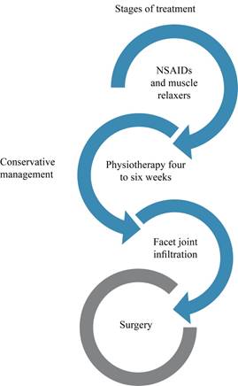
Although the research guidelines aim to improve instrumentation techniques and offer increasingly specific treatments for each of the evolutionary stages of the disease to obtain better results and reduce the incidence of complications, non-surgical management should be the initial action in most spondylolisthesis with and without neurological symptoms.1
Conservative management consists of a regimen of one to two days of rest, followed by a short period of anti-inflammatory drugs and by physical therapy.2 Frymoyer3 established a treatment plan more than two decades ago, which is still used today; this therapy program includes anti-inflammatory drugs, aerobic exercise that improves arterial circulation in the compression zone, weight control, and management of osteoporosis. Regarding anti-inflammatory therapy, the objective is to act directly on the intervertebral joints and the nerve root, reducing the inflammatory mediators released by mechanical compression and therefore reducing pain. Acetaminophen is considered the drug of the first choice, which is preferred over non-steroidal anti-inflammatory drugs (NSAIDs) because it has the same analgesic efficacy to risk-benefit, but without the gastrointestinal and cardiovascular side effects of NSAIDs, this makes it a drug better tolerated by elderly patients.
The next pain management option, in case of a failure within the first four to six weeks, is an infiltration,4 which, is recommended if patients fail a four to six-week course of physical therapy. Epidural corticosteroid injection with local anesthetic is injected over the region of the listhesis to relieve back pain, radicular pain, and neurogenic claudication. In long-term follow-ups in patients undergoing epidural steroid injection, no long-term benefit was demonstrated in degenerative disc disease, herniated disc, radicular low back pain, or spinal stenosis5,6,7,8,9 although, a significant improvement was observed with short-term benefits with pain relief, functional improvement, and decreased operating rates.10,11
Based on a systematic review, it reduced pain by 64 to 81%, disability by 60 to 63% and depression by 56% in patients with low back pain and leg pain and also improved walking tolerance. Even one year after the procedure, pain was reported to be lower than the baseline in a small population of patients. The factors associated with better outcomes after corticoid injection are higher pain scores at baseline, radicular symptoms for fewer than six months, and age less than 70 years. Since degenerative spondylolisthesis develops as a result of inflammatory arthritic and degenerative changes rather than segmental instability, this inflammatory process, could be relieved by epidural injection as a result of targeted delivery of the steroid at the level of spondylolisthesis.12
Facet joint injection is a procedure of injecting local anesthetics and steroids into facet joints for low back pain by facet joint sprain or degenerative changes. It has relatively less side effects and is simpler in terms of techniques than intraspinal treatments due to its direct access to facet joints through paraspinal muscles.13 Studies reported that facet join infiltration is effective not only in axial back pain by facet joints but also in lumbar spinal stenosis.14,15 Hwang SY et al. reported a retrospective study for facet joint infiltration effects on lumbar spinal stenosis patients at risk of surgery hemorrhage due to several medical conditions. Facet joint infiltration was effective in 25 (59.5%) out of 42 patients. On MRI (magnetic resonance imaging), it was more effective in patients with mild-to-moderate central canal stenosis. In this study, it was assumed that steroids can be injected into the epidural space through facet joints. The authors injected 1 ml into each joint and additionally 2-4 ml contrast media or 0.9% normal saline to induce the rupture of the facet joint capsule and the drug efflux into the epidural space. However, there was no correlation between the discharge of contrast media and treatment effect.14 Its short-term benefit could be temporally control of pain only to allow the patient to carry out a better physiotherapy regimen.
Physiotherapy is one of the most used methods in the non-surgical management of symptoms associated with spondylolisthesis. Therapeutic protocols include different modalities for pain management, such as the use of a corset, exercises, ultrasound therapy, electrical stimulation, and modifications of daily activity.16,17,18 Physiotherapy treatments are aimed at reducing pain, restore ranges of mobility, function, improve the balance of the core muscles, strengthen and stabilize the spine.19,20 The use of a stationary bicycle promotes flexion of the spine and decompression of the dural sac, allowing a greater amount of exercise to be performed before presenting the symptoms of neurogenic claudication, as it is a static exercise it avoids the impact on the joints. Other options available are swimming, walking, and exercising on elliptical machines.2
The largest study reported to date comparing conservative versus surgical management is the SPORT study,21,22 for its acronym in English (spine patient outcome research trial), published in 2013 where they follow up for two and four years to 395 patients undergoing surgery and 210 patients with conservative management. In the results, they show that all patients who underwent surgery had a greater improvement than those who were given conservative management and that the subgroups who benefits the most from surgery are: patients under 67 years of age, women, patients without the acid peptic disease, reflex asymmetry, neurogenic claudication, opioids users, patients who do not use antidepressants, disappointment with the symptoms, and those who have a high expectation of surgery. Weinstein et al.23 found that patients with degenerative spondylolisthesis and stenosis treated surgically showed improvement in pain and function during a follow-up period of two years compared to patients who underwent conservative management (Figure 1).
Surgical treatment
If improvement is not archived with the conservative management, surgical treatment must be performed, it brings better outcomes when everything has failed in patients with symptomatic spondylolisthesis, the question to perform a surgical procedure or not is a patient led decision around his symptoms and quality of life, the indications for surgical treatment are:1
Persistent or recurrent lumbar or extremity pain, neurogenic claudication with reduced quality of life, or failure in conservative treatment for a minimum of three months.
Progressive neurological déficit.
Sex, bladder, or neurogenic intestine.
Simultaneously with the etiological description and knowledge of the causes, the surgical treatment of degenerative lumbar spondylolisthesis is developed, initially without the use of instrumentation, seeking only for the root decompression. With the development of pedicle instrumentation and the recognition of degenerative lumbar spondylolisthesis as a specific nosological entity, various approaches and surgical techniques were developed to restrict mobility and/or fuse the affected segments to treat instability and nerve roots compression.24
Posterior interbody fusion (PIF) was initially described in 1925 by Campbell and implemented in 1953 by Cloward in degenerative spondylolisthesis.24 This technique combines direct and indirect root decompression with the fusion between the vertebral bodies by placing an autologous bone graft. Cloward developed the technique using iliac bone grafts after discectomy and later studied the impact of bone grafts on postoperative sagittal balance, reporting a limited rate of complications.25
Internal fixation with a transpedicular screw was described by King in 1944 and associated with interbody fusion in an attempt to avoid nonunion in spondylolisthesis, together with the development of the interbody cage by Roy-Camille contributed greatly to the advancement in current fixation and arthrodesis techniques.26,27 Posterior lumbar interbody fusion (PLIF) is the traditional technique, is achieved by performing a fenestration in the laminae and partially resecting the facet, retracting later the dural sac and nerve roots to access the intersomatic space. In 1982, Harms and Rollinger developed the transforaminal lumbar interbody fusion (TLIF) technique,28 which has an advantage over PLIF, by avoiding over-retraction of the dural sac and nerve roots as the implant entry zone is at through the foramen, facilitating access to the intersomatic space so potentially avoiding injury in these structures,29 later in 1988 Steffee and Sitowski associated posterior arthrodesis with posterior fixation.1 The advantage of these approaches is that posterior access is the most common and familiar technique for spine surgeons, and decompression and fusion procedures can be performed through the same approach. In 2008, Yan et al.30 compared PLIF versus TLIF for single-level fusion in grade I-II degenerative spondylolisthesis. They performed interbody fusions with posterior transpedicular instrumentation and a minimum follow-up of two years, reported no cases of migration and, all patients achieved fusion, also the complication profiles were similar between groups, with radiculitis and screw loosening, while Liu reported a significantly higher rate of dural tear in PLIF (12 vs 3.9%, p = 0.030), postoperative nerve root dysfunction (9.6 vs 1.9%, p = 0.018) and reoperation (10, 4 vs 1.9%, p = 0.018).31
Regarding clinical improvement, both techniques are reported with good or excellent scores in function, with an average pain improvement of four points (p ≤ 0.001).30,31 The percentage of slip significantly improved between preoperative and initial postoperative radiographs in both groups an average of 30.1-31.4%. Liu et al.31 reported that patients in the PLIF group had significantly longer surgery times (242 ± 67 vs 188 ± 46 min, p = 0.037), higher intraoperative blood loss (483 ± 403 vs 308 ± 385 ml, p = 0.035) and higher blood transfusion rates (19.2 vs 4.9%, p = 0.001). Finally, both fusion techniques were successful in significantly increasing intervertebral space and foraminal height. In particular, there were no significant differences between the two radiographic measurements.
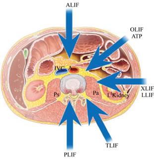
The different treatment options for spondylolisthesis have been extensively studied to identify which offer better clinical results and a lower rate of complications and reoperations, as the anterior approach for interbody fusion (ALIF) provides the best fusion rate due to the wide bone surface of the vertebral platforms compared to the one provided by posterior techniques. Indirect compression can be achieved with these techniques due to the ligamentotaxis effect exerted by the interbody cage. The muscular damage in this technique is minimal, complications, when they appear, tend to be more serious, the ureteral and intestinal injury, damage to the great vessels, and alterations in ejaculation in men are the most described.32
Lateral lumbar interbody fusion (LLIF) is recognized as a less invasive surgical method, performed through an anterolateral transpsoas approach.33 LLIF has been used as an alternative to conventional anterior approaches and can be used from the L1-L2 to L4-L5 segment. There are sub-variants to this technique called extreme lateral (XLIF) and oblique (OLIF) interbody fusion, where the site of entry of the interbody cage varies to a lateral and oblique position, respectively. These techniques present less bleeding and surgical time, shorter hospital stay and lighter postoperative pain than the posterior approach. Among the complications reported is pain on flexion and extension of the hips due to manipulation of the iliac psoas, paresthesia, and motor alterations due to injury to the ilioinguinal, iliohypogastric, lateral femoral cutaneous and genitofemoral nerves, other less frequent injuries include the large vessels trauma and post-incisional hernias.32
The PLIF and LLIF in degenerative spondylolisthesis were compared in a study published by Pawar et al. in 2015,34 reported that the surgery time was similar between the groups, but the average blood loss was significantly lower in the LLIF than the group PLIF (438 vs 750 min, p < 0.01), the incidence of dural tear was lower with LLIF (0 vs 5 p = 0.014). In the LLIF group, foraminal height, intervertebral space height, and lumbar lordosis were restored. No permanent iatrogenic neurological deficits were reported in either group. The LLIF group significantly decreased disability as measured by the Oswestry disability index, but without significant differences in other clinical outcome scores between groups.
Norton et al. reported that patients undergoing interbody fusion through an anterior or lateral approach are significantly less likely to develop intraoperative blood loss anemia but present a higher risk of visceral injury compared to those who underwent PLIF/TLIF35 (Figure 2).
In the comparison between LLIF and minimally invasive TLIF in the treatment of one or two levels of grade I-II of degenerative spondylolisthesis, it was found that blood loss was lower in the LLIF group than in TLIF. The average surgery time and length of hospital stays did not differ between groups. As a complication, there was a weakness in hip flexion, which was observed in the LLIF group in 31% of the patients and resolved within six months in all cases. The sensory or distal motor deficits reported were transient, and no significant difference was identified between the groups. The LLIF fusion rate was 100%, and the TLIF 96%, one pseudarthrosis required reoperation and was the only one reported in the follow-up of the two groups. Pain, disability, and quality of life scores were significantly improved from baseline in both groups. Radiographically, the disc height improved significantly in both groups in all evaluations, however, there was a greater postoperative increase in the central area of the spinal canal in the TLIF, and the LLIF group presented subsidence at the two-year follow-up.
Lumbar fusion techniques in degenerative spondylolisthesis should be individualized to the clinical and imaging characteristics of each patient. These publications present data that indicate that lateral and transforaminal fusion have fewer complications compared to PLIF32 (Table 1).
Table 1: Benefits and disadvantages of the different surgical approaches.
| Benefits | Disadvantages | |
|---|---|---|
| ALIF | • Less bleeding | • Lower lumbar segments only |
| • Big surface for cage placement | • Visceral or vessels damage | |
| • Less muscular damage | • Retrograde ejaculation | |
| • Better lordosis restoring | • Post incisional hernias | |
| LLIF | • Less bleeding | • Hip flexion pain |
| • Big surface for cage placement | • Visceral or vessels damage | |
| • Less muscular damage | • Less lordosis restoring | |
| • Fast surgical timing | • Post incisional hernias | |
| • Upper and lower segments | ||
| PLIF | • Only one approach | • Most dural tear incidence |
| • Almost none visceral or vessels damage | • Dural sac retraction | |
| • All lumbar segments | • More bleeding | |
| • More muscular damage | ||
| • Less surface for arthrodesis | ||
| • Less lordosis restoring | ||
| • Laminae and partial or complete facet resection | ||
| TLIF | • Only one approach | • Complete facet resection |
| • Minimal dural sac retraction | • Nerve root lesion | |
| • Almost none visceral or vessels damage | • Dural tears | |
| • All lumbar segments | • More bleeding | |
| • More muscular damage | ||
| • Less surface for arthrodesis | ||
| • Less lordosis restoring |
Among the surgical techniques considered for the management of degenerative spondylolisthesis; Decompression without fusion is considered the less invasive technique than fusion with and without instrumentation. This technique reduces the morbidity and mortality associated with spinal fusion in older patients.36,37,38,39,40 The one-year readmission rate of patients undergoing lumbar decompression with and without fusion is 9.7 and 7.2%, respectively.41 It has been found that 69% of patients report satisfactory results with decompression without fusion, and 31% present unsatisfactory results. A report with 10-year follow-up, of a group of patients with an average age of 67 years, with a diagnosis of grade I-II degenerative spondylolisthesis a decompression was performed, 69% of the patients reported excellent results and concluded that non-fusion decompression procedures provide adequate results in a select group of elderly patients with low-grade spondylolisthesis.42 Another study of patients with spinal stenosis who underwent laminotomy or laminectomy included a subgroup of patients with degenerative spondylolisthesis without finding data of instability in post-laminotomy patients by preserving the dynamics of the segment by maintaining the integrity of the posterior capsule-ligament complex vs three postoperative laminectomy patients who developed instability.43
Other studies report unfavorable results after decompression without fusion. Modhia U et al.41 reported that 45% had good results of decompression without fusion, and 55% had poor or unsatisfactory results. In contrast, 63% who underwent decompression with posterolateral fusion in situ had satisfactory results. Their study suggests that decompression has better results when a non-instrumented fusion of the segment is added.44
A problem related to non-instrumented in situ fusion is the inability to restore normal lumbar lordosis, particularly when there are involving multiple segments. It has been shown that an in situ fusion that produces kyphosis or hypo lordosis increases the mobility of the adjacent joints and this may be a factor that contributes to the degeneration of the adjacent segment after fusion.45 The manifestations in the degeneration of the adjacent segment can present as symptomatic or asymptomatic degeneration, stress or compression fractures of the adjacent vertebra due to bone fragility secondary to osteoporosis that some patients have, for which some authors recommend the use of decompression and fusion without instrumentation and not fixation with the use of implants, always evaluating the needs and expectations of each patient and remembering that as far as possible, the purpose of surgical treatment is to release nerve compression, reduce listhesis to restore sagittal balance, perform a fixation to stabilize and place a bone graft to achieve arthrodesis of the segment. In an attempt to avoid the degeneration of the adjacent segment, Rosales-Olivarez et al.46 perform studies between the posterolateral fusion technique with the INO plate and circumferential fusion with the INO plate and intersomatic screw plus in patients with a diagnosis of degenerative spondylolisthesis. In their results, both groups improved listhesis, function, and pain. The INO plate + posterolateral fusion (PLF) favors with flexibility and reduces intervertebral height loss in grade 1 or two pre-surgical listhesis, while the use of the INO plate + intersomatic screw + PLF reduces listhesis and decreases the loss of height in listhesis grade 3 or 4. Meanwhile, Juárez-Jiménez et al.47 studied two groups of patients with degenerative lumbar spondylolisthesis operated with the circumferential arthrodesis technique. In 23 patients, a dynamic stabilization system was placed in the overlying segment (group L), they show in the results observed at five years that the ligamentoplasty does not prevent the degeneration of the adjacent segment.
As results are increasingly in favor of surgery for the treatment of degenerative spondylolisthesis, research is focusing on the amount of surgery needed. The literature supports that fusion is necessary to achieve the best and long-lasting results; however, the debate now seems to have focused on the best way to achieve it. Zdeblick48 and colleagues compared non-instrumented posterolateral fusion and two different types of instrumentation, the results revealed a fusion rate of 65% seen with non-instrumented fusions, a fusion rate of 77% with the use of semi-rigid instrumentation, and 95% with the use of rigid fixation.
The addition of anterior column support or 360-degree fusion has many theoretical benefits. Authors propose that anterior spine support improves fusion rates by increasing the surface area available for fusion, offers indirect decompression, and helps restore normal lumbar lordosis. Intersomatic fusion has become a popular method in the treatment of spondylolisthesis, with used ranges from 14% in 1999 to 37% in 2011.49 There are several ways to achieve anterior support, each with its own benefits and a unique set of complications21 (Figure 3).
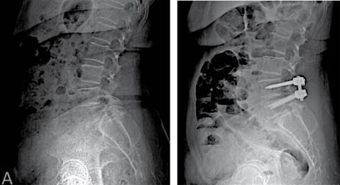
Figure 3: A) X-Ray appreciates L4L5 spondylolisthesis. B) X-Ray obtained after surgery, with L4L5 360o fixation.
When planning a surgical procedure, the degree of osteoligamentary resection necessary to achieve decompression, the degree of listhesis, the segmental instability, the degree of disc degeneration, the severity of the pain, the spinopelvic balance, the inherent surgery risks and the characteristics of the patient must be taken into account to make the best decision. The surgical strategy must be individualized to achieve an adequate fusion with the minimum of possible risks.50 The purpose of interbody fusion is to improve the rate of success in surgery. However, improving the rate of fusion with these methods has no direct relationship with the degree of clinical improvement.51 Another important factor while planning surgery is if it involves a complex procedure, are considered a complex procedure those that involves more than two levels or a 360o arthrodesis. These procedures report greater morbidity, a higher number of serious complications, and a higher rate of rehospitalization in the first 30 days after surgery, as well as a more expensive cost compared to patients who underwent a simple decompression or a decompression with a simple fusion.
Conclusion
Surgery offers several potential benefits in the treatment of degenerative spondylolisthesis, but the existing data do not strongly support its benefit in all patients. They don’t conclusively identify an optimal technique. A decision must be made based on the experience of the surgeon, the clinical and imaging parameters for the selection of the most appropriate approach and fusion method.

