


 nueva página del texto (beta)
 Inglés (pdf)
 Artículo en XML
 Referencias del artículo
 Enviar artículo por email
 Citado por SciELO
  Similares en
SciELO
Similares en
SciELO 
 Permalink
Background
Diabetes mellitus (DM) world prevalence ranges from 4 to 6.5%, with an exponential increase in the last decade.1 Spain does not escape this reality; the prevalence of diabetes in our country is 13.8%, with almost 6% ignoring they have this serious disease.2
The World Health Organization (WHO) calls diabetes the epidemic of the 21st century.2 DM complications cause a major impact on health and quality of life of patients, with an elevated cost to health systems and society around the world.3
One of the most debilitating complications of diabetes mellitus is Charcot neuroarthropathy (CNA). Defined as an inflammatory condition, affects the foot and ankle with varying degrees of bone destruction and deformity, the most classic one known as «rocker bottom» deformity4 (Figure 1).

The diagnosis of CNA in the active phase can be done clinically, confirming the presence of distal neuropathy accompanied by a swollen, red and painful foot.5,6 In the non-active phases of CNA structured foot and ankle deformities increase by 7 times the possibility of amputation in these population, and 12 times when there is an ulcer.7
Eichenholtz classification8 is used to define Charcot foot clinical stages. Brodsky9 classification, in the other hand, allows us to locate the lesion anatomically, midfoot being the most commonly affected area. Recent literature states that existing clinical classifications do not provide a clear pathway for treatment decision and proposes a simpler classification using the terms active and non-active Charcot.4
The goal of CNA treatment, both orthopedic and surgical, is to obtain an ulcer free, stable plantigrade foot, without OM and able to ambulate.10 Achieving these goals notably reduces the rate of amputations.11
Some studies suggest that radiological measures may predict the development of ulcers; Wukich12 defined a limit of 27 degrees of alteration Meary’s line in the lateral X-ray of the foot as a predictor of ulcers.
Classically, surgical treatment was reserved for patients in whom the orthopedic treatment had failed;13 recent publications suggest that in selected patients, early surgical treatment may offer a good option achieving correction and stabilization of the deformity.14
Due to the poor bone quality of these patients, Sammarco15 proposed the «superconstructs» concept, applying large diameter hardware across non affected joints, acting as a frame.
Pinzur16 proposes the «single stage surgery» that can treat both bone and soft tissue infection, and correct foot deformity through a system of circular static fixation. With external fixation techniques, authors have managed to avoid amputation in the majority of their patients.17
Material and methods
In our foot and ankle unit the treatment of CNA follows a protocol (Table 1); according to this treatment algorithm, patients with plantigrade stable foot are treated conservatively with adequate footwear and adapted insoles if Charcot foot is in the non-active phase. In the active phase, the total contact cast (TCC) or diabetic specific walker boots should be used.
Table 1: Charcot neuroarthropathy treatment protocol used in our center.
| Plantigrade foot. Active | Total contact cast |
| Plantigrade foot. Non active | Adequate footwear and adapted insoles |
| Non-plantigrade foot. No history of ulcers and OM, good soft tissues and good bone stock | Internal fixation «superconstruct» |
| Non-plantigrade foot. History or presence of ulcers and OM, bad soft tissues and poor bone stock | «Single stage surgery» with circular external fixation |
| Failed internal fixation | «Single stage surgery» with circular external fixation |
Meanwhile, non-plantigrade patients are treated surgically if they don’t respond to orthopedic measures. If there is no presence or history of ulcers and OM, good soft tissues and good bone stock, internal fixation using superconstructs may be a treatment option. But we must admit that patients with these characteristics are difficult to find in our area.
When there is poor bone stock, soft tissue compromise, an active ulcer and bone infection or recent history of it, we do a «single stage surgery» with circular external fixation.
Surgical treatment in our center is performed, using the «single stage surgery» technique with the circular external fixator that we learned directly from Dr. Michael Pinzur at Loyola University Medical Center in Chicago.
Surgical technique: We use the circular external fixator Distraction Osteogenesis Ring static System® (DePuy Synthes). We proceed to describe the surgical technique for midfoot Charcot, even though it can be used for the lesser common hindfoot Charcot. After spinal or regional anesthesia, the contralateral foot is protected to prevent decubitus injuries during surgery. The tourniquet is placed at thigh level and used only during the first half of the surgery, being released before applying the fixator. The patient is supine; sterile surgical drapes, that include the entire limb, are used.
Step 1. Correction of the equinus deformity: Percutaneous tenotomy of the Achilles tendon, through three incisions allows the hindfoot to return to a more physiological position (Figure 2). If during the preoperative planning equinus deformity is believed to be caused by gastrocnemius shortening we use the Strayer technique to address it.

Step 2. Remodeling osteotomy and infected tissue resection: According to the anatomical location of the deformity apex in the midfoot, and the presence of abduction or adduction deformity, we use a medial or lateral approach to perform the osteotomy, which is extended without a doubt if there is risk of compromising the skin during the procedure. In some complex cases, we have found a double approach to be more comfortable. Subperiosteal plane dissection around the entire circumference of the midfoot creates a safe working space that preserves tendons and neurovascular elements of the area. Adequate exposure is achieved using Hohmann elevators (Figure 3).

Preoperative planning determines the shape and size of the necessary osteotomy in order to achieve the ultimate goal, a plantigrade foot with proper alignment and resection of the infected tissue. This bone resection relaxes the soft tissues (by reduction of the total length of the foot) and contributes to the healing of existing ulcers.
Once the bone wedge has been resected we proceed to temporary fixation with two 3 mm diameter Steinman pins. They are introduced through the dorsum of the foot toward the plantar hindfoot avoiding the dorsal neurovascular bundle. The image intensifier controls the correct position of the foot and the lack of plantar bony prominences.
Step 3. Ulcer excision: If there is a plantar ulcer it should be excised and submitted for microbiological study. There is an option of closing the plantar area with separated nonabsorbable sutures without excessive skin tension. If closing sutures are not possible, it is advisable to close by secondary intention with or without negative pressure systems.
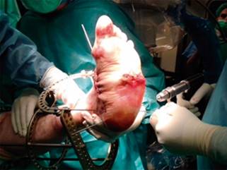
Step 4. Applying the circular frame: The circular static fixator is applied to the patient with 1.8 mm reduction wires. We use two wires on the calcaneus with a 30 degrees angle between each other (Figure 4). The structure at risk here is the retromalleolar medial neurovascular bundle. Once the hindfoot is set, one wire should be applied distal to the arthrodesis area in the midfoot. This last wire is fixed just behind its ideal place, this will focus compression in the arthrodesis area once we apply tension to it.
Subsequently, two wires will be positioned in each of the two tibial rings, with a 30 degree angle between them. If the area to compress is the ankle we can do it simply by descending the distal ring to the foot plate. It is essential to know precisely the anatomical arrangement of the neurovascular elements in each level of placement of the transfixing wires.
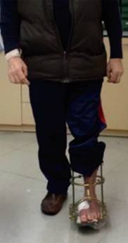
Step 5. Postoperative protocol: Usually, patients start partial weight bearing at 48-72 hours after surgery (Figure 5). Wound and pin site care are done as usual. The patient is discharged from the hospital 48-72 hours after surgery. Follow-up visits and X-rays are done every two to three weeks. Frame removal is performed at 8-12 weeks depending on clinical and X-ray evolution. After removal of the frame, a weight-bearing total contact cast (TCC) is used 4 to 6 weeks. After this, they make a transition to diabetic footwear with adequate insoles. These patients should be followed monthly by trained personnel to prevent recurrence of active Charcot or new ulcerations.
Results
According to our protocol and the use of the circular external fixator we are able to obtain an ulcer-free foot, successful treatment of the OM, correction of the deformity, avoided amputation and prevented recurrence of the ulcer in all our patients. Some complications may arise; superficial infection at pin entrance that is solved with local wound care and, a couple of broken wires that did not need to be replaced.
Discussion
Most Charcot foot injuries can be treated orthopedically.4 The most recently published treatment guide by the International Working Group of Diabetic Foot (IWGDF)18 recommends the use of TCC as the treatment of choice in acute NCA until clinical stabilization, which coincides with our treatment algorithm.
We must avoid amputation at all cost because literature clearly shows that mortality of diabetic patients 5 years after amputation is as high as 60%, being in some cases superior to some types of cancer.19 In some patients, the classical internal fixation techniques may be doomed to failure, due to the poor bone quality, secondary to vitamin D deficiency related osteoporosis, that characterizes diabetic patients.20 Another factor to take into account is that this patient population is prone to pseudarthrosis,21 which applies to the osteosynthesis material to continuous mechanical stress with a high risk of rupture and failure.
In cases of Charcot foot with suspected osteomyelitis and or presence of a plantar ulcer, resection of infected bone with static circular external fixation, with specific antibiotic treatment, guided by microbiological culture, is the best choice of treatment and there are multiple studies that support this technique in these situations.11,13,16,22,23,24
Until now our results regarding cure rate of plantar ulcers, correction of deformity, osteomyelitis eradication and recurrence of injury is consistent with the results obtained by other authors.16,17
There are publications that propose the treatment of Charcot foot with hybrid external fixation systems using classic pins.25 Its disadvantage is the risk of fractures in the site of insertion of the classic big pins24 in diabetic patients with very poor bone quality.
Rogers et al26 refers complications of using static circular fixation systems are frequent but of little clinical relevance. The most common, with an incidence between 10 and 20%, is the superficial infection of pin site that satisfactorily resolves with local treatment or may need oral antibiotic therapy. In our series, the management and prognosis of superficial infection at this level does not vary from the published data.26
Other complications related to ischemia time as compressive neurapraxia, deep vein thrombosis, skin necrosis and wound infection26,27 were not present in our series. The fact that we only use ischemia for the first half of the surgery may play a role in the latest.
For all previous reasons we firmly believe that external circular static fixation is one of the most ad-vantageous treatment options for complex Charcot foot. It has some clear benefits when compared to classic internal fixation in these patients (Table 2). That is why we decided to adopt this technique as treatment of choice in our center.
Table 2: Benefits of circular external fixation compared to classic internal fixation.
| Circular external fixation | Internal fixation | |
|---|---|---|
| Requires ample surgical dissection | No |
Yes |
| Tolerates osteomyelitis | Yes | No |
| Immediate weight bearing | Yes | No |
| Risk of peri-implant fracture | No | Yes |
| Requires good bone stock | No | Yes |
| Pseudarthrosis may affect fixation system | No | Yes |
| Need for removal of osteosynthesis material | No | May be necesary |
Charcot foot is a very complex disease, not only because of the injury itself but because of characteristics inherent to the host in whom it develops. The objectives of the orthopedic surgeon must be to provide an adequate and patient-tailored treatment. The correct assessment of the patient in multidisciplinary diabetic foot units and a strict compliance with treatment protocols, will allow us to give each patient the opportunity to prevent a major amputation.
The weight bearing total-contact cast in acute CNA and superconstructs in very selected patients are literature based valid treatment options contemplated within the algorithm that we use in our center. «Single stage surgery» with circular external fixator will be a useful technique for treating cases of complex Charcot foot with poor bone stock, soft tissue compromise, an active ulcer or bone infection or recent history of it. The use of economic resources for treating these patients after surgery have been significantly lower after «single stage surgery», which is an important fact for deciding treatment of choice nowadays.
«Compliance with ethical standards»
Conflict of interest: The authors declare that they have no conflict of interest.
Funding: There is no funding source.
Ethical approval: This article does not contain any studies with human participants or animals performed by any of the authors.
Informed consent: Informed consent was obtained from all individual participants included in the study.

