SciELO - Scientific Electronic Library Online

 
vol.29 número6Clasificación de las fracturas supracondíleas en niños: Gartland, AO y Holmberg. ¿Cuál tiene el mayor acuerdo interobservador?Uso de un nuevo implante: U-Force N6 a nivel L5 S1 para evitar la inestabilidad lumbar tras discectomía simple índice de autoresíndice de materiabúsqueda de artículos
Home Pagelista alfabética de revistas  

Servicios Personalizados

Revista

Articulo

Indicadores

Links relacionados

  • No hay artículos similaresSimilares en SciELO

Compartir


Acta ortopédica mexicana

versión impresa ISSN 2306-4102

Acta ortop. mex vol.29 no.6 Ciudad de México nov./dic. 2015


Original articles

Changes in the radiological measurements of the tibiofibular syndesmal area in patients with Weber C ankle fractures who were treated with open reduction, internal fixation, and transyndesmal screw

Cambios en las mediciones radiológicas de la sindesmosis tibioperonea en pacientes con fracturas de tipo C de Weber tratados con reducción abierta, fijación interna, y tornillo transindesmal

S Jasqui-Remba1  * 

A Torres-Gómez2 

GA Salas-Morales3 

A Hernández-Martínez4 

1 Residente de traumatología y ortopedia, Centro Médico ABC, miembro AO , Resident fellow FACS. México

2 FACS MSc Profesor Adjunto. Ortopedia. (UNAM). Centro Médico ABC. México.

3 Coordinador clínico de educación e investigación en salud. México

4 Médico interno de pregrado Universidad Anáhuac. México.


Abstract:

The tibiofibular syndesmosis provides stability to the ankle mortise. The ankle syndesmosis is compromised in all Weber C type injuries. The radiographic method described by Merle D'Aubigné considers the bony relationships as a measure of syndesmotic widening. We sought to investigate whether the patients with a C type ankle fracture treated with ORIF and placement of a transyndesmal screw have an increment of the tibiofibular space and decrease of the tibiofibular overlap after the transyndesmal screw is removed. Our sample included 52 patients with Weber C ankle fractures treated by ORIF and transyndesmal screw at a level II trauma center. We measured the tibiofibular clear space and tibiofibular overlap in each phase of the treatment. The transyndesmal screw was removed at day 55.56 (± 21.83). We found an increase of the tibiofibular overlap of 0.20 mm (± 2.29, p = 0.532); and 0.21 mm (± 0.97, p = 0.146) in the tibiofibular clear space. The changes of 2.38% in the tibiofibular overlap and 5.29% in the tibiofibular clear space between the postoperative and post-removal periods were not statistically significant. After removal of the syndesmal screw, there is a slight radiographic broadening of the syndesmosis; however, it is small and statistically not significant.

Key words: Ankle; syndesmosis; measurement; surgery; screw; fixation

Resumen:

La sindesmosis tibioperonea distal proporciona estabilidad a la mortaja del tobillo, viéndose comprometida en todas las lesiones tipo C de Weber. El método radiográfico descrito por Merle D'Aubigné usa las relaciones óseas como una medida del ensanchamiento de la sindesmosis. Investigamos si los pacientes con una fractura de tobillo tipo C tratados con osteosíntesis y colocación de un tornillo transindesmal tienen un incremento del espacio y disminución de la superposición tibioperonea, después de que el tornillo se retira. 52 pacientes con fracturas de tobillo Weber C tratadas mediante osteosíntesis y colocación del tornillo transindesmal en un centro de trauma de nivel II. Medimos el espacio libre tibioperoneo y la superposición en cada fase del tratamiento. El tornillo se retiró el día 55.56 (± 21.83). Encontrando un aumento de la superposición tibioperonea de 0.20 mm (± 2.29, p = 0.532); y 0.21 mm (± 0.97, p = 0.146) en el espacio libre. El aumento del 2.38% en el solapamiento tibioperoneo y 5.29% en el espacio libre entre el postoperatorio y los períodos posteriores al retiro no fueron estadísticamente significativos. Después del retiro del tornillo sindesmal, hay una ligera ampliación radiográfica de la sindesmosis; sin embargo, es pequeña y estadísticamente no significativa.

Palabras clave: Tobillo; sindesmosis; medición; cirugía; tornillo; fijación

Introduction

Ankle fractures are one of the most common injuries treated by orthopedic surgeons. (1),(2),(3),(4),(5),(6)

There are several existing classification methods for these injuries in keeping with biomechanical aspects (e.g. Launge-Hansen) (7) and with the anatomy of the fracture.

The two main anatomical classification methods are the number of malleoli involved (be they unimalleolar 2/3, bimalleolar 1/4, or trimalleolar 7%), (8) and the anatomical region of the fracture in relation to syndesmosis (be it infrasyndesmal, transyndesmal or suprasyndesmal). (9)

Both of these methods present us with good intra- and interobserver reproducibility. (10) Weber C type injuries represent 14 to 47% of ankle fractures. (11)

The tibiofibular syndesmosis is made up of the anterior tibiofibular ligament, the posterior tibiofibular ligament, the transverse tibiofibular ligament, the interosseous ligament and the interosseous membrane. (12) It provides stability to the mortise, along with its tolerance and resistance to rotatory and axial translation forces. (3)

In 1972, Danis and Weber demonstrated that in the case of infrasyndesmal fractures, the syndesmosis is not damaged. However, in the case of transyndesmal fractures, 50% show damage to the syndesmosis, and in supra-syndesmal injuries, there was a 100% damage rate to the syndesmosis. (13) It is known that 100% of Weber type C fractures have rupture of the syndesmosis, even without apparent radiographic alteration. (14)

There are various ways to test the stability of the syndesmosis: clinical stress tests in external rotation, (15) pain caused by direct compression (16),(17) and peroneal translation, (15) as well as X-ray examination. The X-rays may show a "clear tibiofibular space" less than 6 mm in size in AP and mortise views, or the overlapping of the anterior tuberosity of the tibia over the fibula. This radiographic measurement depends on the rotation of the foot relative to the X-ray beam direction. (18)

The main measurements in the AP and mortise X-ray projections were described by Merle D'Aubigné (Figure 1):

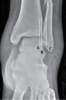
Figure 1: Merle D'Aubigné measurements; A. anterior tubercle of the tibia; B. border of the fibula; C. posterior tubercle of the tibia. 

1. The "clear tibiofibular space", which is represented by the distance between the distal lateral border of the tibia and the medial border of the fibula. Normal: < 6 mm. (19)

2. The "tibiofibular overlap" is the distance between the medial wall of the fibula and the incisal surface of the tibia. Normal: > 6 mm or 42%. (19)

3. The "constant distance" between the two distal tibial tubercles. (18),(19)

The biomechanical instability caused by the syndesmosis is, without doubt, one of the most important predisposing factors for arthrosis of the ankle. (16),(20),(21),(22),(23),(24),(25)

There is a wide variety of tests, surgical interventions, and treatments (26) which ensure optimum care when maintaining the syndesmosis closed and stable when the fixation screw is removed.

The purpose of this study was to evaluate the changes in X-rays of the tibiofibular syndesmosis region (the tibiofibular clear space and the tibiofibular overlap) from the postoperative period to the transyndesmal screw removal in Weber C ankle fracture patients treated with open reduction, internal fixation and placement of a transyndesmal screw.

Material and methods

This is a retrospective study carried out in a cohort of patients with Weber C ankle fractures. They were surgically treated at "Hospital Regional del IMSS # 2 Villa Coapa", a level II trauma center in Mexico City. We included patients between 20 and 60 years of age. All had a transyndesmal fixed Weber C ankle fracture and complete radiographic registers in the preoperative phase of treatment, immediate postoperative before the transyndesmal screw withdrawal, and after its withdrawal (Figure 2) in the period between January 2014 and August 2014. We excluded patients with tibial pilon fractures, exposed fractures, complex injuries or direct trauma. The total sample was of 52 patients. The average age was 44.37 years (± 15.83); 30 were female (57.69%) and 22 male (42.31%) (Table 1).

Figure 2: Analyzed phases of treatment. 

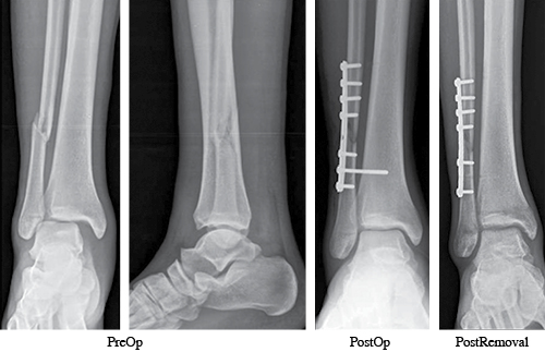
Table 1: Characteristics of the sample. 

Characteristic Value
Age 44.37 ± 15.83
Female sex 30 (57.69%)
Male sex 22 (42.31%)

Values expressed as mean ± SD; absolute frequencies (%).

We studied the radiographic registers of subjects who met the selection criteria. We registered the dates of the fracture, surgery and screw withdrawal, as well as age and gender. Measurements of tibiofibular clear space, tibiofibular overlap and intertubercular distance in each of the x-rays were also recorded.

The measurements were done in the IMPAX 6.4.0.5024 software system, using its included digital millimetric ruler.

Continuous variables were subjected to normality tests (Kolmogorov-Smirnov). All variables had a normal distribution and are thus described as mean, standard deviation, minimum, and maximum. The categorical variables were described as absolute and relative frequencies. Changes in the measurements during different stages of treatment that were considered: preoperative to postoperative; postoperative to pre-screw withdrawal; pre-screw withdrawal to post-screw withdrawal. The primary stage of treatment measured was the postoperative to post-screw withdrawal. The changes in the measurements were analyzed with a paired t-test. The management strategy of missing values was applied "by analysis". Data analysis was performed with IBM SPSS Statistics for Windows (V 16.0, Chicago). A two-tailed p-value of ≤ 0.05 was considered significant.

Results

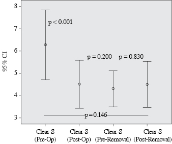
The mean time for the withdrawal of the trans-sindesmal screw was 55.56 days (± 21.83, 23-135) (Table 2). Taking into consideration the Merle-D'Aubigné measurement changes between the postoperative and after the withdrawal of the transyndesmal screw, we observed an increase of the tibiofibular overlap of 0.20 mm (± 2.29, p = 0.532); 0.21 mm (± 0.97, p = 0.146) in the tibiofibular clear space and 0.41 mm (± 2.08, p = 0.177) in the intertubercular distance (Table 3, Figure 3 and Figure 4).

Table 2: Mean time for removal of the syndesmotic screw. 

Value
Time for removal of SS (days) 55.56 (± 21.83, 23-135)

Values expressed as mean (± SD, min-max). SS = syndesmotic screw.

Table 3: Radiographic measurements - Merle D'Aubigné. 

Values expressed as mean (± SD). *Percent of change relative to initial value. TF = Tibiofibular; Clear-S = Clear Space; iT = inter-Tubercular. †T-test for related groups. Means and SDs are different in some cases due to the fact that missing data was pairwise suppressed during analysis.

Figure 3: Changes in the tibiofibular overlap. 

Figure 4: Changes in clear space. 

Discussion

The relevance of fixing the syndesmosis as the most important predisposing factor of stability has been demonstrated by Weening and Bhandari. (27) Maintaining a reduced syndesmosis after the removal of the screw is equally important; thus the importance of the present study.

In this work, we found that after removing the trans-sindesmal screw, no radiographic variations were found, nor anything statistically significant in any of Merle D'Aubigné's measurements. The percentage of change in the radiographic measurements between the postoperative and the withdrawal of the trans-sindesmal screw was small: 2.38% change in the tibiofibular overlap, and 5.29% in the tibiofibular clear space. These changes were not statistically significant. The postoperative radiographic measurements may vary within a safe range. This finding does not necessarily have clinical relevance and does not mean that the original positioning of the screw was wrong or insufficient. It is also probable that in the operating theatre we tend to tighten a bit too much the syndesmosis with the screw.

Previous studies have assessed and compared multiple variables such as differences between surgical procedures, (28) the material with which the syndesmosis is fixed (metal versus titanium (29) versus suture, bioabsorbable or not (3),(30),(31)), the number of cortices, (32) the amount of screws, (33),(34) positioning, (35) the location of the screws, (35),(36),(37),(38),(39),(40),(41) their size, (42) and whether the screw is removed or not. (43),(44) In a previous analysis, Jordan, Talarico, and Schuberth analyzed the effect of the transyndesmal screw removal, as well as its effect on the syndesmosis. They concluded that there is not a significant difference with the withdrawal or in the follow-up. (45)

It is important to consider the variability of different X-ray technicians as projections may vary depending on several factors, including the patient's pain. Millimetric variations in the measurements may be due to these factors. Also, as we know, other important elements exist when it comes to stability, such as deltoid ligament repair. (46)

Given the fact that our inclusion criteria were ample, the results presented in this paper have a high degree of external validity (generalizability) to patients between 20 and 60 years of age with a transyndesmal fixed Weber C closed ankle fracture.

Syndesmosis injuries represent a challenge for orthopedic surgeons in the assessment of the injury, its management, stabilization, and repair.

There are still some issues to be addressed: the ideal number or cortices to be crossed with the syndesmal screw, whether to remove or not the syndesmotic screw, and the best timing for its removal.

With the results of this study, we were able to discover that after the withdrawal of the transyndesmal screw, there are no significant changes in X-ray measurements of the tibiofibular region.

It is clear that there is a difference between clinical and radiographic outcomes, and there is not always a correlation. The observed fact of a slight radiographic aperture of the syndesmosis does not necessarily mean ligamentous deficiency that translates to clinical stability.

In this study, we did not consider the inter-rater variability of radiographic measurements. Although the radiographic technique is standard and we can assume it is constant in all cases; we are aware that even slight variations in the position of the limb (i.e. rotation) may result in alteration of the radiographic measurements due to a variation of the X-ray beam (parallax error). There is a measurement, the intertubercular space, which in theory ought to remain constant due to its anatomical nature. However, we observed a mean difference of 0.41 mm (3.3%) between initial and final radiographic evaluations. We consider this variation as the result of a compound of inter-rater difference and measurement error. This fact might also have had an impact on the other measurements.

The small differences between the postoperative and post-removal periods of the tibiofibular overlap and tibiofibular clear space of 0.20 mm and 0.21 mm, respectively, might not be due to a failure in the syndesmal fixation. We believe that these slight variations could be a consequence of the normal process of screw removal and measurement error.

Acknowledgments

All the authors declare that there is no potential conflict of interest referring to this article.

References

1. Bauer M, Bengnér U, Johnell O, Redlund-Johnell I. Supination-eversion fractures of the ankle joint: changes in incidence over 30 years. Foot Ankle. 1987; 8(1): 26-8. [ Links ]

2. Praemer A, Furner S, Rice DP. American Academy of Orthopaedic S: Musculoskeletal conditions in the United States. Park Ridge, Ill.: American Academy of Orthopaedic Surgeons; 1992: vii, 199. [ Links ]

3. Dattani R, Patnaik S, Kantak A, Srikanth B, Selvan TP. Injuries to the tibiofibular syndesmosis. J Bone Joint Surg Br. 2008; 90(4): 405-10. [ Links ]

4. Van Staa TP, Dennison EM, Leufkens HG, Cooper C. Epidemiology of fractures in England and Wales. Bone. 2001; 29(6): 517-22. [ Links ]

5. Shibuya N, Davis ML, Jupiter DC. Epidemiology of foot and ankle fractures in the United States: an analysis of the National Trauma Data Bank (2007 to 2011). J Foot Ankle Surg. 2014; 53(5): 606-8. [ Links ]

6. Vosseller JT, Karl JW, Greisberg JK. Incidence of syndesmotic injury. Orthopedics. 2014; 37(3): e226-9. [ Links ]

7. Lauge-Hansen N. Fractures of the ankle. II. Combined experimental-surgical and experimental-roentgenologic investigations. Arch Surg. 1950; 60(5): 957-85. [ Links ]

8. Rockwood CA Jr, Green DP, Bucholz RW. Rockwood and Green's fractures in adults. 6th ed./editors, Robert W. Bucholz, James D. Heckman, Charles M. Court-Brown; associate editors, Kenneth J. Koval, Paul Tornetta III, Michael A. Wirth. Philadelphia: Lippincott Williams & Wilkins; 2006. [ Links ]

9. Hughes JL, Weber H, Willenegger H, Kuner EH. Evaluation of ankle fractures: non-operative and operative treatment. Clin Orthop Relat Res. 1979; (138): 111-9. [ Links ]

10. Malek IA, Machani B, Mevcha AM, Hyder NH. Inter-observer reliability and intra-observer reproducibility of the Weber classification of ankle fractures. J Bone Joint Surg Br. 2006; 88(9): 1204-6. [ Links ]

11. Jensen SL, Andresen BK, Mencke S, Nielsen PT. Epidemiology of ankle fractures. A prospective population-based study of 212 cases in Aalborg, Denmark. Acta Orthop Scand. 1998; 69(1): 48-50. [ Links ]

12. Close JR. Some applications of the functional anatomy of the ankle joint. J Bone Joint Surg Am. 1956; 38-A(4): 761-81. [ Links ]

13. Weber BG. Die Verletzungen des oberen Sprunggelenkes. Zweite, überarbeitete und ergänzte Auflage. Vienna: Hans Huber Bern Stuttgart; 1972: 241. [ Links ]

14. Hermans JJ, Wentink N, Beumer A, Hop WC, Heijboer MP, Moonen AF, et al. Correlation between radiological assessment of acute ankle fractures and syndesmotic injury on MRI. Skeletal Radiol. 2012; 41(7): 787-801. [ Links ]

15. Ogilvie-Harris DJ, Reed SC. Disruption of the ankle syndesmosis: diagnosis and treatment by arthroscopic surgery. Arthroscopy. 1994; 10(5): 561-8. [ Links ]

16. Hopkinson WJ, St Pierre P, Ryan JB, Wheeler JH. Syndesmosis sprains of the ankle. Foot Ankle. 1990; 10(6): 325-30. [ Links ]

17. Cotton FJ. Dislocations and joint fractures. Philadelphia: WB Saunders Company; 1911: 654. [ Links ]

18. Harper MC, Keller TS. A radiographic evaluation of the tibiofibular syndesmosis. Foot Ankle. 1989; 10(3): 156-60. [ Links ]

19. Miller MD. Review of orthopaedics. 6th ed. Philadelphia, PA: Elsevier/Saunders; 2012: xvi, 856. [ Links ]

20. De Souza LJ, Gustilo RB, Meyer TJ. Results of operative treatment of displaced external rotation-abduction fractures of the ankle. J Bone Joint Surg Am. 1985; 67(7): 1066-74. [ Links ]

21. Pettrone FA, Gail M, Pee D, Fitzpatrick T, Van Herpe LB. Quantitative criteria for prediction of the results after displaced fracture of the ankle. J Bone Joint Surg Am. 1983; 65(5): 667-77. [ Links ]

22. Egol KA, Pahk B, Walsh M, Tejwani NC, Davidovitch RI, Koval KJ. Outcome after unstable ankle fracture: effect of syndesmotic stabilization. J Orthop Trauma. 2010; 24(1): 7-11. [ Links ]

23. Boytim MJ, Fischer DA, Neumann L. Syndesmotic ankle sprains. Am J Sports Med. 1991; 19(3): 294-8. [ Links ]

24. Veltri DM, Pagnani MJ, O'Brien SJ, Warren RF, Ryan MD, Barnes RP. Symptomatic ossification of the tibiofibular syndesmosis in professional football players: a sequela of the syndesmotic ankle sprain. Foot Ankle Int. 1995; 16(5): 285-90. [ Links ]

25. Roberts RS. Surgical treatment of displaced ankle fractures. Clin Orthop Relat Res. 1983; (172): 164-70. [ Links ]

26. Van den Bekerom MP, Lamme B, Hogervorst M, Bolhuis HW. Which ankle fractures require syndesmotic stabilization? J Foot Ankle Surg. 2007; 46(6): 456-63. [ Links ]

27. Weening B, Bhandari M. Predictors of functional outcome following transsyndesmotic screw fixation of ankle fractures. J Orthop Trauma. 2005; 19(2): 102-8. [ Links ]

28. Van den Bekerom MP, Hogervorst M, Bolhuis HW, van Dijk CN. Operative aspects of the syndesmotic screw: review of current concepts. Injury. 2008; 39(4): 491-8. [ Links ]

29. Beumer A, Campo MM, Niesing R, Day J, Kleinrensink GJ, Swierstra BA. Screw fixation of the syndesmosis: a cadaver model comparing stainless steel and titanium screws and three and four cortical fixations. Injury. 2005; 36(1): 60-4. [ Links ]

30. Thornes B, Walsh A, Hislop M, Murray P, O'Brien M. Suture-endobutton fixation of ankle tibiofibular diastasis: a cadaver study. Foot Ankle Int. 2003; 24(2): 142-6. [ Links ]

31. Cottom JM, Hyer CF, Philbin TM, Berlet GC. Transosseous fixation of the distal tibiofibular syndesmosis: comparison of an interosseous suture and endobutton to traditional screw fixation in 50 cases. J Foot Ankle Surg. 2009; 48(6): 620-30. [ Links ]

32. Høiness P, Strømsøe K. Tricortical versus quadricortical syndesmosis fixation in ankle fractures: a prospective, randomized study comparing two methods of syndesmosis fixation. J Orthop Trauma. 2004; 18(6): 331-7. [ Links ]

33. Xenos JS, Hopkinson WJ, Mulligan ME, Olson EJ, Popovic NA. The tibiofibular syndesmosis. Evaluation of the ligamentous structures, methods of fixation, and radiographic assessment. J Bone Joint Surg Am. 1995; 77(6): 847-56. [ Links ]

34. Rudi TP, Murphy WM. AO principles of fracture management. Stuttgart and New York: Thieme; 2000. [ Links ]

35. Kukreti S, Faraj A, Miles JN. Does position of syndesmotic screw affect functional and radiological outcome in ankle fractures? Injury. 2005; 36(9): 1121-4. [ Links ]

36. McBryde A, Chiasson B, Wilhelm A, Donovan F, Ray T, Bacilla P. Syndesmotic screw placement: a biomechanical analysis. Foot Ankle Int. 1997; 18(5): 262-6. [ Links ]

37. Miller RS, Weinhold PS, Dahners LE. Comparison of tricortical screw fixation versus a modified suture construct for fixation of ankle syndesmosis injury: a biomechanical study. J Orthop Trauma. 1999; 13(1): 39-42. [ Links ]

38. Peter RE, Harrington RM, Henley MB, Tencer AF. Biomechanical effects of internal fixation of the distal tibiofibular syndesmotic joint: comparison of two fixation techniques. J Orthop Trauma. 1994; 8(3): 215-9. [ Links ]

39. Seitz WH, Bachner EJ, Abram LJ, Postak P, Polando G, Brooks DB, et al. Repair of the tibiofibular syndesmosis with a flexible implant. J Orthop Trauma. 1991; 5(1): 78-82. [ Links ]

40. Thordarson DB, Hedman TP, Gross D, Magre G. Biomechanical evaluation of polylactide absorbable screws used for syndesmosis injury repair. Foot Ankle Int. 1997; 18(10): 622-7. [ Links ]

41. Yde J, Kristensen KD. Inferior tibiofibular diastasis treated by staple fixation. J Trauma. 1981; 21(6): 483-5. [ Links ]

42. Thompson MC, Gesink DS. Biomechanical comparison of syndesmosis fixation with 3.5- and 4.5-millimeter stainless steel screws. Foot Ankle Int. 2000; 21(9): 736-41. [ Links ]

43. Bell DP, Wong MK. Syndesmotic screw fixation in Weber C ankle injuries -should the screw be removed before weight bearing? Injury. 2006; 37(9): 891-8. [ Links ]

44. Hamid N, Loeffler BJ, Braddy W, Kellam JF, Cohen BE, Bosse MJ. Outcome after fixation of ankle fractures with an injury to the syndesmosis: the effect of the syndesmosis screw. J Bone Joint Surg Br. 2009; 91(8): 1069-73. [ Links ]

45. Jordan TH, Talarico RH, Schuberth JM. The radiographic fate of the syndesmosis after trans-syndesmotic screw removal in displaced ankle fractures. J Foot Ankle Surg. 2011; 50(4): 407-12. [ Links ]

46. Burns WC, Prakash K, Adelaar R, Beaudoin A, Krause W. Tibiotalar joint dynamics: indications for the syndesmotic screw -a cadaver study. Foot Ankle. 1993; 14(3): 153-8. [ Links ]

1Level of evidence: IV (Serie de casos)

2Este artículo puede ser consultado en versión completa en http://www.medigraphic.com/actaortopedica

*Mailing address: Salomón Jasqui Remba Av. Bosques No. 1515 Torre maple 702, México D.F. E-mail: drsjasqui@gmail.com

Creative Commons License This is an open-access article distributed under the terms of the Creative Commons Attribution License