


Serviços Personalizados
Journal
Artigo
 Inglês (pdf)
 Artigo em XML
 Referências do artigo
 Enviar este artigo por email
Indicadores
-
 Citado por SciELO
 -
 Acessos
Links relacionados
-
 Similares em
SciELO
Similares em
SciELO
Compartilhar

 Permalink
Botanical Sciences
versão On-line ISSN 2007-4476versão impressa ISSN 2007-4298
Bot. sci vol.91 no.4 México Dez. 2013
Fitoquímica
Inhibition of growth and urease of Helicobacter pylori by Korean edible seaweed extracts
La inhibición del crecimiento y de la ureasa de Helicobacter pylori por extractos de algas marinas comestibles coreanas
Bo-Bae Lee1,5, Jae-Suk Choi1,5, Hye Eun Moon1, Yu-Mi Ha1, Myung Sook Kim2, Kwang Keun Cho3, and In Soon Choi1,4,6
1RIS Center, Industry-Academic Cooperation Foundation, Silla University, Busan, Republic of Korea.
2Department of Biology, Jeju National University, Jeju-si, Jeju-do, Republic of Korea.
3Department of Animal Resources Technology, Cyeongnam National University of Science and Technology, Jinju, Cyeongnam, Republic of Korea.
4Department of Biological Science, Silla University, Busan, Republic of Korea.
5Contributed equally.
6Corresponding author: ischoi@silla.ac.kr
Received: August 1st, 2012
Accepted: November 8th, 2012
Abstract
Of 27 Korean seaweed species screened for potential anti-H. pylori activity, seven (25.9%) showed strong inhibitory activity based on the agar diffusion method. The strongest activity was observed for ethanol extracts from Ishige okamurae. At 1 mg/disk, the inhibition zone of I. okamurae extract was 9.0 mm, and the minimum inhibitory concentration was 12 µg/ml based on the broth microdilution assay. Based on the free urease assay system, the 80% methanol extracts from I. okamurae had 75.4% inhibition at 0.1 mg/ml. To identify the primary active compounds, I. okamurae powders were successively fractionated according to polarity into five classes of constituents including saccharides, lipids, phenolics, alkaloids, and nitrogen compounds. The I. okamurae phenolic compounds had significant antimicrobial activity (12 µg/ml minimum inhibitory concentration), while the nitrogen compound extract significantly inhibited H. pylori urease activity (80.84% at 1 mg/ml). We evaluated the I. okamurae ethanol and 80% methanol extract for acute toxicity in BALB/c mice. Over the 2-week observation period, no death occurred in any mouse administered a dose of 5 g/kg body weight. These results suggest that I. okamurae extract can be used to develop therapeutic agents for chronic gastritis and peptic ulceration.
Key words: antimicrobial activity, Helicobacter pylori, Ishige okamurae, seaweed, urease.
Helicobacter pylori is a Gram-negative, micro-aerophilic, spiral-shaped bacterium that infects up to 50% of the global population. Several investigations have shown that H. pylori cause many gastroduodenal diseases, such as gastritis, gastric and duodenal ulcers, and cancer (Marshall and Warren, 1984; Parsonnet et al., 1991). Helicobacter pylori can survive in the stomach by releasing urease, which is an extracellular, cell-bound enzyme with a molecular weight of approximately 580 kDa that accounts for up to 5-10% of the total cell protein. This enzyme converts urea to ammonia, which counteracts and neutralizes the stomach acid, creating an environment that protects H. pylori (Hassani et al., 2009). Helicobacter pylori is sensitive to various antibiotics such as macrolides, clarthromycin, metronidazole, amoxicillin, and tetracycline (Nagata et al., 1995). However, clinical trials with these antibacterial agents alone have predominately failed to eradicate H. pylori (Chiba et al., 1992). For long-term eradication, triple therapy consisting of two antimicrobial agents and a proton pump inhibitor, such as lansoprazole, is considered highly efficient, with 80% of patients showing clearance of the pathogen (Graham et al., 1992), albeit there are side-effects such as diarrhea. Recently, there has been an increase in the number of cases of metronidazole- and/or clarithromycin-resistant strains of H.pylori (Midolo et al., 1996). Thus, it is important to identify novel therapeutic agents, other than antibiotics, that are both highly effective and safe.
Seaweeds produce various secondary metabolites with different activities, making them a good source of bioactive compounds. Numerous studies have examined the biological activities of compounds from seaweeds against human pathogens, but few reports have explored their effects against Helicobacter pylori, an important etiological agent of chronic gastritis, peptic ulcers, and gastric cancer. Therefore, this study examined the anti-H. pylori activity, and urease inhibition activity of seaweed extracts from 27 species of edible seaweed found along the coast of Korea.
Materials and methods
Seaweed extracts. A total of 27 seaweed species were collected from various locations in South Korea between June 2000 and April 2006. Seaweed tissues were washed with tap water to remove salt, epiphytes, and sand, and then dried for 1 day at room temperature. The dried tissues were ground into a powder using a coffee grinder for 5 min. To extract ethanol-soluble components, 1 L ethanol was added to 20 g of each powder and extracted for one day. This was repeated three times, and the combined extracts were evaporated to dryness. To prepare the 80% methanol extract, the same procedure was performed. Stock solutions were prepared by adding 1 ml ethanol or 80% methanol to 100 mg of each dried extract. Stock solutions were filtered through a 0.22 uM filter and stored at -20 °C until use (Jin et al., 1997).
Culturing of microorganisms. Helicobacter pylori (KCTC 12083) obtained from the Korean Collection for Type Cultures (KCTC; Daejeon, Korea) was maintained in Brucella broth base agar (Sigma B3051) with 5% horse serum (GIB-CO) at 37 °C under microaerobic conditions (10% CO2) in a CO2 incubator (Heal Force, Shanghai, China).
Disk diffusion method. Antimicrobial activity was determined using the disk diffusion method, following the guidelines of the National Committee for Clinical and LaboratoryStandards (NCCLS) for M2-A8 (NCCLS, 2003a). The microbe was incubated in Brucella medium with 10% horse serum for 24 h, under microaerobic conditions, and then adjusted to approximately 2.0x10⁸ CFU/ml. The solution (1 ml) was spread onto the Brucella agar plate. Uniform-sized (8 mm diameter) filter-paper disks were impregnated with seaweed extracts and then placed on the surface of an agar plate seeded with the organism to be tested. The plates were incubated at 37 °C for 72 h under microaerobic conditions. Antimicrobial activity was defined by measuring the diameter of the growth inhibition zone (mm). Positive controls were also used simultaneously. All disk diffusion tests were performed in triplicate, independently.
Determination of MIC values. The antimicrobial activity was determined using the broth microdilution assay, following the guidelines NCCLS for aerobic bacteria M7-A6 (NCCLS, 2003b) in 96-well U-shaped microplates. Helicobacter pylori inocula were prepared from 24 h broth cultures, and suspensions were adjusted to 0.5 McFarland standard solution turbidity. The seaweed extracts were first diluted to the highest concentration (12.5 mg/ml) to be tested, and then serial two-fold dilutions were made in a concentration range from 0.19 µg to 12.5 mg/ml. The 96-well plates were prepared by dispensing 100 µl inoculum and 100 µl of each sample into wells. The first well, containing 100 µl Brucella broth with no compound and 100 |ul inoculum on each strip, was used as a negative control. The second well, containing 90 µl Brucella broth, 10 µl ethanol, and 100 µl inoculum on each strip was used as a vehicle control. The final volume in each well was 200 µl The plates were incubated at 37 °C for 48 h under microaerobic conditions. The MIC value was defined as the lowest concentration that yielded no bacterial cell growth. Positive controls were also used simultaneously. All MIC tests were performed in triplicate, independently.
Urease preparation. Urease was obtained by collecting whole-plate-dense Helicobacter pylori colonies using a sterile plastic loop, which were resuspended vigorously by vor-texing in 5 ml 20 mM sodium phosphate buffer (pH 7) for 30-40 s. Thereafter, the suspension was washed twice with the same buffer. The resuspended cells were centrifuged at 9,000 x g for 5 min at 4 °C. The supernatant was used for the urease inhibition assay (Tabak et al., 1999). The protein content of the enzyme solution was measured using the Bradford method (Bradford, 1976). Dye reagent (40 µl) (BioRad protein assay kit II; Bio-Rad Laboratory, Hercules, CA) was added to 160 µl supernatant. After incubation at 25 °C for 30 min, the absorbance at 595 nm was measured.
Urease inhibition assay. Urease inhibition was measured using 100 µl enzyme solution and 300 µl 20 mM sodium phosphate buffer (pH 7) containing 100 mM urea, which was incubated at 37 °C for 7 min. Next, 100 µl 1 N sulfuric acid was added to stop the reaction. For ammonia determination, the indophenol method was used. Phenol-nitroprus-side reagent and alkali reagents (2.5 ml each) were added to the reaction mixture. After incubation at 65 °C for 20 min, the absorbance at 630 nm was measured (Woo et al., 1998). Percent inhibition was determined using the following equation:
% inhibition = (activity without inhibitors - activity with inhibitors/activity without inhibitors) x 100. Positive controls were also used simultaneously. All urease inhibition assays were performed in triplicate, independently.
Constituent separation. For constituent separation, seaweed powders (20 g) were extracted three times with 1 L methanol water (4:1). Crude extracts were evaporated under vacuum and then successively fractioned according to polarity into different classes including saccharides, lipids, phenolics, alkaloids, and nitrogen compounds (Harborne, 1998).
Determination of total phenolic acid and nitrogen compounds. Total phenolic and nitrogen compound contents of the samples were determined with Folin-Ciocalteau reagent and ninhydrin reagent, respectively. Total phenolic content was expressed as milligrams of gallic acid equivalents per one gram of sample, and total nitrogen compound content was expressed as milligrams of glycine equivalents per one gram of sample.
Acute toxicity test. To confirm the safety of the ethanol and 80% methanol extracts of Ishige okamurae for the development of therapeutic agents, BALB/c mice (8-10 weeks old; 20-25 g body weight) were used for acute toxicity tests (Cho et al., 2007). The animals were kept at room temperature (24 ± 1 °C) on a 12 h light/dark cycle with free access to food and water. For the acute toxicity test, mice were fasted for 6 h with water provided ad libidum. Both of the seaweed extracts were evaporated under vacuum at 35 °C using a rotary evaporator and then extracts (5 g/10 ml 5% Tween 80/kg body weight) were administered orally to mice (n = 5). The animals were observed for any abnormal behavior for 3 h, and mortality was noted for up to two weeks. A group of animals treated with Tween-80 alone served as the control. Animal experiments were performed in accordance with the US NIH Guidelines for the Care and Use of Laboratory Animals.
Statistical analysis. All experiments were performed at least three times independently. The significance of the results was calculated using Student t-test, and results were taken to be statistically significant at a P < 0.01 and b P < 0.05 as compared control.
Results
Screening for antimicrobial activity. Of the 27 seaweed species screened for potential antimicrobial activity against Helicobacter pylori, only seven species (25.9%) showed activity based on the disk diffusion method (Table 1). Among these, Capsosiphon fulvescens, Ishige okamurae, I. sinicola, Meristotheca papulosa, and Ulva pertusa exhibited considerable activity against bacterial growth (>10 mm at 3 mg/ disk).
Ethanol extracts from the Chlorophyta Codium fragile, Enteromorpha compressa, and E. linza; the Phaeophyta Ecklonia kurome, E. stolonifera, Eisenia bicyclis, Hizikia fusiformis, Laminaria japonica, Petalonia binghamiae, Sargassum thunbergii, Sargassum sp., and Undariapinnatifida; and the Rhodophyta Bangia atropurpurea, Chondaria crassicaulis, Chondracanthus intermedia, Chondrus ocellatus, Gracilaria verrucosa, Grateloupia filicina, Hypnea charoides, and Porphyra yezoensis exhibited low antimicrobial activity based on the disk diffusion method. As a positive control, the antimicrobial activity of amoxicillin and tetracycline against Helicobacter pylori at 0.5 µg/disk produced a 25.7 ± 1.5 and 12.7 ± 2.1 mm inhibition zone, respectively.
The antimicrobial activities of the seven selected species were evaluated by measuring the MIC values against Helicobacter pylori (Table 1). The MIC for Ishige okamurae was 12 | g/ml, showing the highest activity, followed by I. sinicola, showing 49 µg/ml. For the remainder of the samples, the MICs were above 100 µg/ml. As a positive control, the MICs of amoxicillin and tetracycline against H. pylori were 0.0060 and 0.047 µg/ml, respectively.
Inhibition of Helicobacter pylori urease activity. Because urease has a critical role in the pathogenesis of gastric diseases by H. pylori, we evaluated the inhibition activity of ethanol and 80% methanol extracts from the seven selected species against H. pylori urease (Table 2). Based on the H. pylori urease system, the inhibitory effect of 80% methanol extracts were greater than ethanol extract, excluding 1 mg/ml Monostroma nitidum, which had a similar inhibitory effect. Among the seven selected species, 80% methanol extracts from Ishige okamurae showed the highest inhibitory activity of 75.41% and 92.82% at 100 µg/ml and 1 mg/ml, respectively, followed by I. sinicola with inhibitory activities of 61.93% and 91.88%, respectively. For the remainder of the samples, the inhibitory effect was below 20%. Thus, I. okamurae was selected and used for further studies. As a positive control using a reference drug, the inhibitory effect of the urease inhibitor, acetohydroxamic acid was 83.55% at 1 mg/ml.
Fractionation of Ishige okamurae extracts. To identify the primary active compounds, I. okamurae powders were successively fractionated according to polarity into five classes of constituents including saccharides, lipids, phenolics, alkaloids, and nitrogen compounds (Tables 3, 4). The dried powder (20 g) of I. okamurae that was collected in Sacheon, Namhae, was extracted three times with 1 L methanol-water (4:1), and the crude extract was evaporated, which yielded a dark brown gummy residue. The fraction acidified to pH 2 and extracted with chloroform yielded a moderately polar mixture of phenolic compounds (425.2 mg) with significant antimicrobial activity (Table 3). The remaining aqueous acid layer was basified to pH of 10 with ammonium hydroxide, and extracted with chloroform-methanol (3:1, twice) followed by chloroform. Next, the aqueous basic layer was evaporated and extracted with methanol to produce a dark brown nitrogen compound extract (2,144.7 mg), which had an inhibitory effect on Helicobacter pylori urease activity (Table 4).
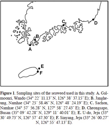
MIC values of phenolics of Ishige okamurae collected from various sites. Antimicrobial activities may vary by season and habitat (Moreau et al., 1988; Stirk et al., 2007; Vidyavathi and Sridhar, 1991). To investigate variation in the inhibitory effect on Helicobacter pylori growth, we determined the MIC values of phenolics of I. okamurae that were collected at different sampling sites and sampling times, such as Sachon, Namhae (June 2000), Sinyang, Jeju (February 2006, April 2012), and U-do, Jeju (July 2009) (Figure 1, Table 3). Of these phenolics, the phenolic extract of I. okamurae collected from U-do, Jeju, in July 2009 had the highest MIC (12 µg/ml); followed by Sachon, Namhae, in June 2000 (24 µg/ml); Sinyang, Jeju, in April 2012 (98 µg/ml); and Sinyang, Jeju, in February 2006 (391 µg/ml).
Inhibition of Helicobacter pylori urease activity by nitrogen compounds of Ishige okamurae collected from different sites. To investigate variation in the inhibition of H. pylori urease activity, we examined the inhibitory effect of nitrogen compounds on the urease activity of I. okamurae collected from different sampling sites and sampling times (Table 4). Of these nitrogen compounds, samples from Sinyang, Jeju, in February 2006 had the highest inhibitory effect (80.84%);, followed by Sachon, Namhae, in June 2000 (38.08%); Udo, Jeju, in July 2009 (30.05%); and Sinyang, Jeju, in April 2012 (23.62%).
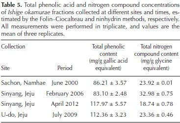
Total phenolic acid and nitrogen compounds in Ishige okamurae fractions. To confirm the content of phenolics and nitrogen compounds fractionated according to polarity, we used the Folin-Ciocalteau and ninhydrin methods to evaluate the total phenolic and total nitrogen contents, respectively, in I. okamurae fractions collected at different sites and times. As seen in Table 5, I. okamurae samples collected from Sinyang, Jeju, in April, 2012 had the highest total phenolic acid content (117.97 mg/g); followed by those collected from U-do, Jeju in July, 2009 (112.36 mg/g); Sachon, Namhae in June, 2000 (86.21 mg/g); and Sinyang, Jeju in February, 2006 (83.10 mg/g). Samples collected from Sinyang, Jeju in February 2006 had the highest nitrogen compound content (32.98 mg/g); followed by those from Sachon, Namhae in June, 2000 (23.92 mg/g); U-do, Jeju in July, 2009 (23.36 mg/g); and Sinyang, Jeju in April, 2012 (18.74 mg/g).
Acute toxicity. Although, Ishige okamurae is commonly used as a foodstuff in Korea and China (Oh et al., 1990), we evaluated the acute toxicity of ethanol and 80% methanol extracts of I. okamurae in mice. Over the 2-week observation period, no death occurred in any mice administered a dose of 5 g/kg body weight. Mice administered seaweed extract reacted by wandering briefly and returned to normal behavior after ~10 min. According to the World Health Organization (1992), a herbal medicine is considered toxic if the LD50 is lower than 5 g/kg body weight. Thus, these extracts would be classified as non-toxic. Therefore, our data suggest that these extracts can be safely used by humans at moderate doses.
Discusssion
Helicobacter pylori is classified by the World Health Organization and the International Agency for Research on Cancer as a class 1 carcinogen. Helicobacter pylori is a Gram-negative microaerophilic helical bacillus that inhabits various areas of the human stomach. Infection with this organism is strongly associated with chronic gastritis, peptic ulcer, and gastric carcinoma. The bacterium colonizes the gastric epithelial surface, and withstands the stomach's hostile ambience by microaerophilic growth capability and high urease activity, which accounts for approximately 6% of the soluble protein. Unlike urea-positive bacteria, H. pylori urease is located both in the cytoplasm and on the surface of H. pylori cells, and is reportedly one of the major surface proteins (Bode et al., 1989; Hu and Mobley, 1990). Urease is critical for H. pylori colonization of the gastric mucosa; namely, urease hydrolyzes urea and releases ammonia, which neutralizes acid and allows for survival of the bacterium and initial colonization. Therefore, if urease activity is inhibited, H. pylori could not survive in the presence of gastric acid.
Eradication of the organism from the stomach results in significant remission from the above diseases. Current eradication regimens involve the use of combination therapies (a proton pump inhibitor such as lansoprazole and two antibiotics, most commonly macrolides, clarthromycin, metronidazole, amoxicillin, and tetracycline) with an expected success rate between 80% and 90%. However, increasing resistance to these drugs is a growing global concern. Therefore, it is important to develop an effective method for reducing the level of resistant Helicobacter pylori strains. Previous studies have attempted to develop a novel antimicrobial agent capable of preventing and/or treating H. pylori. Until now, most studies have mainly focused on terrestrial plants (Boyanova and Neshev, 1999; Tabak et al., 1999; Takabayashi et al., 2004; Robles-Zepeda, et al., 2011).
In this study, ethanol extracts of 27 edible seaweed species were prepared and evaluated for their antimicrobial activity against Helicobacter pylori using the disk diffusion method. Among these, seven extracts showed high levels of antimicrobial activity and were further evaluated. Among them, the brown seaweed Ishige okamurae showed the strongest anti-H. pylori activity and urease inhibition activity. This species is widespread on rocks in the upper and middle intertidal zone of rough open coasts and is commonly used as soil fertilizer and a foodstuff in Korea and China (Oh et al., 1990). This seaweed has various beneficial biological activities, such as antioxidant (Heo and Jeon, 2009), anti-inflammatory (Kim et al., 2010), anti-diabetes activities (Min et al., 2011), and can protect against ultraviolet light (Heo et al., 2010).
To identify the active compounds in Ishige okamurae, the seaweed powders were successively fractionated according to polarity into five classes of constituents. Of these fractionates, phenolics and nitrogen compounds had the lowest MIC values and highest inhibitory effect on urease, respectively, showing that different compounds may have different bioactivities. Generally, the seaweed showed variation in cellular chemical composition and biological activity according to season, habitat, and different thalli in seaweed (Lobban and Harrison, 1994). Antimicrobial activities may show seasonal and habitat variation (Moreau et al., 1988; Vidyavathi and Sridhar 1991; Stirk et al., 2007 ). When we investigated inhibition of I. okamurae growth and urease activity collected from different sites and times, phenolics from the seaweed collected at U-do, Jeju, in July 2009 showed the highest MIC values (12 µg/ml). Otherwise, the highest inhibitory effect (80.84%) was found for nitrogen compounds of I. okamurae collected from Sinyang, Jeju in February 2006. In this study, we also found variation in the antimicrobial activity in the seaweed collected at different sites and times.
We found that Ishige okamurae phenolic compounds possessed antimicrobial activity and that nitrogen compounds inhibited urease, which have a critical role in the survival of Heliobacter pylori in the gastric mucosa and in the pathogenesis of this organism. We compared the MIC values and total phenolic acid contents of the phenolic fractions of I. okamurae collected at different sites and times. The highest MIC value (12 µg/ml) was identified in phenolics from seaweed collected at U-do, Jeju in July, 2009. The highest total phenolic acid content (117.97 mg/g) was identified in phenolics from seaweed collected at Sinyang, Jeju in April, 2012. In the remainder of the samples, the MIC values were proportional to the total phenolic acid content.
The mismatch between the MIC values and total phenolic acid contents probably results from the difference between the content of each phenolic compound in the phenolics fraction and its antimicrobial activity. Further research is needed to elucidate the profiles, contents, and antimicrobial activities of phenolic compounds in the phenolics fraction of Ishige okamurae collected at different sites and times.
We compared the urease inhibitory effect and total nitrogen compound contents of the nitrogen-compound fractions of Ishige okamurae collected at different sites and times. The greatest inhibitory effect (80.84%) and the highest total nitrogen compound content (32.98 mg/g) was found in nitrogen compounds from I. okamurae collected from Sinyang, Jeju in February 2006. For the remainder of the samples, the inhibitory effects were proportional to the total nitrogen compound content.
In conclusion, our results suggest that the seaweed Ishige okamurae may be useful as a treatment for gastrointestinal disorders caused by Helicobacter pylori. These results can be used as a basis for further characterization and elucidation of the active compounds responsible for this antimicrobial activity.
Acknowledgments
This work was supported by the Global Healthcare Industry RIS Center, Ministry of Knowledge Economy, Republic of Korea. KKC was also supported by Cooperative Research Program for Agriculture Science and Technology Development (Project No. PJ007605), Rural Development Administration and Priority Research Centers Program through the National Research Foundation of Korea (NRF) funded by the Ministry of Education, Science and Technology (20090093813), Republic of Korea. Two anonymous reviewers provided helpful suggestions to improve the manuscript.
Literature cited
Bode G., Malfertheiner P., Nilius M., Lehnhardt G. and Ditschuneit H. 1989. Ultrastructural localisation of urease in outer membrane and periplasm of Campylobacter pylori. Journal of Clinical Pathology 42:778-779. [ Links ]
Boyanova L. and Neshev G. 1999. Inhibitory effect of rose oil products on Helicobacter pylori growth in vitro: preliminary report. Journal of Medical Microbiology 48:705-706. [ Links ]
Bradford M.M. 1976. A rapid and sensitive method for the quantitation of microgram quantities of protein utilizing the principle of protein-dye binding. Analytical Biochemistry 72:248-254. [ Links ]
Chiba N., Rao B.V., Rademaker J.W. and Hunt R.H. 1992. Metaanalysis of the efficacy of antibiotic therapy in eradicating Helicobacter pylori. The American Journal of Gastroenterology 87:1716-1727. [ Links ]
Cho J.Y., Kang J.Y., Khan M.N.A., Park N.H., Kim S.K. and Hong Y.K. 2007. Anti-inflammatory activites of Undaria pinnatifida and Laminaria japonica (Phaeophyta). Journal ofFisheries Science and Technology 10:127-132. [ Links ]
Graham D.Y., Lew G.M., Malaty H.M., Evance D.G., Evance D.J. Jr, Klein P.D., Alpert L.C. and Genta R.M. 1992. Factors influencing the eradication of Helicobacter pylori with triple therapy. Gastroenterology 102:493-496. [ Links ]
Harborne J.B. 1998. Phytochemical Methods: A Guide to Modern Techniques of Plant Analysis. 3rd Ed. Chapman and Hall, London. [ Links ]
Hassani A.R., Ordouzadeh N., Ghaemi A., Amirmozafari N., Ha-mdi K. and Nazari R. 2009. In vitro inhibition of Helicobacter pylori urease with non and semi fermented Camellia sinensis. Indian Journal of Medical Microbiology 27:30-34. [ Links ]
Heo S.J and Jeon Y.J. 2009. Evaluation of diphlorethohydroxy-carmalol isolated from Ishige okamurae for radical scavenging activity and its protective effect against H2O2-induced cell damage. Process Biochemistry 44:412-418. [ Links ]
Heo S.J., Ko S.C., Kang S.M., Cha S.H., Lee S.H., Kang D.H., Jung W.K., Affan A., Oh C. and Jeon Y.J. 2010. Inhibitory effect of diphlorethohydroxycarmalol on melanogenesis and its protective effect against UV-B radiation-induced cell damage. Food and Chemical Toxicology 48:1355-1361. [ Links ]
Hu L.T. and Mobley H.L. 1990. Purification and N-terminal analysis of urease from Helicobacter pylori. Infection and Immunity 58:992-998. [ Links ]
Kim K.N., Heo S.J., Yoon W.J., Kang S.M., Ahn G., Yi T.H. and Jeon Y.J. 2010. Fucoxanthin inhibits the inflammatory response by suppressing the activation of NF-xB and MAPKs in lipo-polysaccharide-induced RAW 264.7 macrophages. European Journal of Pharmacology 649:369-375. [ Links ]
Jin H.J., Kim J.H., Sohn C.H., DeWreede R.E., Choi T.J., Towers G.H.N., Hudson J.B. and Hong Y.K. 1997. Inhibition of Taq DNA polymerase by seaweed extracts from British Columbia, Canada and Korea. Journal of Applied Phycology 9:383-388. [ Links ]
Lobban C.S. and Harrison P.J. 1994. Seaweed Ecology and Physiology. Cambridge University Press, Cambridge. [ Links ]
Marshall B.J. and Warren J.R. 1984. Unidentified curved bacilli in the stomach of patients with gastritis and peptic ulceration. Lancet 1(8390):1311-1315. [ Links ]
Midolo P.D., Lambert J.R. and Turnidge J. 1996. Metronidazole resistance: a predictor of failure of Helicobacter pylori eradication by triple therapy. Journal of Gastroenterology and Hepa-tology 11:290-292. [ Links ]
Min K.H., Kim H.J., Jeon Y.J. and Han J.S. 2011. Ishige okamurae ameliorates hyperglycemia and insulin resistance in C57BL/ KsJ-db/db mice. Diabetes Research and Clinical Practice 93:70-76. [ Links ]
Moreau J., Pesando D., Bernard P., Caram B. and Pionnat J.C. 1988. Seasonal variations in the production of antifungal substances by some dictyotales (brown algae) from the French Mediterranean coast. Hydrobiologia 162:157-162. [ Links ]
Nagata K., Takagi E., Tsuda M., Nakazawa T., Satoh H., Nakao M., Okamura H. and Tamura T. 1995. Inhibitory action of lan-soprazole and its analogs against Helicobacter pylori: Inhibition of growth is not related to inhibition of urease. Antimicrobial Agents and Chemotherapy 39:567-570. [ Links ]
National Committee for Clinical Laboratory Standards (NCCLS). 2003a. Performance standards for antimicrobial disk susceptibility tests. Approved Standard. 8th Ed. Document M2-A8. NCCLS, Wayne. [ Links ]
National Committee for Clinical Laboratory Standards (NCCLS). 2003b. Methods for antimicrobial susceptibility tests for bacteria that grow aerobically. Approved Standard. 6th Ed. Document M7-A6. NCCLS, Wayne. [ Links ]
Oh Y. S., Lee I.K. and Boo S.M. 1990. An annotated account of Korean economic seaweeds for food, medical and industrial uses. The Korean Journal of Phycology 5:57-71. [ Links ]
Parsonnet J., Friedman G.D., Vandersteen D.P., Chang Y., Vogel-man J.H., Orentreich N.D.E.E. and Sibley R.K. 1991. Helico-bacter pylori infection and the risk of gastric carcinoma. The New England Journal of Medicine 325:1127-1131. [ Links ]
Robles-Zepeda R.E., Veläzquez-Contreras C.A., Garibay-Escobar A., Gälvez-Ruiz J.C. and Ruiz-Bustos E. 2011. Antimicrobial activity of Northwestern Mexican plants against Helicobacter pylori. Journal of Medicinal Food 14:1280-1283. [ Links ]
Stirk W.A., Reinecke D.L. and van Staden J. 2007. Seasonal variation in antifungal, antibacterial and acetylcholinesterase activity in seven South African seaweeds. Journal of Applied Phycology 19:271-276. [ Links ]
Tabak M., Armon R. and Neeman I. 1999. Cinnamon extracts' inhibitory effect on Helicobacter pylori. Journal of Ethnophar-macology 67:269-277. [ Links ]
Takabayashi F., Harada N., Yamada M., Murohisa B. and Oguni I. 2004. Inhibitory effect of green tea catechins in combination with sucralfate on Helicobacter pylori infection in Mongolian gerbils. Journal of Gastroenterology 39:61-63. [ Links ]
Vidyavathi N. and Sridhar K.R. 1991. Seasonal and geographical variations in the antimicrobial activity of seaweeds from the Mangalore Coast of India. Botanica Marina 34:279-284. [ Links ]
World Health Organization. 1992. Research Guidelines for Evaluating the Safety and Efficacy of Herbal Medicine. Regional Office for Western Pacific, Manila. [ Links ]
Woo T.W., Chang M.S., Chung Y.K., Kim K.B., Sohn S.K., Kim S.G. and Choi W.S. 1998. Inhibitory action of YJA20379, a new proton pump inhibitor on Helicobacter pylori growth and urease. Archives of Pharmacal Research 21:6-11. [ Links ]

