SciELO - Scientific Electronic Library Online

 
vol.78 número4Tratamiento farmacológico de la hiperplasia prostática benigna. Revisión de la bibliografía¿Cuál es la mejor opción de tratamiento quirúrgico en varones con síntomas de la vía urinaria inferior? índice de autoresíndice de materiabúsqueda de artículos
Home Pagelista alfabética de revistas  

Servicios Personalizados

Revista

Articulo

Indicadores

Links relacionados

  • No hay artículos similaresSimilares en SciELO

Compartir


Revista mexicana de urología

versión On-line ISSN 2007-4085versión impresa ISSN 0185-4542

Rev. mex. urol. vol.78 no.4 Ciudad de México jul./ago. 2018  Epub 25-Jun-2021

https://doi.org/10.24245/revmexurol.v78i4.2216 

Artículos de revisión

Penile Doppler ultrasound in post-radical prostatectomy evaluation: A review and recommendations

Ultrasonido Doppler de pene en la evaluación posterior a prostatectomía radical. Revisión bibliográfica y recomendaciones

Juan Fernando Uribe-Arcila1 

Luis Javier Aluma-Sánchez2 

1M. D., Servicio de Urologia, Hospital Pablo Tobon Uribe, Medellín, Colombia.

2M. D., Servicio de Urologia, Hospital Pablo Tobon Uribe, Imbanaco, Cali, Colombia


Abstract

Changes in sexual function are common in patients following radical prostatectomy for prostate cancer. Sexual rehabilitation after radical prostatectomy requires a complex process in which the diagnosis is refined, and an accurate treatment program is chosen. Penile Doppler ultrasound is a commonly used method for diagnosing erectile dysfunction and it is very useful in certain cases after surgery to improve the assessment of arterial evaluation, venous leaks, and quality of the cavernous tissue and fascia. A literature search was conducted using the databases from Google and PubMed to identify original and review articles that examined the uses of penile Doppler ultrasound in post-radical prostatectomy evaluation or post-surgery rehabilitation. Search terms included: Erectile dysfunction post radical prostatectomy, sexual function post radical prostatectomy, Penile evaluation postprostatectomy, Diagnosis of erectile function after radical surgery, Penile Doppler ultrasound AND prostatectomy, Penile Doppler ultrasound AND sexual rehabilitation. The initial search resulted in 415 articles. After applying additional filters, 46 studies were included in the present review. Backgrounds of the most relevant guidelines were cited: Standard practice in sexual medicine, Standard operating procedure in sexual medicine, International Consultation on Sexual Medicine, and the EAU and AUA guidelines. Information on the use of penile Doppler ultrasound before surgery is extremely inconsistent in the literature. The recommendations for a successful evaluation of post-radical prostatectomy patients were included. Sexual rehabilitation after radical prostatectomy requires a complex process. There is great inconsistency in the literature with respect to the definition of what is considered normal erectile function before surgery and what may be considered normal erection after radical prostatectomy. The cost of penile Doppler ultrasound is a modest component of the penile post-radical prostatectomy rehabilitation process. Current evidence does not support the systematic use of penile Doppler ultrasound, but it must be included in the management algorithm of the patient undergoing radical prostate surgery so that erectile function can be properly evaluated.

Keywords: Prostate cancer; Sexual Dysfunction; Erectile dysfunction; Sexual rehabilitation; Penile Doppler ultrasound; Radical Prostatectomy

Resumen

Los cambios en la función sexual son comunes en pacientes con cáncer de próstata intervenidos de prostatectomía radical. La rehabilitación sexual después de la prostatectomía implica un proceso complejo para establecer el diagnóstico y tratamiento específico. El ultrasonido Doppler de pene es un método común para diagnosticar la disfunción eréctil y útil en ciertos casos después de la cirugía para evaluar el estado de las arterias, tejido cavernoso y la fascia, además de identificar fugas venosas. Se realizó la búsqueda bibliográfica en las bases de datos de Google y PubMed para identificar artículos que examinaron la ecografía Doppler peneana en la evaluación de pacientes intervenidos de prostatectomía radical o rehabilitación posquirúrgica. Términos de búsqueda incluidos: disfunción eréctil posprostatectomía radical, evaluación peneana posprostatectomía, función eréctil después de cirugía radical y rehabilitación sexual. En la búsqueda se obtuvieron 415 artículos. Después de aplicar filtros adicionales se incluyeron 46 estudios. Se citaron los antecedentes de las pautas más relevantes: intervenciones quirúrgicas en medicina sexual, consulta internacional de medicina sexual y estrategias de la EAU y la AUA. La información del ultrasonido Doppler peneano antes de la cirugía es extremadamente inconsistente en la bibliografía. Se incluyeron las recomendaciones para la evaluación exitosa de pacientes con prostatectomía radical. La rehabilitación sexual después de la prostatectomía radical requiere un proceso complejo. Existe inconsistencia en la bibliografía respecto de la definición de la función eréctil normal antes de la cirugía y lo que se considera erección normal después de la prostatectomía radical. El costo del ultrasonido Doppler peneano es un componente modesto del proceso de rehabilitación de la prostatectomía. La evidencia actual no respalda el uso sistemático de la ecografía Doppler peneana, pero debe incluirse en el algoritmo de tratamiento de pacientes intervenidos de cirugía radical de próstata, para que la función eréctil pueda evaluarse adecuadamente.

Palabras clave: Cáncer de próstata; disfunción sexual; disfunción eréctil; rehabilitación sexual; ultrasonido Doppler de pene; prostatectomía radical

TAKE-HOME MESSAGES

  • Penile Doppler ultrasound is one of five specialized diagnostic tests for erectile dysfunction, grade B recommendation.

  • Patients that undergo surgery can expect to experience significant postoperative changes, and erection is one of the most important health-related quality of life outcomes.

  • Current evidence does not support the systematic use of penile Doppler ultrasound, but it must be included in the management algorithm of the patient undergoing radical prostate surgery for the proper evaluation of erectile function.

INTRODUCTION

Changes in sexual function after radical prostatectomy in patients with prostate cancer are unpredictable and impossible to avoid.1,2 Patients who undergo surgery (open, laparoscopic, or robotic) can expect to experience significant changes post-surgery, and erection is one of the most important health-related quality of life outcomes influencing patient choice of treatment and post-surgery satisfaction. Numerous articles have been published in the literature on post-radical treatments, but inexplicably there is little information on diagnostic options. Penile Doppler ultrasound is a common method for diagnosing erectile dysfunction. The response to intracorporeal injections of vasoactive substances during that procedure is useful in the evaluation of arterial and venous-occlusive function.3,4 Sexual rehabilitation after radical prostatectomy requires a complex process in which the diagnosis is refined and an accurate treatment program is chosen. 5 Sexual function, mainly erectile dysfunction, after radical prostatectomy, is an obvious challenge for the urologist.6,7 Part I of the present review paper provides a comprehensive inventory of the available guidelines; Part II addresses the ítems that are necessary for performing an optimal ultrasound examination as part of the complex process of post-radical prostatectomy erection recovery; and Part III is aimed at defining sharable recommendations.

EVIDENCE ACQUISITION

A literature search was conducted using Google and the National Library of Medicine’s PubMed databases to identify original and review articles, either published or e-published, on the uses of penile Doppler ultrasound in post-radical prostatectomy evaluation or post-surgery rehabilitation, up to February 2018. The search terms included: Erectile dysfunction post-radical prostatectomy, sexual function post radical prostatectomy, Penile evaluation post-prostatectomy, Diagnosis of erectile function after radical surgery, Penile Doppler ultrasound AND prostatectomy, Penile Doppler ultrasound AND sexual rehabilitation. The reference list of the articles retrieved, as well as relevant reviews, were also studied. The initial search produced 415 articles and, after applying additional filters, 46 studies were included in the present review.

BACKGROUND

The recommendations of the most relevant guidelines are cited with respect to the use of penile Doppler ultrasound after radical prostatectomy. Unfortunately, urologic guidelines are conflicted in terms of recommendations.8

Standard practice in sexual medicine (Seftel 2006): Penile Doppler ultrasound is an optional part of examination. There is no specifically mentioned post-radical prostate evaluation.9

Standard operating procedure (2013): Penile Doppler ultrasound is an objective and reliable diagnostic method for documenting penile hemodynamics. It requires skilled personnel and modern equipment that may be cost-prohibitive in certain settings. Objective vascular testing that provides a physiologic diagnosis may help direct appropriate therapy (Sikka, et al).10 Penile Doppler ultrasound is not mentioned in the chapter about preservation of erectile function outcomes after radical prostatectomy (Mulhall, et al.).11

International Consultation on Sexual Medicine (2015): Color duplex Doppler penile ultrasonography (CDDPU) is one of five specialized diagnostic tests for erectile dysfunction, grade B recommendation (Dennerstein, et al.),12 (Salonia, et al.).6 Diagnostic penile Doppler ultrasound is not specifically mentioned for post radical prostate evaluation (Incrocci, et al.).13

EAU guidelines (Hatzimouratidis et al., 2016):14 Erectile dysfunction is common after radical prostatectomy, irrespective of the surgical technique used. LE: 2B. Specific diagnostic tests should be included in the initial evaluation only in the presence of: primary erectile dysfunction; young patients with a history of pelvic or perineal trauma that could benefit from potentially curative revascularization surgery; patients with penile deformities that might require surgical correction; patients with complex psychiatric or psychosexual disorders; and patients with complex endocrine disorders. Specific tests may be indicated at the request of the patient or his partner, or for medicolegal reasons (e.g., implantation of penile prosthesis to document end-stage erectile dysfunction, sexual abuse). LE: 4B.

AUA Guidelines (Board of Directors 2012; 2017 reaffirmed): 15 The Consensus Statement on indications for a penile ultrasound examination include but are not limited to: evaluation of erectile dysfunction; evaluation of Peyronie’s disease; others. Penile Doppler ultrasound is not mentioned in the chapter about preservation of erectile function outcomes after radical prostatectomy.

PREOPERATIVE ULTRASOUND EVALUATION

The preoperative assessment of a candidate for radical prostatectomy is the first compulsory step in preventing postoperative ED. That recomendable attitude has been called "the good start".16 Patient factors (including age, baseline erectile function, personal antecedents, and status of comorbid conditions), evaluation of the clinical and pathologic characteristics of the prostate cancer (e.g., Gleason and NMR findings), nerve-sparing selection (unilateral vs bilateral nerve sparing), type of surgery (e.g., intrafascial vs interfascial vs extrafascial surgeries), surgical techniques (e.g., open, laparoscopic, and robotic-assisted radical prostatectomy), and surgeon factors (e.g., surgical volume and surgical skill). The evaluation of each of those items contributes to achieving a better long-term result and they are the key significant contributors to erectile function recovery.17 Penile shortening and/or deformities may appear or become aggravated after radical surgery. The patient should be informed of that potential side effect. 1 The literature reviewed does not support the recommendation of the systematic use of preoperative ultrasound evaluation with respect to radical prostate surgery, but there are at least three situations in which its performance is recommended:

The patient has a preoperative condition that justifies the evaluation with ultrasound. (e.g., Peyronie’s disease).

There is a risk of legal problems (e.g., related to the job or social position of the patient).

The patient claims to have excellent erections and you have doubts (e.g., due to advanced age or comorbidities).

POSTOPERATIVE ULTRASOUND EVALUATION

Penile evaluation

Nodules, plaques, penile shortening, and de novo deformities (hourglass and other), have been described after radical surgery.18 Although penile Doppler ultrasound enables the intracavernosal evaluation of each of those lesions, it is essential to first observe the penis in flaccidity and then in erection to describe the anatomic findings. An important component of this phase is to measure the penis in a flaccid state and then in erection. That first phase of external inspection and response to vasoactive medication has been called the 'erection test'. During that part of the examination, whoever performs it can make note of the different penile findings, unrelated to ultrasound, which can be very useful in the prognosis and treatment proposed post radical prostatectomy.

Arterial evaluation

Normal reference values during penile Doppler ultrasound are: Peak systolic velocity (PSV: 25 cm/ sec or more), End diastolic velocity (EDV: 5 cm/sec or less), Acceleration time (AT: 0.11 sec or less), and Resistance index (RI: 0.85 or more).19-21 Abnormal arterial findings after radical prostatectomy have two possible origins:

Abnormal findings prior to surgery: A less tan 75% increase in baseline cavernosal artery diameter, abnormal PSV (less than 25 cm/ sec), and AT (more than 0.11 sec), usually bilateral, are suggestive of arterial disease, but those findings could also be related to arteriosclerosis or other common arterial pathologies, prior to surgery. In some cases, congenital arterial malformations (aneurysms, fistulas) may be found, of which the patient was previously unaware, affecting his erectile recovery.

Abnormal findings after surgery: Penile Doppler ultrasound is a direct and indirect method for diagnosing vascular lesions acquired after radical prostatectomy. To decrease the risk of false positives, it is necessary to verify arterial flow at the perineal level, when penile flows are persistently low. In addition to measuring flow, the asymmetry between the arteries should be verified.

Venous evaluation

Veno-occlusive disease is suggested by abnormal EDV (more than 5 cm/ sec) and RI (less than 0.85). Regardless of the surgical technique, the removal of the prostate may result in an almost obligatory period of dormancy of the nerves that govern the functional aspects of erection. That situation may lead to a loss of daily and nocturnal erections associated with persistent failure of cavernous oxygenation and secondary erectile tissue damage resulting from the production of pro-apoptotic factors and pro-fibrotic factors within the corpora cavernosa.22,23 Those changes are coupled with postoperative erectile dysfunction of varying degrees and the development of venous leakage, which indicates a poor prognosis for erectile function recovery.24,25

Venous leakage was first described in the nineteenth century (Raymond and Duncan, 1895)26,27 and more than one hundred years later, venous leakage diagnosis still needs to mature, even though it is key to the functioning of the penis as a three-chamber pump. Trost et al.28 mention the difficulty of finding relevant literature about venous surgery and how data analyses cannot be carried out due to the lack of standardization in patient selection, hemodynamic testing, surgical technique, follow-up, and surgical outcome. The text mentions the articles of Florez and Mulhall (2011),29Cayan (2008),30 and Rao and Donatucci (2001),31 with their respective and highly variable results. Other theories and surgical techniques for venous leakage have been proposed by the Korean urologist GL Hsu (2006, 2010).32-34 Despite the heterogeneity in the data, there is sufficient clinical evidence supporting ultrasound evaluation, even though venous leaks acquired post fibrosis are considered part of the physiopathology of post-radical prostate erectile dysfunction and are mentioned by different authors in their algorithms. Perhaps the best known is that described by Hatzimouratidis,35 presented below (Figure 1):

Figure 1 Algorithm of the physiopathology of postradical prostate erectile dysfunction. 

The incidence of venous leakage increases in proportion to the time interval after surgery, supporting those pathophysiologic mechanisms as the cause of disturbance and providing the rationale for early penile rehabilitation before penile fibrosis occurs. Penile Doppler ultrasound can identify two different types of venous leakage:

Previous leaks: usually congenital, that make the erectile recovery of the patient after radical prostatectomy difficult.

Acquired leaks: intracavernous leaks, usually associated with areas of cavernous fibrosis. They are the most frequent type after radical prostatectomy.

Fascial and cavernous tissue evaluation

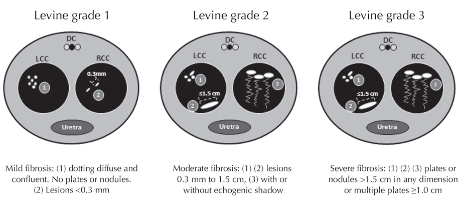
The evaluation of the fascia and the cavernous tissue after radical surgery is fundamental due to the concepts of the venous leakage/fibrosis relationship previously expressed36,37 and because there may be a peak in the prevalence of Peyronie's disease post radical prostatectomy. That pathology includes a variety of states of severe penile fibrosis that compromise the tunica albuginea, but they, alone, do not cover the wide range of other types of penile fibrosis.38-41 To clarify the panorama of what post-radical prostatectomy Penile Doppler ultrasound can find in the intracavernous tissue, it is necessary to consider another fibrous pathology called Chronic Traumatic Cavernosophaty (CTC), which, unlike Peyronie’s disease, does not compromiso the tunica albuginea.I,42-46 An echographic classification grade of calcification was published by Laurence Levine47,48 and revised in Campbell’s Urology 11th ed. (2016).49 Calcification areas were defined as hyperechoic regions with the presence of acoustic shadows using Levine’s three echographic grades: Grade 3 (plates > 1.5 cm in any dimensión or multiple plates ≥1.0 cm); Grade 2 (lesions 0.3 mm to 1.5 cm); and Grade 1 (lesions <0.3 mm). In accordance with the Levine classification, Grade 1 is mild, Grade 2 is moderate, and Grade 3 is severe) (Figure 2).50 Other classifications have been used according to ultrasonographic patterns: a formed, solitary, hyperechoic lesion with no acoustic shadow (group A), moderately hyperechoic multiple scattered calcified lesions with acoustic shadows (group B), dense calcified hyperechoic plaque with acoustic shadow (group C).51

DC: dorsal complex; LCC: left corpus cavernosum; RCC; right corpus cavernosum.

Figure 2 Levine classification for penile fibrosis. 

Neurologic evaluation

Penile erection is a neurovascular event requiring intact neural and vascular pathways. Most surgeries are supposed “nerve-sparing” surgeries. That type of surgery spares the cavernous nerves that run bilaterally along the prostate. Despite nerve-sparing surgery, the cavernous nerves are stretched and bruised intraoperatively and may take 18 to 24 months post-surgery to heal. Men may fail to have natural erections during that period of time, which can lead to penile tissue atrophy, fibrosis, structural alterations, and venous leak, as cited above.52 Penile Doppler ultrasound is not the appropriate test for evaluating neurologic status after radical surgery.6 However, it is possible to obtain indirect data of potential damage to the neurovascular bundle. A finding that should always be included in the report is the appearance of acute pain in the suprapubic region, after the use of a vasoactive drug related to severe damage of the unilateral or bilateral neurovascular bundle.

COST ANALYSIS

The greater reluctance to including penile Doppler ultrasound as a routine item in the postoperative evaluation protocol of radical prostatectomy patients is the increase in cost, together with the assumption of few benefits from its systematic use. The cost of treatment of radical prostatectomy-related erectile dysfunction can be projected to increase but, compared with the technology used to manage prostate cancer (Robotics, PET scan, etc.), the cost of penile Doppler ultrasound is actually modest.

RECOMMENDATIONS

  • — Use the appropriate ultrasound equipment (the correct transducer, and ideally, HD resolution).

  • — Select the appropriate patient.

  • — Complete all the steps: Penile, arterial, venous and tissue evaluation.

  • — Use a classification to report penile fibrosis (we recommend the Levine classification).

  • — If you are an expert in penile Doppler ultrasound and receive patients from other colleagues: give a thorough report and provide generous recommendations.

CONCLUSIONS

Sexual rehabilitation after radical prostatectomy requires a complex process. There is great inconsistency in the literature as to the definition of what is considered normal erectile function before surgery and what a man may consider normal erection after radical prostatectomy. One possibility is to use validated psychometric instruments with recognized cutoff points for normalcy and severity during the preoperative and postoperative evaluations.17 Although there is no consensus, our analysis confirmed that penile Doppler ultrasound after radical prostatectomy can be a diagnostic alternative. Current evidence does not support the systematic use of penile Doppler ultrasound, but it must be included in the management algorithm of the patient undergoing radical prostate surgery for the proper evaluation of erectile function. Further studies with adequate follow-up and larger samples should be conducted to reach a comprehensive conclusion.53

REFERENCES

1 Elliott S, Matthew A. Sexual Recovery Following Prostate Cancer: Recommendations From 2 Established Canadian Sexual Rehabilitation Clinics. Sex Med Rev. 2017 Nov 30. pii: S2050- 0521(17)30114-2. doi: 10.1016/j.sxmr.2017.09.001. [Epub ahead of print] Review. PubMed PMID: 29199096. [ Links ]

2 Skolarus TA, Wolf AM, Erb NL, Brooks DD, Rivers BM, Underwood W 3rd, Salner AL, Zelefsky MJ, Aragon-Ching JB, Slovin SF, Wittmann DA, Hoyt MA, Sinibaldi VJ, Chodak G, Pratt-Chapman ML, Cowens-Alvarado RL. American Cancer Society prostate cancer survivorship care guidelines. CA Cancer J Clin. 2014 Jul-Aug; 64(4):225-49. doi: 10.3322/caac.21234. Epub 2014 Jun 10. [ Links ]

3 Altinkilic B, Hauck EW, Weidner W. Evaluation of penile perfusion by color-coded duplex sonography in the management of erectile dysfunction. World J Urol. 2004;22:361-4. [PubMed] [ Links ]

4 Mueller SC, Lue TF. Evaluation of vasculogenic impotence. Urol Clin North Am. 1988;15:65- 76.[PubMed] [ Links ]

5 Gandaglia G, Suardi N, Cucchiara V, et al. Penile rehabilitation after radical prostatectomy: does it work? Transl Androl Urol 2015;4:110-123. [ Links ]

6 Salonia A, Adaikan G, Buvat J, Carrier S, El-Meliegy A, Hatzimouratidis K, McCullough A, Morgentaler A, Torres LO, Khera M. Sexual Rehabilitation After Treatment For Prostate Cancer-Part 2: Recommendations From the Fourth International Consultation for Sexual Medicine (ICSM 2015). J Sex Med. 2017 Mar;14(3):297-315. doi: 10.1016/j.jsxm.2016.11.324. PubMed PMID: 28262100. [ Links ]

7 Rabbani F, Stapleton AM, Kattan MW, Wheeler TM, Scardino PT. Factors predicting recovery of erections after radical prostatectomy. The Journal of urology. 2000; 164(6):1929-34. [PubMed: 11061884] [ Links ]

8 Blecher G, Almekaty K, Kalejaiye O, Minhas S. Does penile rehabilitation have a role in the treatment of erectile dysfunction following radical prostatectomy? F1000Res. 2017 Oct 31;6:1923. doi: 10.12688/f1000research.12066.1. eCollection 2017. Review. PubMed PMID: 29152231; PubMed Central PMCID: PMC5664996. [ Links ]

9 Seftel A. Diagnosis of erectile dysfunction; Chapter 6. In: Porst H, Buvat J. Standard practice in sexual medicine. Blackwell publishing, 2006. [ Links ]

10 Sikka SC, Hellstrom WJG, Brock G, and Morales AM. Standardization of vascular assessment of erectile dysfunction. J Sex Med 2013; 10:120-129. [ Links ]

11 Mulhall JP, Bivalacqua TJ, and Becher EF. Standard operating procedure for the preservation of erectile function outcomes after radical prostatectomy. J Sex Med 2013;10:195-203. [ Links ]

12 Hatzichristou D, Kirana PS, Banner L, Althof SE, Lonnee-Hoffmann RA, Dennerstein L, Rosen RC. Diagnosing Sexual Dysfunction in Men and Women: Sexual History Taking and the Role of Symptom Scales and Questionnaires. J Sex Med. 2016 Aug;13(8):1166-82. doi: 10.1016/j.jsxm.2016.05.017. Review. PubMed PMID: 27436074. [ Links ]

13 Bennett N, Incrocci L, Baldwin D, Hackett G, El-Zawahry A, Graziottin A, Lukasiewicz M, McVary K, Sato Y, Krychman M. Cancer, Benign Gynecology, and Sexual Function--Issues and Answers. J Sex Med. 2016 Apr;13(4):519-37. doi: 10.1016/j.jsxm.2016.01.018. Epub 2016 Mar 25. Review. PubMed PMID: 27045256. [ Links ]

14 Hatzimouratidis K, Giuliano F, Serefoglu EC, Moncada I, Salonia A, Muneer A, Verze P, et al. European Association of Urology. Guidelines of Male Sexual Dysfunction. 2016. [ Links ]

15 American Institute of Ultrasound in Medicine Practice (AIUM). Parameter for the Performance of an Ultrasound Examination in the Practice of Urology. Urologic Ultrasound Utilization. 2011 [ Links ]

16 Capogrosso P, Salonia A, Briganti A, Montorsi F. Postprostatectomy Erectile Dysfunction: A Review. World J Mens Health. 2016 Aug;34(2):73-88. doi: 10.5534/wjmh.2016.34.2.73. Epub 2016 Aug 23. Review. PubMed PMID: 27574591; PubMed Central PMCID: PMC4999493. [ Links ]

17 Salonia A, Burnett AL, Graefen M, Hatzimouratidis K, Montorsi F, Mulhall JP, Stief C. Prevention and management of post prostatectomy sexual dysfunctions. Part 1: choosing the right patient at the right time for the right surgery. Eur Urol. 2012 Aug;62(2):261-72. doi: 10.1016/j.eururo.2012.04.046. Epub 2012 May 3. Review. PubMed PMID: 22575909. [ Links ]

18 Fode M, Serefoglu EC, Albersen M, Sønksen J. Sexuality Following Radical Prostatectomy: Is Restoration of Erectile Function Enough? Sex Med Rev. 2017 Jan;5(1):110-119. doi: 10.1016/j.sxmr.2016.07.005. Epub 2016 Aug 24. Review. PubMed PMID: 27566909. [ Links ]

19 Berookhim BM. Doppler Duplex Ultrasonography of the Penis. J Sex Med. 2016 Apr;13(4):726-31. doi: 10.1016/j.jsxm.2016.02.161. Epub 2016 Mar 25. PubMed PMID:27045266. [ Links ]

20 Suresh A, Balachandran A, Indira N, Ramprakash HV. Role of penile color doppler in the evaluation of erectile dysfunction. Int J Sci Stud. 2015;3(7):23-32. [ Links ]

21 Golijanin D, Singer E, Davis R, Bhatt S, Seftel A, Dogra V. Doppler evaluation of erectile dysfunction - part 2. Int J Impot Res. 2007;19(1):43-48. [ Links ]

22 Mulhall JP, Bella AJ, Briganti A, McCullough A, Brock G. Erectile function rehabilitation in the radical prostatectomy patient. J Sex Med 2010;7:1687-98. [ Links ]

23 Hatzimouratidis K, Burnett AL, Hatzichristou D, McCullough AR, Montorsi F, Mulhall JP. Phosphodiesterase type 5 inhibitors in postprostatectomy erectile dysfunction: a critical analysis of the basic science rationale and clinical application. Eur Urol 2009;55:1334-47. [ Links ]

24 Iacono F, Giannella R, Somma P, Manno G, Fusco F, Mirone V. Histological alterations in cavernous tissue after radical prostatectomy. J Urol 2005;173:1673-6. [ Links ]

25 Tal R, Valenzuela R, Aviv N, et al. Persistent erectile dysfunction following radical prostatectomy: the association between nerve sparing status and the prevalence and chronology of venous leak. J Sex Med 2009;6:2813-9. [ Links ]

26 Wooten JS. Ligation of the dorsal vein of the penis as a cure for atonic impotence. Texas Med J. 1902; 18:325-328. [ Links ]

27 Wespes E, Schulman C. Venous impotence: pathophysiology, diagnosis and treatment. J Urol 1993; 149: 1238±1245 [ Links ]

28 Trost LW, Munarriz R, Wang R, Morey A, Levine L. External Mechanical Devices and Vascular Surgery for Erectile Dysfunction. J Sex Med. 2016 Nov;13(11):1579-1617. doi: 10.1016/j.jsxm.2016.09.008. PubMed PMID: 27770853. [ Links ]

29 Flores S, Tal R, O’Brien K, et al. Outcomes of crural ligation surgery for isolated crural venous leak. J Sex Med 2011; 8:3495-3499. [ Links ]

30 Cayan S. Primary penile venous leakage surgery with crural ligation in men with erectile dysfunction. J Urol 2008; 180:1056-1059. [ Links ]

31 Rao DS, Donatucci CF. Vasculogenic impotence. Arterial and venous surgery. Urol Clin North Am. 2001 May;28(2):309-19. Review. PubMed PMID: 11402583. [ Links ]

32 Hsu GL, Chen HS, Hsieh CH, Lee WY, Chen KL, Chang CH. Salvaging penile venous stripping surgery. J Androl. 2010 May-Jun;31(3):250-60. doi:10.2164/jandrol.109.008409. PubMed PMID: 19745217. [ Links ]

33 Hsu GL, Chen HS, Hsieh CH, Ling PL, Wen HS, Liu LJ, Chen CW, Liu MW. Insufficient response to venous stripping surgery: is the penile vein recurrent or residual? J Androl. 2006 Sep-Oct;27(5):700-6. PubMed PMID: 16775251. [ Links ]

34 Hsu GL, Chen HS, Hsieh CH, Lee WY, Chen KL, Chang CH. Clinical experience of a refined penile venous stripping surgery procedure for patients with erectile dysfunction: is it a viable option? J Androl. 2010 May-Jun;31(3):271-80. doi: 10.2164/jandrol.109.008532. PubMed PMID: 19926885. [ Links ]

35 Hatzimouratidis K, Burnett AL, Hatzichristou D, McCullough AR, Montorsi F, Mulhall JP. Phosphodiesterase type 5 inhibitors in postprostatectomy erectile dysfunction: a critical analysis of the basic science rationale and clinical application. Eur Urol. 2009 Feb;55(2):334-47. doi: 10.1016/j.eururo.2008.10.028. Epub 2008 Oct 21. Review. PubMed PMID: 18986755. [ Links ]

36 Shafik A, Shafik I, El Sibai O, Shafik AA. On the pathogenesis of penile venous leakage: role of the tunica albuginea. BMC Urol. 2007 Sep 5;7:14. PubMed PMID: 17803807; PubMed Central PMCID: PMC1995196. [ Links ]

37 Uribe Arcila JF. Fuga venosa dorsal profunda como causa de disfunción eréctil primaria y secundaria no estándar (serie de 57 casos). Urol Colomb. 2016. Available in: http://dx.doi.org/10.1016/j.uroco.2016.08.002. [ Links ]

38 De le Peyronie F. Sur quelques obstacles quis’ opposent à l’éjaculation naturelle de la semence. Mem Acad Roy Chir 1743, 1: 425-34. [ Links ]

39 Brenot PH: Male impotence. A historical perspective. p. 46-53. Ed. L´Esprit du Temps. Paris, 1994. [ Links ]

40 Hauck EW, Weidner W. François de la Peyronie and the disease named after him. Lancet. 2001 Jun 23;357(9273):2049-51. PubMed PMID: 11438159. [ Links ]

41 Dunsmuir WD, Kirby RS. Francois de la Peyronie (1678-1747): the man and the disease he described. Br J Urol 1996; 78: 613-22. [ Links ]

42 Chung E, Ralph D, Kagioglu A, Garaffa G, Shamsodini A, Bivalacqua T, Glina S, Hakim L, Sadeghi-Nejad H, Broderick G. Evidence-Based Management Guidelineson Peyronie's Disease. J Sex Med. 2016 Jun;13(6):905-23. doi: 10.1016/j.jsxm.2016.04.062. PubMed PMID: 27215686. [ Links ]

43 Ferretti L, Fandel TM, Qiu X, Zhang H, Orabi H, Wu AK, Banie L, Wang G, Lin G, Lin CS, Lue TF. Tunica albuginea allograft: a new model of LaPeyronie's disease with penile curvature and subtunical ossification. Asian J Androl. 2014 Jul-Aug;16(4):592-6. doi: 10.4103/1008-682X.125900. PubMed PMID: 24759578; PubMedCentral PMCID: PMC4104088. [ Links ]

44 Laurence A. Levine, and Stephen Larsen. Cap 31. Diagnosis and Management of Peyronie Disease. En: Alan J. Wein, Louis R. Kavoussi, Alan W. Partin, Craig A. Peters. Campbell-Walsh Urology: 11th Edition. Elsevier, Philadelphia, 2016. pp. 722-748. [ Links ]

45 Omalu BI, DeKosky ST, Minster RL, Kamboh MI, Hamilton RL, Wecht CH. Chronic traumatic encephalopathy in a National Football League player. Neurosurgery. 2005 Jul;57(1):128-34; discussion 128-34. PubMed PMID: 15987548. [ Links ]

46 Turner RC, Lucke-Wold BP, Logsdon AF, Robson MJ, Dashnaw ML, Huang JH, Smith KE, Huber JD, Rosen CL, Petraglia AL. The Questto Model Chronic Traumatic Encephalopathy: A Multiple Model and Injury Paradigm Experience. Front Neurol. 2015 Oct 20;6:222. doi: 10.3389/fneur.2015.00222. eCollection 2015. PubMed PMID: 26539159; PubMed Central PMCID: PMC4611965. [ Links ]

47 Levine LA, Coogan CL. Penile vascular assessment using color duplex sonography in men with Peyronie’s disease. J Urol 1996;155(4):1270-3. [ Links ]

48 Levine LA, Greenfield JM. Establishing a standardized evaluation of the man with Peyronie’s disease. Int J Impot Res 2003;15(Suppl. 5):S103-12. [ Links ]

49 Laurence A. Levine, and Stephen Larsen . Cap 31. Diagnosis and Management of Peyronie Disease. En: Alan J. Wein, Louis R. Kavoussi, Alan W. Partin, Craig A. Peters. Campbell-Walsh Urology: 11th Edition. Elsevier, Philadelphia , 2016. pp. 722-748. [ Links ]

50 Yuruk E, Serefoglu EC. Re: Peyronie’s disease plaque calcification--prevalence, time to identification, and development of a new grading classification. J Sex Med. 2014 May;11(5):1351. PubMed PMID: 24877175. [ Links ]

51 Bekos A, Arvaniti M, Hatzimouratidis K, Moysidis K, Tzortzis V, Hatzichristou D. The natural history of Peyronie's disease: an ultrasonography-based study. Eur Urol. 2008 Mar;53(3):644-50. Epub 2007 Jul 17. PubMed PMID: 17673362. [ Links ]

52 Nelson CJ, Lacey S, Kenowitz J, Pessin H, Shuk E, Roth AJ, Mulhall JP. Men's experience with penile rehabilitation following radical prostatectomy: a qualitative study with the goal of informing a therapeutic intervention. Psychooncology. 2015 Dec;24(12):1646-54. doi: 10.1002/pon.3771. Epub 2015 Feb 24. PubMed PMID: 25707812; PubMed Central PMCID: PMC4765804. [ Links ]

53 Liu C, Lopez DS, Chen M, Wang R. Penile Rehabilitation Therapy Following Radical Prostatectomy: A Meta-Analysis. J Sex Med 2017;14:1496-1503. [ Links ]

Received: May 2018; Accepted: July 2018

Correspondencia: Juan Fernando Uribe Arcila medicinasexual@une.net.co

Este artículo debe citarse como

Uribe-Arcila JF, Aluma-Sánchez LJ. Penile Doppler ultrasound in post-radical prostatectomy evaluation: A review and recommendations. Rev Mex Urol. 2018 julio-agosto;78(4): 335-343.

1

Making a disease analogy with other specialties, CTC is identical to a pathology called Chronic Traumatic Encephalopathy (CTE), as demonstrated by the findings described by the Nigerian forensic pathologist, Bennet Omalu. CTE has a typical neuropathologic picture with diffuse amyloid plaques, scattered neurofibrillary tangles, and tau positive neuritic filaments in neocortical areas, clinically causing severe cognitive problems. The disorder was initially found in American football players but has since been found in other athletes and in individuals whose professions involve repetitive micro trauma.

Creative Commons License This is an open-access article distributed under the terms of the Creative Commons Attribution License