


 texto en
  Inglés (pdf)
 Artículo en XML
 Referencias del artículo
 Enviar artículo por email
 Citado por SciELO
  Similares en
SciELO
Similares en
SciELO 
 Permalink
Introduction
Cryptosporidium spp. is one of the most common zoonotic parasites in humans. In livestock, mainly cattle, it is often associated with profuse watery diarrhea, significant weight loss and mortality in adults and calves, which significantly affects the productivity of the dairy industry (Delafossea et al., 2015; Thompson et al., 2016). Studies conducted in various regions of the world have shown that the prevalence of Cryptosporidium spp. in dairy herds considerably varies (Degerli et al., 2005; Silverlas et al., 2012; Garro et al., 2016); however, its prevalence frequently exceeds 20 %, showing a ubiquitous and endemic distribution in dairy farms (Cai et al., 2017; Feng & Xiao, 2017). Cattle infection in farms is caused by the ingestion of Cryptosporidium oocysts, commonly excreted by unhealthy cattle, in amounts that can reach up to 4.15 x 10⁷ oocysts per gram of feces (Fayer et al., 1998).
Once in the environment, Cryptosporidium spp. oocysts can contaminate water, food, fomites and surfaces; and they are able to survive in these environments for weeks or months, since hard outer shell of the oocysts protects them from adverse physical and chemical factors, which increases the likelihood of infection, compared to other microorganisms (Budu-Amoako et al., 2012; Swaffer et al., 2014; Sterk et al., 2016).
Cryptosporidium spp. is routinely detected by microscopic methods in fresh feces of infected cattle; however, in the last 25 years, molecular techniques such as Polymerase Chain Reaction (PCR), fragment sequencing and analysis, have been increasingly used to identify species and genotypes of Cryptosporidium, because microscopic techniques are often unable to distinguish a microorganism among various species, since it shares morphological characteristics and size with other microorganisms, causing false positive results (Thompson et al., 2016; Yap et al., 2016); in addition, its molecular characterization has helped to understand the dynamics of infection in dairy cattle and the eventual transmission to humans (Swaffer et al., 2014; Garro et al., 2016; Thompson & Ash, 2016). For example, C. parvum, C. bovis, C. ryanae and C. andersoni have been shown to commonly infect heifers and milking cows without clinical manifestations. C. parvum has also been recognized to be exclusively related to calves; while for cattle, C. bovis, C. ryanae are the dominant species; and C. andersoni commonly infects adult animals (Fayer et al., 2007; Karanis et al., 2010; Garro et al., 2016). However, C. parvum is the most widely distributed species in cattle of dairy herds, mainly when dairy farming is intensive (Xiao & Feng, 2008).
The Lagunar Region (LR), also known as the “lagoon region”, is a metropolitan area from northern Mexico that covers the cities of Torreón, Matamoros, Francisco I. Madero and San Pedro in the state of Coahuila and Gomez Palacio, Ciudad Lerdo, Mapimí and Tlahualilo in Durango (Figure 1). It is the main area of dairy cattle production in Mexico, consisting of more than 4 x 10⁵ dairy cows and an annual milk production of 2,433 million liters, which each year contributes to about 21 % of the milk production throughout the country (Salazar-Sosa et al., 2007; Fortis-Hernández et al., 2009; SAGARPA, 2011). However, despite its economic importance, the evaluation of prevalence of Cryptosporidium spp. In this region remains a neglected task and little has been done to assess its prevalence in dairy cattle (Maldonado et al., 1998; Vazquez-Flores, 2000; Castillo-García et al., 2009). The present investigation presented an exploratory nature with a descriptive scope, and was aimed at determining the prevalence and distribution of Cryptosporidium spp. in adult cattle and freshly weaned calves of 39 dairy herds from the LR, as well as determining Cryptosporidium species that were present in the farms analyzed.

Material and Methods
Description of the study area
The LR is located in the Chihuahuan Desert at 1110-1200 m above sea level, at the limit of Coahuila and Durango between 25° 34’ 18’’ and 26° 06´ 31´´ north latitude and 102° 45´ 08´´ and 104° 06’ 53´´ west longitude. It includes 15 municipalities, 10 in Durango and 5 in Coahuila (Figure 1). The annual rainfall varies from 100 to 200 mm, a potential evapotranspiration of 2,396 mm and an average temperature of 22.1 °C. From March to June, it has annual average seasonal winds of 6.5 km h-1. From April to May, dust storms may occur with gusts of wind up to 78 km h-1, the historical maximum reported for the area (Figueroa-Viramontes et al., 2015). The LR have the highest production of dairy cattle in Mexico, with more than 4 x 10⁵ cattle heads, which in turn produces approximately 9.25 x 10⁵ ton of dry-based manure per year (Salazar-Sosa et al., 2007; Fortis-Hernández et al., 2009). The latter is able to fertilize agricultural fields of the dairy industry for forage production and it is a common practice in the LR (Figueroa-Viramontes et al., 2015).
Samples collection
For the present study, 78 samples of cattle feces (39 from adult cattle and 39 from freshly weaned calves) were taken randomly in 39 dairy herds, where 33 of them have more than 600 cows in production. These samples were collected in the municipalities of Matamoros (3 herds), Francisco I. Madero (14 herds), Torreón (1 herd), Tlahualilo (2 herds), Lerdo (13 herds) and Gómez Palacio (6 herds) (Figure 1), between July 30th and August 2nd, 2012. The 39 dairy herds represented 10 % of the total of 380 herds established in the LR, according to data from SAGARPA (2011).
Approximately 40 g of feces were taken directly from animals’ rectum by using sterile 50 mL tubes. Samples were properly labeled and placed in coolers at 5-10 °C and then transported to the Parasitology Laboratory of the Faculty of Agriculture and Zootechnics of the Juárez University from the State of Durango for analysis in the first 24 hours after collection.
Isolation of Cryptosporidium oocysts from cattle feces
Oocysts were isolated by a modification of the sedimentation method with formalin-ether (Levine et al., 1988). Briefly, one gram of each stool sample was resuspended in 3.5 mL of sterile distilled water and filtered through a wet sterile gauze. Filtered sample was centrifuged at 1,100 xg for 5 minutes in triplicate and the supernatant was removed by decantation. Sediment was resuspended by adding 5 mL of 10 % formalin and after 5 minutes, 4 mL of ether was added to adjust the volume to 10 mL. The mixture was vortexed and centrifuged again at 1,100 xg for 5 minutes. The supernatant was decanted and samples were taken from the pellet to obtain two smears for microscopic observation; the rest of the pellet was stored at -20 °C for later molecular analysis. For microscopic oocysts detection, the collected smears were stained with the modified method of Ziehl-Neelsen (Henriksen & Pohlenz, 1981; Casemore et al., 1985) and observed with a bright field microscope under a 100 X objective lens. The identification of red and egg-shaped oocysts of approximately 4 to 6 μm in diamter was recorded as positive samples (Henriksen & Pohlenz, 1981).
Identification of Cryptosporidium spp. by PCR and sequencing
Previously obtained frozen pellets were thawed and fractions of 1 mL were subjected to several centrifugation steps at 800 xg for 5 min, and the pellet of each sample was resuspended in a final volume of 5 mL. For DNA extraction, the phenol-chloroform method was used with 2 % cetyl trimethylammonium bromide adapted from Doyle & Doyle (1987) with some modifications. Briefly, samples were put in a boiling water bath for 5 min and allowed to cool at room temperature. 1 mL of each sample was taken and centrifuged at 10,000 xg for 5 min, this step was repeated three times. The pellet was resuspended in 400 μL TE 1X buffer pH 8 and 50 μL lysozyme (10 mg/mL, Sigma, USA) were added, it was mixed and incubated at 37 °C for 1 h. 75 μL of 10% SDS (Sigma, USA) and 5 μL of proteinase K (10 mg/mL, Sigma, USA) were added. The mixture was agitated and incubated at 65 °C for 10 min in a dry bath. 100 μL of 5M NaCl were added. The mixture was agitated and 100 μL of a CTAB / NaCl; solution (2 % CTAB; 10Mm Tris-HCl pH 8; 20mm EDTA pH 8; 1.4M NaCl) preheated at 65 °C were added. The mixture was agitated and incubated at 65 °C for 10 min in a dry bath. The mixture was heated in boiling water for 5 min and 750 μL of chloroform/isoamyl alcohol (24:1) was added. The mixture was agitated and centrifuged at 13,000 xg for 15 min. The supernatant was recovered in a 1.5 mL tube and 0.6 vol of isopropanol were added and incubated at -20 °C for 30 min. It was centrifuged at 13,000 xg for 15 min and supernatant was discarded, 1 mL of cold ethanol was added and centrifuged at 13,000 xg for 10 min, the supernatant was carefully discarded and the excess ethanol was evaporated at room temperature for 20 min. Finally, the DNA obtained was re-suspended in 30 μL of 1X TE buffer and stored at -20 °C until its use. The integrity of the DNA obtained was analyzed by electrophoresis in a 8% agarose gel. PCR reactions were performed to amplify a 345 bp fragment of the Heat shock protein gene (Hsp 70) by using 25 to 50 pmol of each primer; sense 5´-TCCTCTGCCGTACAGGATCTCTTA-3´ and antisense 5´-TGCTGCTCTTACCAGTACTCTTATCA-3´ (Di Giovanni & Le Chevallier, 2005). 200 mM of a mixture of dNTPs (GIBCO-BRL), 2 mM MgCl2, PCR buffer 1 X (200 mM Tris-HCl pH 8.0 and 500 mM KCI), 1 U Taq-DNA polymerase enzyme were used, plus 200 ng DNA and sterile milliQ water to reach a final volume of 25 μL. In an endpoint-PCR thermal cycler, a cycle of denaturation was programmed at 95 °C for 10 min, followed by 30 cycles of denaturation at 95 °C for 30 seconds, primers alignment at 60 °C for 1 minute, extension at 72 °C for 30 seconds, and a final extension cycle of 10 minutes at 72 °C. The reaction was stabilized at 4 °C for further analysis by electrophoresis. PCR products were separated by electrophoresis in 1 % agarose gels for 1 hour at 100 V and then stained with GelRedTM (NucleicAcid®) and visualized in a UV transilluminator. 25 out of the PCR products of the positive samples were sent to Macrogen Co, in Seoul, Republic of Korea, for sequencing in a 3730XL DNA sequence analyzer (Thermo Fisher Scientific Inc). Finally, the nucleotide sequences of the Hsp70 gene amplified fragment were compared in the database of the National Center for Biotechnological Information (NCBI); and finally, with the Basic Local Alignment Search Tool (BLAST), 4 sequences were analyzed, which showed differences in the previous analysis to determine alignments and differences in their sequences.
Results and Discussion
Fifty of the 78 samples analyzed by the modified method of Ziehl-Neelsen gave positive results for Cryptosporidium spp. oocysts, with a prevalence of 64 % for all cattle sampled. However, molecular analysis detected 56 positive samples of the 78 analyzed (Table 1), showing a prevalence of 71.79 % (34/39 [87.17 %] in weaned calves, and 22/39 [56.41 %] in adults). It is important to mention that 100 % of the analyzed farms showed presence of Cryptosporidium spp., in addition, all the samples considered as positive by microscopic analyses were confirmed by PCR and only 5 of the analyzed calves showed no infection by Cryptosporidium spp. oocysts.
Table 1 Prevalence of Cryptosporidium spp. in the analyzed samples for both groups of sampled bovines by Ziehl-Neelsen and PCR.
| Sampled groups | Analyzed samples | Positive results Cryptosporidium spp. by Zhiehl-Neelsen | Positive results Cryptosporidium spp. by PCR | Prevalence of Cryptosporidium spp. (PCR) (%) |
|---|---|---|---|---|
| Calves | 39 | 20 | 22a | 87.17 |
| Milked Cows | 39 | 30 | 34a | 56.41 |
| Total | 78 | 50 | 56 | 71.79 |
aThere was no statistical significance in prevalence between milked cows and calves (p>0.05).
Identifying a prevalence of Cryptosporidium spp. in 71.79 % of cattle analyzed in the LR represents an alarming fact, since this rate is significantly higher than the one observed in previous studies conducted in Mexico, where it has been suggested that the prevalence of Cryptosporidium spp. in cattle can range from 22 % to 67 % (Maldonado et al., 1998; Vazquez-Flores, 2000; Castillo-García et al., 2009), with a higher infection level for calves, as presented in this study (Fayer et al., 1997, Castillo-García et al., 2009). It has been found that the prevalence of Cryptosporidium spp. in cattle is variable, and that this is closely linked to animals’ age, sanitary management in production farms, as well as the access to the sanitary services and water quality (Fayer, 2004; Zanaro and Garbossa, 2008 ; Pulido-Medellín et al., 2014). In other regions of the world, the prevalence of Cryptosporidium spp. in cattle has been reported from 30 % to more than 70 %; for example, a prevalence of 72.75 % has been reported in Spain (Lentze et al., 1999; Cardona et al., 2011; Silverlas et al., 2012); in Turkey, 31.4 % (Degerli et al., 2005); in Colombia, 53.3 % (Vergara & Quilez, 2004) and 48 % (Pulido-Medellín et al., 2014); and in Venezuela a prevalence of 43 % to 75 % has been reported (Valera et al., 2001). In other Latin American countries such as Brazil, the prevalence of Cryptosporidium spp. is low compared to Mexico. For example, Meireles et al. (2011), found that 10.7 % of cattle in cowsheds from São Paulo, Brazil contained the presence of Cryptosporidium spp. Paz e Silva et al. (2013), determined a prevalence of 14 % in cattle also in São Paulo, Brazil, in which calves showed a prevalence of 26 % and adults only in 2 %. Lombardelli et al. (2019), found a prevalence of 25.5 % of Cryptosporidium parvum in the central region of Argentina, determining a positive correlation among animals with diarrhea compared to those that did not present the symptom. For its part, Palacios-Ordóñez (2017), identified the prevalence of Cryptosporidium spp. in calves from 0 to 4 months and its relationship with the contamination of water bodies in San Fernando canton region, Azua province, Ecuador. In this investigation, it was determined that 93.3 % of the calves contained the presence of Cryptosporidium and in 92.2 % of the water samples the presence of the parasite was also found, in concentrations of 5 oocysts/100 mL. This research suggests that fattening animals can be considered responsible for the presence of the parasite in water bodies and for the incidence in children in the study area, who presented an incidence of 14.3 %.
In adult cattle, 17 of the 39 samples tested were negative. Studies conducted by Castro-Hermida et al. (2002); Fayer et al. (2007) and Castillo-García et al. (2009), suggest that age is not a risk factor for Cryptosporidium spp. infection, which was confirmed in the present study, where the statistical analysis did not show any significant differences (p>0.05) between age groups (Table 1). It is important to mention that the majority of negative results in the analyzed calves came from dairy farms with 2 to 10 cattle, where calves were raised with their mothers. Cacciò & Widmer (2014), suggested that the number of animals in the farm determines the incidence of Cryptosporidium infection, with a higher frequency in unhealthy animals present in large flocks, due to a high load of fecal pathogens. In the present study, most of the samples came from intensive dairy production systems, where frequently the number of animals exceeds 600, and in which it is common practice to separate the animals from their mothers a few hours after birth. Calves are held in wooden cages where they are with other animals of the same age to feed them with a bottle, commonly used to feed more than one animal, and where Good Management and Cleaning Practices are not generally applied, increasing the probability to transmit Cryptosporidium spp. and other microorganisms (Fayer & Xiao, 2008; Cacciò & Widmer, 2014). Studies conducted by Castro-Hermida et al., (2002), showed that Cryptosporidium parvum infection decreases when lactating animals were kept together with their mothers, in contrast to those in contact with animals of the same age.
For the molecular identification of Cryptosporidium spp. in the stool samples analyzed, PCR products of Hsp 70 gene were selected and sequenced from 25 samples that were positive and they were compared with data published in the GenBank of the National Center for Biotechnology Information (NCBI). This analysis showed 100 % identity with Cryptosporidium parvum. Only isolate 21 showed a 91 % similarity with Hsp 70 gene (Figure 2), which suggests that C. parvum may be responsible for the cryptosporidiosis of calves in the LR, which differs with other studies, where up to four Cryptosporidium species causing cryptosporidiosis in cattle have been identified (Amer et al., 2013; Ananta et al., 2014).
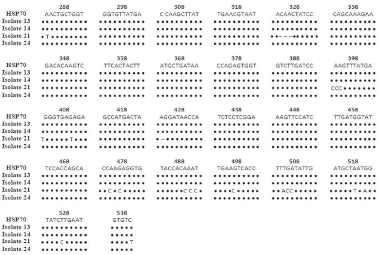
Figure 2 Sequence analysis of the Hsp 70 gene of four selected isolates (13, 14, 21, 24) obtained from the LR during the actual study. Dots show homology between isolated nucleotides and GenBank references. Sequence deletions and nucleotide substitutions are represented by hyphens and nitrogenous bases, respectively.
Conclusions
In the present study, a general prevalence of 71.79 % of Cryptosporidium was determined in dairy herd animals of the Lagunar Region (LR), where freshly weaned calves showed the highest prevalence, with 87.17 %; while the adults evaluated had a prevalence of 56.41 %. Cryptosporidium parvum was the species that was present in 100 % of the 25 samples sequenced and compared in NCBI GenBank, so it can be considered as an endemic species in the LR. Results suggest that the age of cattle is not a factor that determines Cryptosporidium infection, since its prevalence in calves did not show significant differences compared to adults. The high prevalence of C. parvum in cattle of dairy herds represents a risk for the dairy industry in the LR, as well as public health if manure is applied as a fertilizer in agricultural fields. It is suggested to continue monitoring the presence of microorganisms of interest such as Cryptosporidium, which provide evidence for a better management of cattle in dairy herds in the LR, as well as to reduce milk-associated sanitary and public health risks.

