


 texto en
  Inglés (pdf)
 Artículo en XML
 Referencias del artículo
 Enviar artículo por email
 Citado por SciELO
  Similares en
SciELO
Similares en
SciELO 
 Permalink
Introduction
In monocotyledons, vascular bundles maintain their individuality and they are distributed across the entire fundamental tissue of the stem. Vascular bundles can form one or two rings around the stem and leave a hole at the center (e.g., Zinnia elegans L.), or they can also be arranged in an isolated and dispersed form in the parenchyma (atactostele) (e.g., Tulipa orphanidea). Atactoestele is the most common anatomical arrangement of the vascular bundles in monocotyledons. It has also been reported that atactostele significantly improves the water transport in monocotyledons when compared to dicotyledons whose vascular bundles are found oriented around to the medulla (Esau, 1985; Twumasi et al., 2005; Alonso, 2011; Soykan & Meriç, 2012).
Different anatomical aspects of the stem, including the area of xylem per vascular bundle, number, and diameter of the vessel elements, could impact water flow, and perhaps vase life. For instance, in rose cv. “Polo”, despite the significant difference in the number of vessel elements observed (i.e., 242.6, 314.9 and 388.7 per mm2) across different sections of the stem (33, 41, and 54 cm from the floral button), no significant differences in the fresh weight or vase life were observed (De-La-Cruz-Guzmán et al., 2016). In contrast, it has been demonstrated that vessel elements with a small diameter significantly increase the water flow and the vase life. For example, shoots of Zinnia elegans have vessel elements with 60 and 65 μm of diameter, and showed a significant improvement in the water flow and an increase by two days in its vase life when comparing with shoots containing vessel elements with wider diameter. This difference has been explained by the fact that vessel elements with wider diameter are less resistant to water flow but more susceptible to cavitation (van Leperen et al., 2002; Twumasi et al., 2005; van Doorn, 2012).
In floral stems, the vase life is related to the fresh weight and the absorption rate, which occurs when stems are placed in water or preservatives solutions such as Chrysal clear®, which improve water flow and provide energetic compounds for the elongation and opening of flower buds (van Meeteren et al., 2001; Hernández-Fuentes et al., 2006).
Hence, hydration and vase life depend, among other factors, on the preservative solution and/or the distribution of vascular bundles, number, diameter, and area of the vessel elements in different levels of the floral stem. Quantitatively describing and explaining these characters would allow to understand the relations between the anatomy of the floral stem and vase life. In alstroemeria cv. Rebecca there are not anatomical descriptions on different levels of the stem axis available. Therefore, the goal of this work was evaluate the descriptive and quantitative anatomy at 2, 20, 40 and 60 cm of the inflorescence of alstroemeria cv. Rebecca and relate it to the fresh weight, the absorption rate and vase life of floral stems trimmed at the same lengths.
Materials and Methods
Alstroemeria was propagated by rhizomes, which were purchased in San Gregorio Atzompa, Puebla, Mexico. For this study, altroemeria rhizomes propagation was carried out from August to December 2016 in the greenhouse of the Unidad de Morfologia y Funcion, from the Facultad de Estudios Superiores Iztacala, UNAM. Three-year-old plants with rhizomes were wrapped in kraft paper and dark plastic and then transported to the laboratory, where they were stored at 20 °C for 24 h. Subsequently, plants were unpacked, and the rhizomes were washed and submerged in 1 % tecto® 60 solution for 30 min. Planting was carried out on a 6 m long bench by 0.8 m of width and 0.3 m of depth filled with tezontle with granulometry ≤ 5 mm. 330 liters of water were uniformly added into the tezontle bench to reach 100 % humidity, equivalent to 30 cbar. During cultivation the humidity was adjusted to 100 % by the addition of Steiner solution, a universal nutrient solution (Steiner, 1961), when the tensiometer indicated 56 cbar, equivalent to 80 % humidity. pH and electrical conductivity of the solution were 6.1 and 2.0 dSm-1, respectively. No symptoms of nutritional disorders (nutrient deficiency and/ or toxicity), neither incidence of pest or diseases were observed in any of the phenological phases of alstroemeria.
In the flowering phase, twelve stems with dehiscent buttons at their apex were harvested and separated to form four treatments, each one containing three biological replicates. Treatments consisted in floral stems trimmed at four levels from the inflorescence: 2, 20, 40 and 60 cm. Small pieces of 0.5 cm were obtained from the base of each level stem and fixed in FAA (10 % formaldehyde; 5 % acetic acid; 50 % ethanol; 35 % distilled water) for one week. Subsequently, these sections were washed in water and sequentially dehydrated in 30 to 100 % ethanol, followed by treatment with 100 % xylene. Upon dehydration, small pieces were embedded in liquid paraffin. Semi-thin sections (20 μm) were prepared using a rotation microtome model Leica RM2125 RTS and subsequently stained with safranine “O” and green fast FCF (Ruzin, 1999).
To evaluate vase life, twenty alstroemeria stems, each one containing three dehiscent buttons at the apex, were harvested from the experimental system described in the previous section, and immediately transported to the laboratory. Stems were trimmed 2, 20, 40 and 60 cm from the inflorescence, and four treatments, each one containing 5 biological replicates, were established. Seven leaves located at the basal part were removed from the stems belonging to the treatments 20, 40 and 60 cm only. Stems from all treatments were individually weighed. Subsequently, stems belonging to the 2 cm treatment were transferred into vases containing 50 mL of 10 % Chrysal clear® solution, whereas stems belonging to the 20, 40 and 60 cm treatment were transferred into vases containing 250 mL of the same preserving solution. Vases containing stem of each treatment were randomly distributed in an area of the laboratory with 10 μmoles m-2 s-1 light, 20 ± 3 °C, 45 ± 7.5 % relative humidity, and photoperiod of 12 h. 10 % Chrysal clear® solution was replaced every 5 days, and at the same time, each stem was trimmed 1.0 cm at its basal part.
Assessments
Descriptive and quantitative anatomy. For anatomical description, the epidermis was
located in each cross-section, and four zones, Z1, Z2, Z3, and Z4, were established.
For quantitative analysis, cross-sections of each replicate and treatment were
analyzed; each cross section was radially divided into eight equal parts, and
finally, two opposite quadrants of 1 mm2 were selected. In each quadrant,
the following parameters were evaluated: a) number of vascular bundles per
mm2 and per stem cross-section, b) vascular bundles area, c) xylem
area, d) number and area of the vessels, e) vulnerability index:
In the vase. Fresh weight (FW) and solution absorption rate (SAR). Fresh weight of each flower stem, as well as the weight of the preserving solution contained in the vase (in the absence of floral stems) was daily recorded by using a digital balance (Velab® ES-1000H, with 0.01 g of accuracy). Percentage of FW and SAR was expressed in mL g-1 d-1 (Rezvanypour & Osfoori, 2011).
Floral buttons Length. Length of the floral buttons, measured from the base to the apex, was daily evaluated with a digital vernier brand Scala® with 0.01 mm precision.
Floral diameter and vase life (VL). When the buttons were dehiscent in the apex, the floral diameter was daily recorded with the same vernier, measuring the top diameter of each floral button. The VL was determined by counting the number of days that the stems remained in the vase without showing symptoms of senescence (i.e., wilting), falling petals, leaves or foliage yellowing. The VL assessment concluded when in the floral peduncle, half plus a floral button showed symptoms of senescence.
Statistical analysis. Results were processed with descriptive statistics, including One-Way Analysis of Variance, and comparison tests (Tukey, α ≤0.05). SAS® v. 9.0 Software for Windows was used for all statistical analyses.
Results and Discussion
Descriptive anatomy. In cross section, stems of alstroemeria showed a circular contour with diameters of 1.5 cm in the areas near the inflorescence and 2.0 cm in the furthest. At 2 cm from the inflorescence, a simple epidermis formed by round cells covered by a thin cuticle was observed. In the interior of the stem axis, eight strata of parenchyma and isolated vascular bundles formed the Z1. Z2 was defined by a continuous band of smaller cells with slightly thickened cell walls. Z3 contained scattered vascular bundles, whose diameters were on average by 16.73 μm. Finally, the Z4 was located at the central part of the stem and was exclusively formed by parenchymal tissue (Figure 1A and 1B). As the distance increases, with respect to the inflorescence, epidermal cells remained with no visual changes.
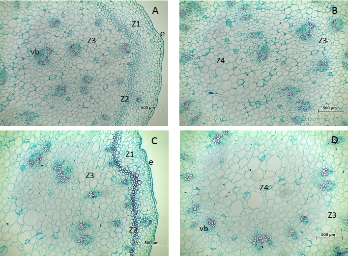
Figure 1 Transversal views on two levels of the floral stem, with respect to the inflorescence, of alstroemeria cv. Rebecca. A, B. 2 cm from the inflorescence; C, D. 20 cm from the inflorescence. Z, zone; e, epidermis; vb, vascular bundles.
From 20 to 60 cm, on the stem axis, the Z1 showed slightly flattened cells and its parenchyma strata decreased up to six, the vascular bundles were scarcer in the basipetal orientation. Z2 showed cells that began to be lignified, until forming sclerenchyma tissue that was evident at 20 cm. This tissue formed a continuous ring that was formed by three to four layers of cells that surround numerous vascular bundles. In Z3, the vascular bundles (vb) decreased in number, but they were abundant in the basipetal orientation. At 20, 40 and 60 cm, the parenchyma cells present in the Z4 were getting bigger towards the center, but spaces, developed by a separation of cell walls, were observed. These spaces were not observed at 2 cm (Figure 1; Figure 2).
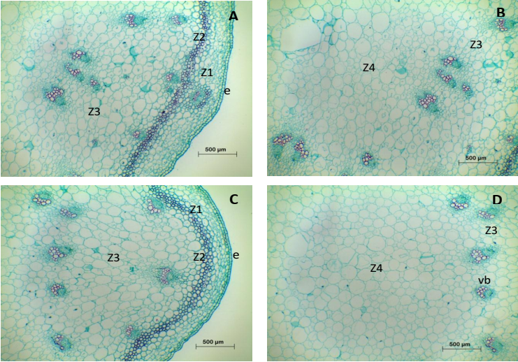
Figure 2 Transversal views at two levels of the floral stem, with respect to the inflorescence, of alstroemeria cv. Rebecca. A, B. 40 cm from the inflorescence; C, D. 60 cm from the inflorescence. Z, zone; e, epidermis; vb, vascular bundles.
Anatomical studies on monocot flower stems show certain differences. For example, in Zinnia elegans the vascular bundles are located around the medulla, while in Tulipa orphanidea the vascular bundles are embedded into it (Twumasi et al., 2005; Soykan & Meriç, 2012). In alstroemeria cv. Rebecca, the distribution of vascular bundles was similar to T. orphanidea, and in accordance with the general pattern described for monocotyledons, particularly with the arrangement of the vascular cylinder anatomy, which is atactostele.
Quantitative anatomy: Number and area of the vascular bundles, area of the xylem. At 2 cm from the inflorescence, the number of vascular bundles per mm2 or per section cross-section of the stem was 6.3 and 80.6, both values represented almost double than those observed at 20, 40 or 60 cm. In the opposite direction, the areas of the vascular bundles and xylem, by vascular bundle, were greater than 20, 40 or 60 cm (Table 1).
Table 1 Vascular bundles and xylem area, in transversal views, of floral stems of alstroemeria cv. Rebecca trimmed to 2, 20, 40 and 60 cm with respect to the inflorescence.
| Floral stem length | Number of vascular bundles | Area (μm2) | |||
|---|---|---|---|---|---|
| (cm) | mm2 | Stem cross section | Vascular bundles | Xylem | |
| 2 | 6.33 az | 80.6 a | 28161 b | 5990.3 b | |
| 20 | 3.33 b | 46 b | 39556 a | 9722.7 a | |
| 40 | 3.50 b | 43 b | 37962 a | 8470.4 a | |
| 60 | 3.17 b | 46 b | 37200 ab | 8603.9 a | |
| DHS | 1.69 | 13.99 | 9419.2 | 2130.8 | |
| CV (%) | 25.31 | 14.35 | 31.57 | 31.26 | |
zThe means followed by different letters, in each column, indicate significant differences (Tukey, α ≤ 0.05). DHS, difference honest significant; CV, coefficient of variation. Each data is the average of six quadrants.
Floral stems of Zinnia elegans and Tulipa orphanidea have on average 24 and 47 vascular bundles by cross section (Twumasi et al., 2005; Soykan & Meriç, 2012), somewhat similar was observed in alstroemeria stems trimmed at 20, 40 or 60 cm. It is worthy to mention that in those studies on Z. elegans and T. orphanidea it was not specified how and at what level the counting was performed. This type of information is crucial because the variation in the number of vessel elements, at different levels of the stem axis, could help explain the differences in absorption rate of the solution and its relation with life in the vase. For example, stems of alstroemeria cv. Rebecca trimmed at 2 cm have the largest number of vascular bundles, minor area of the xylem and vessel elements with smaller diameters compared to stems trimmed at 20, 40 or 60 cm, which have fewer vascular bundles and vessel elements with larger diameters. In stems trimmed at 2 cm, these anatomical characteristics promoted the rate of absorption towards the floral buttons, since small vessel elements have more resistance to water flow, but are less susceptible to cavitation, when comparing with vessel elements with larger diameter, which offer less resistance to flow, but are more susceptible to cavitation (Cohen et al., 2012).
Number and area of vessels, vulnerability index (VI). At 2 cm from the inflorescence, the number of vessel elements was higher, but with significantly smaller areas when comparing to stems trimmed at 20, 40 or 60 cm, in which the number of vessel elements ranged between 36.8 and 51.6 with an average area of 26.7 μm2, respectively. The VI radial or tangential was smaller than 1, without taking into consideration the length of the floral stem (Table 2).
Table 2 Number and area of vessels and radial or tangential vulnerability index, in transversal views, of floral stems of alstroemeria cv. Rebecca trimmed to 2, 20, 40 and 60 cm with respect to the inflorescence.
| Floral stem Length (cm) |
Number of vessels per mm2 |
Vessels area (μm2) |
radial VI | tangential VI |
|---|---|---|---|---|
| 2 | 134.1 az | 15.11 c | 0.13 d | 0.11 d |
| 20 | 51.6 b | 27.37 a | 0.56 c | 0.59 c |
| 40 | 36.8 b | 27.03 ab | 0.77 a | 0.82 a |
| 60 | 40.3 b | 25.61 ab | 0.68 b | 0.72 b |
| DHS | 29.42 | 1.58 | 0.03 | 0.04 |
| CV (%) | 26.22 | 39.19 | 44.06 | 46.39 |
zThe means followed by different letters, in each column, indicate significant differences (Tukey, α ≤ 0.05). VI, vulnerability index; DHS, difference honest significant; CV, coefficient of variation. Each data is the average of six quadrants.
The number of vessel elements can be different among cultivars or even in the same floral stem. For instance, floral stems of the rose cultivars ‘Lovely Red’ and ‘Rouge Baiser’ have 267 and 308 vessel elements per mm2, respectively, while in rose cv. ‘Polo’ the number of vessel elements is 243, 315 or 389, depending on whether they are located at 33, 41 or 54 cm in length with respect to the floral button. This evidence indicates that the vessel elements increase in the basipetal orientation (Cohen et al., 2012; De-la-Cruz-Guzmán et al., 2016).
Floral stems of alstroemeria cv. Rebecca trimmed at 2 cm from the inflorescence tripled their number of vessel elements when comparing to those counted in stems trimmed at 20, 40 or 60 cm, which did not show any significant differences among them. In accordance, it has been reported that water conductivity is significantly improved when stems contain a higher number of vessel elements in small areas, which contribute to being less susceptibility to cavitation (Hargrave et al., 1994).
It has been observed that the area containing vessel elements is usually smaller when it is closer to the flower bud. For instance, in floral stems from rose cv. ‘Polo’ with 25, 35 or 50 cm in length, the area of the vessel elements was 4.4, 12.4 and 12.5 μm2, respectively (Arriaga-Frías et al., 2016). A similar pattern was observed in alstroemeria cv. Rebecca, particularly in floral stems with 2 cm of length, which show a small area containing vessel elements.
Vulnerability index (VI) values greater than 1.0 indicate that cultivars are vulnerable or slightly resistant to water stress, while values smaller than 1.0 indicate that the stems were stressed and are resistant to embolism (Carlquist, 1977; Hacke et al., 2001). In this study, the VI radial or tangential of alstroemeria cv. Rebecca was less than 1.0, which would indicate that floral stems experimented water stress during its cultivation. However, substrate moisture remained ≥ 80 %, therefore, different values of VI were attributed to anatomical variations in the stem and not due to a period of stress in the substrate. A similar response, but without specifying the growth conditions, was observed in stems of rose cv. ‘Polo’ trimmed at 25, 35 and 50 cm from the floral button, whose VI values were of 0.14, 0.83 and 1.24, respectively (Arriaga-Frías et al., 2016). In alstroemeria and in rose cv. ‘Polo’, the IV increases in the basipetal orientation, which indicates that the resistance to embolism is greater in places near the flower bud.
Radial and tangential diameter of the vessel elements. At 2 cm from the inflorescence, the radial or tangential diameter of the vessel elements varied from 2.5 to 45 μm; the highest frequency (80 %) was between 7.6 and 27.5 μm respectively. In the stems with 20, 40 or 60 cm, the radial or tangential diameter fluctuated from 5 to 74 μm, the highest frequency (80 %) of vessel elements, was located between 12.1 and 46.0 μm. The radial or tangential diameters were similar in the four levels of the floral stem (Figure 3).
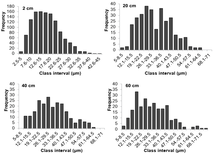
Figure 3 Radial or tangential diameters of vessel elements, in transversal views, of floral stems of alstroemeria cv. Rebecca trimmed to 2, 20, 40 and 60 cm with respect to the inflorescence.
On rose floral stems trimmed at 33, 41 or 54 cm from the floral bud, the radial or tangential diameters of the vessel elements fluctuated from 40 to 220 and from 60 to 300 µm, the bigger distribution (60 %) ranged from 60 to 120 and from 100 to 180 μm, respectively (De-la-Cruz-Guzmán et al., 2016). Regardless of its location in the floral stem, the vessel elements of alstroemeria cv. Rebecca had smaller diameters compared to those of rose cv. ‘Polo’. It has been reported that vessel elements with diameters greater than 75 μm are more susceptible to embolism during periods of stress water (Hargrave et al., 1994; Nijsse et al., 2000). In accordance, the water conductivity is likely more efficient in alstroemeria cv. Rebecca than in rose cv. ‘Polo’.
Fresh weight (FW). On the second day of incubation in vase conditions, fresh weight increased 9, 12, 16 and 22 % in the floral stems trimmed at 60, 40, 20 or 2 cm with respect to the inflorescence, respectively. Until day 4, stems trimmed at 2 cm kept their fresh weight higher. After the seventh day, the FW fluctuated from 94 to 99 % (Figure 4).
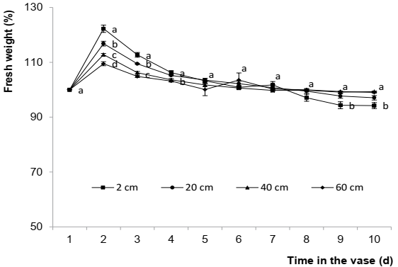
Figure 4 Fresh weight in floral stems of alstroemeria cv. Rebecca trimmed to 2, 20, 40 or 60 cm with respect to the inflorescence. Each data is the average of five repetitions ± standard error. Different letters in each evaluation time indicate significant differences (Tukey, α ≤ 0.05).
It has been reported that stem fresh weight varies across the life in the vase, at the beginning it increases following by a decrease in the latest stages. Additionally, it has been observed that those stems able to constantly keep for longer periods of time, have a longer life in the vase (Ichimura & Shimizu-Yumoto, 2007; Lü et al., 2010). Fresh weight is related to the hydration of the flower stems and it is important because the metabolic processes that provide energy to the petals perform better when hydration is optimal (van Meeteren & van Gelder, 1999; Taiz & Zeiger, 2010). Stems of alstroemeria cv. Rebecca, trimmed at 2 cm from the inflorescence, maintained by 4 d greater fresh weight with respect to those stems trimmed at 20, 40, or 60 cm, because the 2 cm had a greater number of vessel elements with smaller diameters, which contribute to the water flow, being less prone to cavitation. In contrast, the number of vessel elements decreased and its diameter increased in stems trimmed at 20 cm, which made the conduction system prone to cavitation and less efficient in the flow of water. Recently, it has been reported a relationship between the number and diameter of the vessel elements with the water flow associated with fresh weight and vase life in chrysanthemum cv. ‘Hartman’ and rose cv. ‘Topaz’ (De-La-Cruz-Guzmán et al., 2018).
Solution absorption rate (SAR). On day 1, the absorption rate in the floral stems trimmed at 2 cm from the inflorescence was 0.37 mL g-1 d-1, while in stems trimmed at 20, 40 or 60 cm was in average by 0.50 mL g-1 d-1. At day 2, an increase of 34% in the fresh weight was observed in stems trimmed at 2 cm, whereas an increase of 40 % was observed in stems trimmed at 20, 40 or 60 cm. During the first 4 days, the SAR value for stems trimmed at 2 cm remained higher (0.52 mL g-1 d-1) when comparing with the other trimming treatments whose SAR value was in average by 0.42 mL g-1 d-1 with no differences among them. After day 4, SAR fluctuated from 0.20 to 0.25 mL g-1 d-1 regardless of the length of the flower stems.
Floral stems of rose cv. ‘Polo’ trimmed at 33 cm have higher absorption rate than those trimmed at 41 or 54 cm (De-La-Cruz-Guzmán et al., 2016). A similar response was observed in alstroemeria cv. Rebecca, since the floral stems trimmed at 2 cm had a rate of absorption greater than those trimmed at 20, 40 or 60 cm in length.
When floral stems are placed into a vase, the absorption rate increases during the first days and then decreases. A decrease in water consumption can be attributed to cavitation events at the base of the stem, either by the proliferation of bacteria, formation of air bubbles or by synthesis of metabolites produced at the trimming moment (Spinarova & Hendriks, 2005; Fanourakis et al., 2012; Arévalo-Galarza et al., 2012; van Doorn, 2012). Delayed cavitation has been correlated with longer life in the vase. Cavitation can be eliminated when 3 cm of the base of the floral stems are trimmed out, and when the solution contained in the vase is renewed (Arévalo-Galarza et al., 2012; De-La-Cruz-Guzmán et al., 2018).
Button length, floral diameter and vase life (VL). The maximum length of the flower buds, which was presented on the seventh day of permanence in the vase, was 50, 55, 62 and 66 mm for the floral stems trimmed at 60, 40, 20 and 2 cm from the inflorescence respectively. The maximum floral diameter in stems at 2 cm appeared on day 11 and was 48.5 % higher compared to stems trimmed at 60 cm, whose opening average was 44.15 mm and occurred on day 9. Vase life was 2.2 d higher in the stems trimmed at 2 cm compared with the other trimming treatment, which, on average, showed a VL value of 12.2 d (Table 3).
Table 3 Floral diameter (mm) and vase life in stems of alstroemeria cv. Rebecca trimmed to 2, 20, 40 or 60 cm from the inflorescence.
| Floral stem length (cm) | Time in the vase (d) | VL (d) | ||
|---|---|---|---|---|
| 9 | 10 | 11 | ||
| 2 | 64.93 az | 64.01 a | 67.81 a | 14.4 a |
| 20 | 64.18 a | 59.77 a | 57.02 b | 12.6 b |
| 40 | 53.58 b | 52.02 b | 53.13 bc | 12.2 b |
| 60 | 44.43 c | 43.48 c | 44.54 c | 11.8 b |
| DHS | 6.33 | 7.48 | 9.14 | 1.27 |
| CV (%) | 16.41 | 20.22 | 22.08 | 5.54 |
zThe means followed by different letters, in each evaluation time or vase life (VL), indicate significant differences (Tukey, α ≤ 0.05) DHS, difference honest significant; CV, coefficient of variation. Each data is the average of five repetitions.
The preservative compounds added in the vase solution modify the opening and delay the senescence symptoms. For example, with the use of 0.1 mM gibberellic acid, the floral opening in alstroemeria cv. ‘Dancing Queen’ is 6.4 cm and its flowering life is 11.4d. However, when 200 ppm 8-citrate hydroxyquinoline is combined with 2 % sucrose, the floral opening increases 0.8 cm and life of vase 2.2.d (Sea et al., 2012).
In floral stems of alstroemeria trimmed at 2 cm from the inflorescence, the leaf area fluctuated between 10 and 20 cm2, whereas in those trimmed at 20, 40 or 60 cm the fluctuation was between 200 and 450 cm2. This observation suggests that the opening of the floral buttons and vase life were promoted by the Chrysal clear® solution, particularly a concentration of 10 %, and not by the translocation of photosynthates that accumulated in the leaves during its cultivation. In stems with bigger foliage, the energetic compounds contained in the vase solution were distributed in the leaves and in the petals, but in stems with less foliage, this type of compounds was mostly directed towards the flower buds.
Conclusions
In cross section, floral stems of alstroemeria cv. Rebecca have epidermis with cuticle, parenchyma and sclerenchyma band that surrounds most of the vascular bundles.
Vascular bundles are distributed throughout the fundamental tissue of the stem, with arrangement of atactostele (bigger bundles at the center and smaller bundles at the periphery).
At 2 cm from the inflorescence, floral stems of alstroemeria cv. Rebecca have more vessel elements and vascular bundles per mm2 or cross-section, whereas at 20, 40 or 60 cm the number of both of them decreases, but their areas and diameters increase.
Floral stems trimmed at 2 cm from the inflorescence have a vase life of 14.4 d, this is because their vessel elements are less susceptible to cavitation and the distance traveled by the water is smaller. At 20, 40 or 60 cm, vase life decreases 2.2 d, this is because the vessel elements have a larger diameter and were more susceptible to cavitation.
The descriptive and quantitative anatomy, in different levels of the floral stem, allow to explain the differences in hydration and vase life of alstroemeria cv. Rebecca.

