


Servicios Personalizados
Revista
Articulo
 texto en
  Inglés (pdf)
 Artículo en XML
 Referencias del artículo
 Enviar artículo por email
Indicadores
-
 Citado por SciELO
 -
 Accesos
Links relacionados
-
 Similares en
SciELO
Similares en
SciELO
Compartir

 Permalink
Intervención (México DF)
versión impresa ISSN 2007-249X
Intervención (Méx. DF) vol.13 no.26 México jul./dic. 2022 Epub 05-Dic-2023
https://doi.org/10.30763/intervencion.273.v2n26.52.2022
Academic reports
The Conservation of Elasmobranchii Remains in the Sacred Precinct of Tenochtitlan. An Overview

* Museo del Templo Mayor-Proyecto Templo Mayor (MTM-PTM), Instituto Nacional de Antropología e Historia (INAH), México, asanromanp@gmail.com y mariabarajas35@yahoo.com.mx
Vast amounts of animal remains have been recovered from the archaeological excavations in the sacred precinct of Tenochtitlan, among them, those from fish with cartilaginous skeletons stand out. Their specific characteristics represent a challenge in terms of their proper recovery, conservation, and exhibition. In the following pages, an overview of these materials´ properties is presented, followed by a description of the different excavation contexts. A brief historical review of the different procedures applied is also presented, ending with some considerations for future work in this field.
KEY WORDS: chondrichthyes; elasmobranchii; Templo Mayor; Tenochtitlan; Mexica
Las excavaciones arqueológicas en el recinto sagrado de Tenochtitlan han revelado numerosos restos de animales, entre los que destacan notablemente los procedentes de peces con esqueleto cartilaginoso, cuyas características específicas representan un reto para su recuperación, intervención y exposición. En las siguientes páginas presentamos una general de las características de estos materiales óseos, y discutiremos los diferentes contextos en los que fueron encontrados estos materiales óseos, y proporcionaremos una breve revisión histórica de la gama de procedimientos aplicados, junto con algunas reflexiones para trabajos futuros.
PALABRAS CLAVE: condrictios; elasmobranquios; Templo Mayor; Tenochtitlan; mexicas
INTRODUCTION
During 40 years of archaeological excavations in the sacred precinct of Tenochtitlan, it has become clear that that in these types of ritualistic deposits, exogenous fauna from the Basin of Mexico are constantly being found, and which reflect the diversity of animal species represented across Mexica symbolism (López, 1993, p. 143).
The extensive studies by various specialists on ichthyofauna, has brought to light interesting data on the remains of the fish species found in these offerings; among them we see that species with particularly striking colors are common, as well as some poisonous species and even some with morphologies resembling mythological monsters (Díaz-Pardo, 1982; Díaz-Pardo and Teniente, 1991; Guzmán and Polaco, 2000, p. 153; Robles et al., 2018, pp. 21-22). Amongst the diversity of aquatic animals from the Templo Mayor archaeological site’s collections, there are two main types of fish, categorized according to their skeleton type: boney or cartilaginous (Chondrichthyes).
From the conservation-restoration professional perspective, there are very few records describing how archeological conservators have approached the complexity that the recovery and treatment of Chondrichthyes remains involve, not only in the sacred precinct of Tenochtitlan but also on a national level as a whole.
This text shows the conditions in which these elements were found in the Mexica’s ceremonial site, providing a brief reflection on their state of conservation, the different methodologies used for their extraction from the archaeological matrix and, the various factors surrounding their long-term conservation, including analyses, permanent storage, exhibition display, and the diffusion of knowledge.
The sacred precinct of Tenochtitlan
Currently located in the first square of Mexico City, under numerous layers deposited by human activity over many centuries, the sacred precinct of Tenochtitlan was built during the Late Postclassic period (1200 - 1521 A.D.) on a small island in the center of the lake basin. The island’s terrain expanded, gaining space from the water by carrying materials from both the lake bed and the surrounding areas (rocks) and constructed using the stilt system (Mazari, et al., 1989, pp. 149-155).
The low permeability and stilt clay composition of the soil, (López & Tentle, 2012, p. 43-46), has granted constant conditions of relative humidity and temperature in the subsoil of some areas of the former Mexica hegemonic center, favoring the discovery of organic and inorganic materials.
The enormous pressure on the subsoil caused by the maintenance and constant remodeling of the ceremonial center, along with the construction of Colonial, Modern and Contemporary buildings, combined with the bodies of water dehydration, have caused the differential sinking of the ground and generated contexts with low, null, or fluctuating relative humidity (RH) (López, 1989, p. 148).
Following the discovery of particular materials, these conditions must be taken into account to take precautions during the archaeological excavations.
The chondrichthyes
This type of fish is characterized by a cartilaginous skeleton, including elasmobranchii (sharks and rays), and holocephali (chimaeras). (Compagno et al., 2005, p. 5; Moyle & Cech, 2009, p. 226-230, 255-273; Helfman et al., 2009, p. 197-229).
Many Elasmobranchii remains have been recovered from the offerings at the sacred precinct of Tenochtitlan, their excavation and conservation pose a challenge due to their biological characteristics.
Biologically, the skeleton of these fish is made up of a predominant organic phase, composed mainly of collagen, and a mineral phase, in smaller proportion, made up of calcium phosphate in the form of hydroxyapatite. This cartilaginous structural system shows two different calcification forms: the first corresponds to the vertebrae, with a biconical shape and highly calcified segments; the second is formed by a mosaic-like layer that covers a non-crystallized nucleus, in this layer each tesseral calcifies from the center towards the outer edges (Dean & Summers, 2006, p. 165-166). The calcification process occurs throughout the lifetime of the individuals, so that juvenile specimens present less mineralization than adult specimens (Seidel et al., 2017, p. 2-4). This “mosaic” has specific arrangements and densities according to the animal´s anatomical section, responding to the structural needs of each body part.
The teeth, caudal spines, and dermal denticles1-which conform the skin-are composed of dentine and vitrodentine, both formed by fluorapatite crystals and a small amount of collagen (Enach, 2012, p. 290-299; Moyle & Cech, 2009, p. 15-17; Helfman et al., 2009, p. 36-40)
Elasmobranchii in the sacred precinct of Tenochtitlan
The remains of more than a hundred of this type of ichthyofauna have been identified over the course of four decades of exploration in the area occupied by the Mexica’s ceremonial center, corresponding to the discovery of sharks and rays, including sawfish.
According to the bibliography (López, 2005, p. 191-192; Guzmán y Polaco, 2000; Robles, 2017, p. 21-56; Robles et al., 2018), the presence of these fish is linked to the representation of the terrestrial monster known in Nahuatl as Cipactli2, a symbol of the Earth and its abundant production.
The remains of cartilaginous skeletons are identified by the peculiar morphology of the tessellated cartilage, by their biconical vertebrae, or by distinctive anatomical segments, such as sawfish rostral cartilages, shark buccal teeth, stingray caudal spines or dermal denticles (Figure 1).
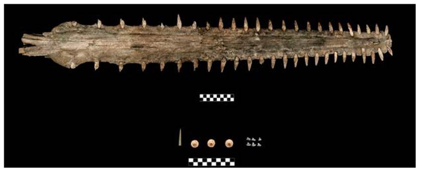
Source: (Photograph: Mirsa Islas y Érika Lucero Robles, 2016; courtesy: Proyecto Templo Mayor [PTM], Mexico).
FIGURE 1 Above: rostral cartilage from sawfish (Offering 141). Below, from left to right: skate caudal spine (Offering 120) and teeth from a shark’s mouth (Offering 141)
The quantification corresponds to the recovery of 31 sharks, 27 rays, and 85 sawfish, according to the archaeologist Érika Lucero Robles (personal communication, March 10th, 2021), keeping in mind that this analysis is under constant revision and that new discoveries are made daily, both on the field and while studying associated and previously unidentified remains.
These vessels have been found in three types of offerings registered at the site: deposited directly in the landfill; placed inside two-piece stone boxes, and finally, the most common, those built with tezontle ashlars and flagstone used in the form of floor and cover (López, 2005, p. 124-131). They have been found in completely dry to flooded contexts, which confirms the great resistance of these materials despite their fragility at the time of discovery.
Considerations on the State of Conservation
Thanks to the varying conditions of the specific contexts that have been excavated, it is known that the state of preservation of these faunal remains depends both on their morphology and their interaction with the environment.
On one hand, the vertebrae always present greater stability, due to their compact shape, and a higher degree of calcification, regardless of the humidity conditions of the site. The same occurs with the caudal spines of rays, their dermal denticles, and their teeth, both rostral-whose chemical composition gives them greater resistance to the environment-as well as those from the lower and upper jaws.
On the other hand, segments composed of tessellated cartilage are much more unstable, and their preservation inside the matrix depends on the loss or conservation of the unmineralized cartilaginous nucleus, or its substitution by the surrounding area´s sediments, as well as the stability of the specific conditions of the ritual deposit.
The elements found in dry contexts filled with sediment are in good preservation conditions and are resistant as a unit. The anatomical sections found in constant conditions of waterlogging or very high relative humidity without filling are generally the most degraded, as is the case with those found in deposits with fluctuating conditions, since the pressure caused by the elements placed over the cartilaginous tissue as well as the presence and flow of water favor the hydrolysis of the protein and the dragging of the mineralized sections. Finally, the sections in conditions of high humidity, but with filling, are extremely fragile and show considerable deformations and fractures (Figure 2).
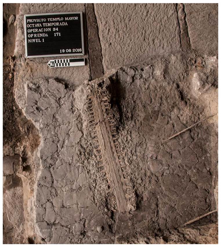
Source: (Photograph: Mirsa Islas, 2016; courtesy: Proyecto Templo Mayor [PTM], Mexico).
FIGURE 2 Offering 171
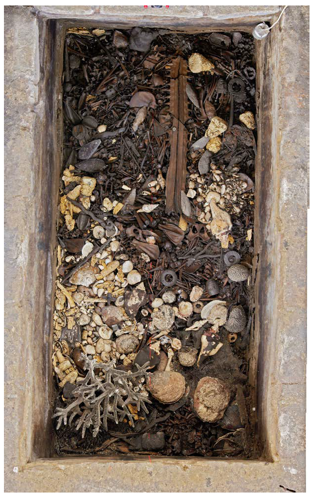
There are also cases in which, thanks to the stable conditions of the context, it is possible to recover sections of the skin of these marine animals. Although it is difficult to notice their presence in situ, a thin, shiny layer above the tessellated cartilage can be identified by a trained eye. These remains are recognizable under the microscope by finding dermal denticles which, although easily shed under fluctuating conditions, can be recovered when analyzing the deposit’s sediments (Barajas et al., 2015, p. 39-50; Barajas et al., 2016; p. 63-67, 159-162 and annex 2; Barajas & Sanromán, 2017, p. 172-178, 188-194, 204-205 and 211-213; Barajas & Sanromán, 2018, p. 173-178, 200-201 and 221-235; Barajas & Sanromán, 2019, p. 222-240; Robles et al., 2018, p. 24; Bolaño et al., 2022) (Figure 3).
Intervention
The conservation procedures that are carried out in the Templo Mayor Project (TMP) focus on providing structural stability to the recovered materials, to ensure both their long-term conservation and the preservation of the “archeological data”, that is, the relevant information for the different scientific analyses carried out by the research team. In this sense, decision-making is based on the criteria of minimum intervention and re-treatability. The treatments described herein are applied in general to this type of elements and can be used as a sort of guide for future findings.
Preventive Measures in the Context
The participation of a field conservator is essential upon the discovery of this type of remains within the ritual deposits since decisions must be taken immediately to ensure the permanence and stability of the cartilaginous skeletons during the excavation and recording process.
First and foremost, it is crucial to always maintain the context’s conditions, recording and studying the humidity and temperature data with digital thermohydrometers, using water containers, plastic covers, or humidifiers, if necessary, in order to maintain stable parameters at the levels at which the remains were found. In addition, it is important to have a cover over the excavation area, to minimize alterations due to insolation; depending on the characteristics of the context, it can be temporary, using tarpaulins and sheets, or permanent, when closed spaces are built to protect the research area.
Excavation and Registry
The excavation of offerings is carried out following arbitrary levels in which the superficial layer of visible objects is defined and freed in a very meticulous and controlled manner, facilitating the detailed registry process and the subsequent analysis of each object. The work around these faunistic remains is performed with soft delicate tools, including bamboo swabs, wooden or bone spatulas and styli, and brushes, to avoid any possible marks on the surface of the cartilaginous tissue, which could cause confusion during taphonomic analysis.
Parallel to the excavation, graphic and photographic records are carried out, using a methodology designed and implemented in 2010 by the team of the Templo Mayor Project (TMP) in which, using scaled zenith photographs of deposits, printed in a proportion of 1:1, the elements that comprise a layer are traced in a plastic sheet placed over the print. The tracings serve as a guide for the digital AutoCAD® drawing; osteoarcheological field analysis are also carried out (Chávez et al., 2011) and the conservation registry, which consists of the elaboration of materials that support what was done by the archaeological team, to avoid the loss of information and ensure a good intervention a posteriori. The latter includes a written record of the conditions upon discovery and preventive treatments used, drawings and detailed photographs of the elements, and even Mylar® or acetate tracing, which will facilitate the intervention processes in the field laboratory.
Likewise, a written record is kept, using a database developed in FileMaker®, in which the team of archaeologists fills in the records for artifacts and organic materials, and the restorers fill in the conservation records.
In situ Intervention
It is sometimes necessary to carry out direct intervention processes simultaneously with preventive conservation and registry actions when the faunal remains are still in situ. The type of procedures carried out depends on the characteristics of the burial matrix, the state of conservation of the cartilaginous material, and the excavation plan.
The superficial cleaning of these remains is carried out using fine tools in a generalized manner over them to delimit the forms and make an adequate record; soft brushes and water applied with a dropper are used for humid contexts, while soft brushes are used in dry ones; occasionally specific actions are performed with swabs slightly moistened with water. The sediment recovered from these processes is registered as a sample for laboratory analysis, where it is possible to recover dermal denticles for future identification.
Another common process is veiling the surface with Japanese paper or non-woven materials to help maintain the humidity of the anatomical segment, or to ensure its unity and prevent its disintegration. The first are secured to the surface of the materials using potable water, using a sprayer when considerable desiccation is observed. For the second ones, various adhesives have been used throughout the years, such as EMA copolymers (ethylene methacrylate copolymers, Paraloid B72® in different concentrations, with polar and nonpolar solvents) and cellulose derivatives (hydroxypropyl cellulose, hydroxypropyl methylcellulose, sodium carboxymethylcellulose), each presenting advantages and disadvantages for the conservation process and a posteriori analysis. This veil is usually planned as part of the extraction that has evolved and varied over time.
Consolidation -for remains composed of tessellated cartilage mosaic- is another process that has been carried out in situ in almost a generalized manner. This is mainly carried out in those cases where the element´s state of conservation is poor, and presents fragility, fissures, fractures, deformations, and general loss of cohesion. Different EMA and PVB resins (polyvinyl butyral PVB, Mowital B60H®) have been used in different concentrations and in solution or emulsion (EMA, Primal AC 33®) whose application has been by dripping, taking care not to contaminate the rest of the context. (Hasbach, 2000, p. 135-136; Sanromán, 2009, pp. 8-12; Escalante & Soto, 2012, p. 113-114; Barajas et al., 2015, p. 132-143; Barajas et al., 2016, p. 63-67, 159-162 and Annex 2; Barajas & Sanromán, 2017, p. 172-178, 188-194, 204-205 and 211-213; Barajas & Sanromán, 2018, p. 173-178, 200-201 and 221-235; Barajas & Sanromán, 2019, pp. 222-240).
Once the veils and consolidates are dry, the team starts to recover the ichthyological remains from the context.
Extraction
This process varies, depending on the anatomical section found in the deposit and its state of preservation, as well as each offering´s specific characteristics.
The sections corresponding to vertebrae, teeth and caudal spines are recovered manually or with fine tools, such as tweezers, since they are better preserved due to their morphological characteristics and calcification or mineralization. Dermal denticles are recovered in the same way but under the stereoscopic microscope due to their dimensions.
On the other hand, the anatomical sections composed of tessellated cartilage present a greater complexity, depending on their preservation conditions. The most common way of lifting these elements is through the creation of blocks and using various types of supports to ensure that the faunal remains maintain their coherence and do not deteriorate further.
A wide range of materials have been used to build these blocks, depending on their availability, temporality, and previous experiences with bone remains and other archeological materials.
The veil is an essential component in the creation of said blocks, which serves as the first insulating layer between the element to be extracted and the filling material of the block. Isolating both the element to be extracted and the rest of the context is vital for the correct extraction of the block, and to avoid contaminating the offering. Materials used in addition to the veil include layers of plastic (usually PE polyethylene), self-adhesive plastics, aluminum foil, and layers of cloth with various adhesives, mainly acrylic resins. The materials that support the block are placed on top of these layers.
Depending on the specific characteristics of each case, plaster bandages, wood or cardboard is used to make the blocks, which are made to contain the filling materials, including: plaster, plaster with polystyrene beads, synthetic adhesives with polystyrene beads, expansive polyurethane resin (PU) and low-density epoxy resins (EP) (Araldit® or Ren Paste®). In almost all cases, these blocks have served as auxiliary support for laboratory work. Some of these form part of the permanent exhibition at the Museo del Templo Mayor (MTM) (Figure 4).
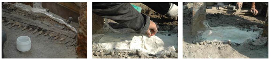
Source: (Photograph: Néstor Santiago, 2015, courtesy: Proyecto Templo Mayor [PTM], Mexico)
FIGURE 4 Offering 165, extraction with plaster bandages
It is important to highlight that the production of this type of block has been recently avoided, with a preference for instead using several layers of veils, providing greater visual control of the element being extracted, along with the actions carried out on the other objects deposited during the extraction.
A great range of auxiliary supports are used to lift the blocks, they are inserted gently between the element and the lower layers of the area, making sure to provide enough support for the cartilaginous remnant and to also completely separate it from the sediment and other materials in the offering. The use of wooden plaques with metallic edges, metallic sheets, and polyethylene or other rigid or flexible plastic sheets is recorded (Figure 5).
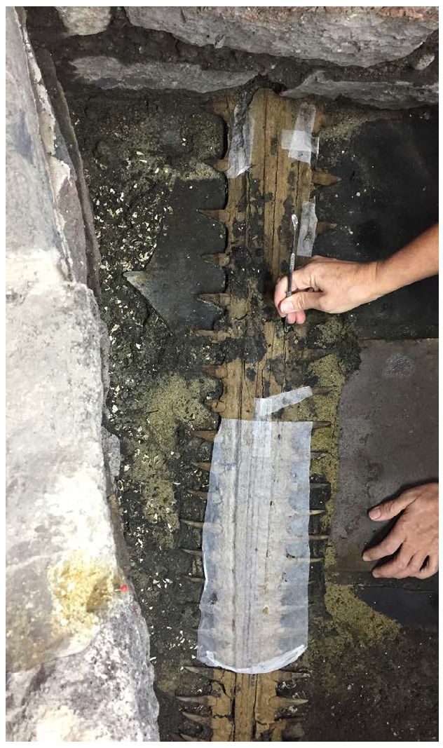
Source: (Photograph: Mirsa Islas, 2017, courtesy: Proyecto Templo Mayor [PTM], Mexico)
FIGURE 5 Offering 174, placement of facing
To ensure a homogeneous and simultaneous action on all the elements, lifting the block requires at least two or three people.
It should be noted that once the block has been lifted, in most cases it is turned over to cover, isolate and fill the underside ensuring the objects are not damaged during subsequent transportation.
More recently, after the extraction of cartilaginous elements is completed, they are then placed on a pillow of polystyrene balls, isolated with self-adhesive plastic and Tyvek®, which serves as a support that can be modified as needed during the intervention procedures (Figure 6).

Source: (Photograph: Mirsa Islas, 2016, courtesy: Proyecto Templo Mayor [PTM], Mexico)
FIGURE 6 Offering 171, Polystyrene balls support
Several considerations must be considered during the extraction of a block, including its final size and weight, how the block will be transported to the field or formal laboratory, as well as the amount of time that will elapse before the direct intervention begins.
In our personal experience, the blocks were moved a very short distance because the field lab was a few meters away from the excavation site; however, ensuring that the whole team is careful and aware when moving these fragile materials, helps avoid unnecessary accidents. Also, intervention should start almost immediately and any issues solved promptly, for example, by placing additional supports or coverings.
Intervention in the Laboratory
The processes that took place in the field are continued in the laboratory, in a controlled environment.
For caudal spines, teeth, and vertebrae, the direct intervention begins by finishing the surface cleaning; using paintbrushes and tap water, though in certain cases a rolled swab, dampened with ethanol or water, is used to control the humidity applied to the material.
In all cases, following the aqueous processes, controlled drying is performed, to avoid any abrupt changes that may cause new alterations.
Tessellated cartilage mosaic and certain vertebrae -mainly from young individuals- need a consolidation process, where low-concentration dispersions of acrylic resins (usually between 2 and 5% in ethanol; greater concentrations in less polar solvents were used previously) are applied locally, or in the case of the vertebrae, by immersion. As this is an irreversible process, systematic sampling helps subsequent analysis.
Sampling is performed jointly with archaeozoologists; it is done by selecting and removing segments, usually those already detached, and others with specific characteristics, and separating the teeth and vertebrae, which will be left untreated. This procedure is the same for specimens that are treated in situ for which, segments that are less impregnated or that have certain diagnostic elements are selected.
Once the consolidation process is finished, according to the anatomical section being worked on, the fragments are adhered using polyvinyl butyral (PVB) based resins, applied with a brush, and allowed to air dry. Subsequently, if needed, gaps can be filled with ceramic paste (kaolin and PVB) or Modostuc® (as commercially known) reinforcing the union areas. When missing portions compromise the anatomical section’s structure and stability, replacements are made using a hard modeling paste3 made with kaolin, PVB, and fiberglass, avoiding distortion of the archeological data; these procedures are common on rostral cartilage from sawfish (Hasbach, 2000, p. 135-136; Escalante & Soto, 2012, p. 113-114; Malváez, 2011, pp. 20-39; Soto, 2013, p. 110-114; Barajas et al., 2015, p. 132-133; Barajas et al., 2016, p. 159-162; Barajas & Sanromán, 2017, p. 172-178, 188-194, 204-205 y 211-213; Barajas & Sanromán, 2018, p. 173-178, 200-201 y 221-235; Barajas & Sanromán, 2019, p. 222-240) (Figure 7).
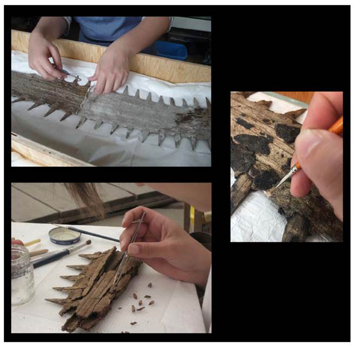
Source: (Photograph: María Barajas, Néstor Santiago and Adriana Sanromán, 2016, 2010 & 2017; courtesy: Proyecto Templo Mayor [PTM], Mexico).
FIGURE 7 Processes in the field laboratory
Finally, color retouching with varnish paints or watercolor is applied to give visual unity between the cartilage, gap fillers and replacements.
Analysis
From the outset, the PTM has had a multidisciplinary team, enabling as precise as possible taxonomic identification since the start of the excavation (López, 1993, p. 32-33). However, once recovered and restored, the ichthyofauna remains must be studied by specialists who, in addition to identifying their species, help us understand their habitat, origin and biological profile, size, age, and any anomalies or deformations, to name just a few. Since the 1970s, there has also been close collaboration with specialists from the “Ticul Álvarez Solórzano” Archaeozoology Laboratory of the Deputy Directorate of Laboratories for Academic Support at the Instituto Nacional de Antropología e Historia (INAH), where biologists Óscar Polaco and Ana Fabiola Guzmán analyzed and identified the majority of the Chondrichthyes from our collection. Currently we also work with biologists Nataly Bolaño and Uriel Mendoza from the Biology Institute (BI) at the Universidad Nacional Autónoma de México (UNAM), both specialized in the study of this type of ichthyofauna (Figure 8).

Source: (Photographs: Mirsa Islas, 2016; photomicrographs: Ing. Mario Monroy [SLAA-INAH, Scanning Electron Microscopy Laboratory], 2017; courtesy: Proyecto Templo Mayor [PTM], Mexico).
FIGURE 8 Analysis with specialists and observation of dermal denticles under the scanning electron microscope
Among the analyses, archaeological interpretation was carried out, helping to understand the association between these faunistic remains and the other materials present in the deposits, allowing us to better know their symbolism, their preparation process previous to their burial, relative dating, etc., as described at the beginning of this text.
Storage, Exhibition, Diffusion and Dissemination
To ensure the preservation of the collection in optimal conditions, once the conservation intervention has finished, and in parallel with the analysis, the permanent packaging is made. For this purpose, custom-made black polypropylene (PP) boxes lined with slabs of polyethylene foam (PE, Ethafoam®) are used. To avoid alterations through movement or vibrations, the foam is carved in the shape of the most susceptible remains, such as sawfish rostral cartilage or sections of tessellated mosaic. These carved areas are then covered with non-woven textile made of high-density polyethylene (HDPE, Tyvek®) fibers to avoid surface abrasion of the faunal remains. The vertebrae, caudal spines and teeth are packed in individual hermetic polyethylene bags that are placed in specific sections within the Ethafoam® slabs. It must be stressed that to avoid confusion or loss of information, each element is always identified with its corresponding excavation data (Sanromán & Barajas, 2022) (Figure 9).
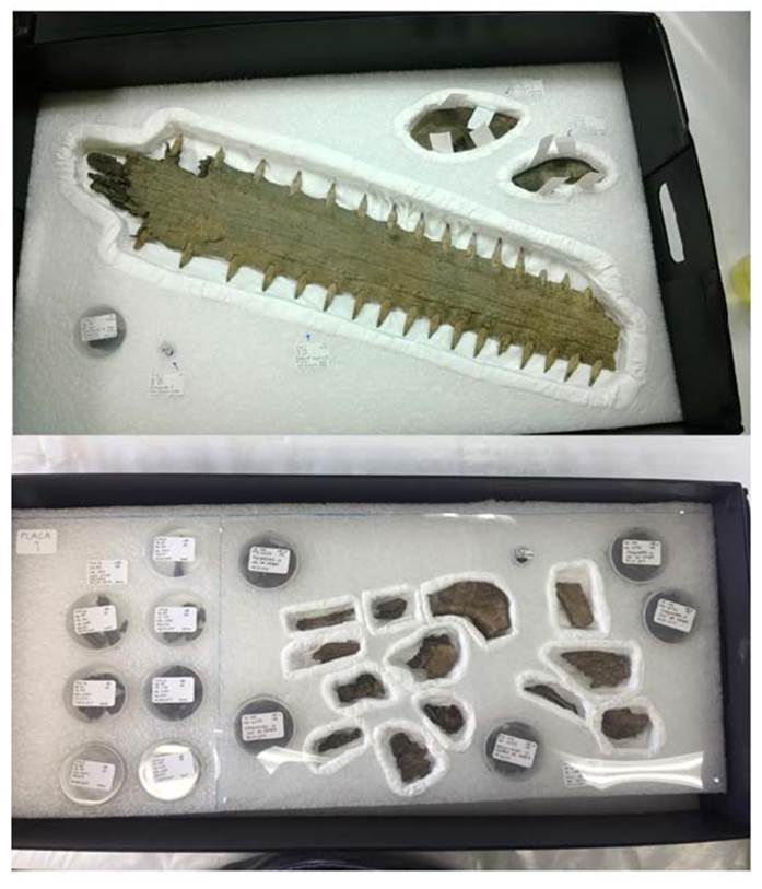
Source: (Photographs: Adriana Sanromán; courtesy; Proyecto Templo Mayor [PTM], Mexico).
FIGURE 9 Packages of different anatomical sections
Once the objects recovered by the PTM are transferred to the Safekeeping Storage for Archeological Collections at MTM, the storage conditions are stable, and the environmental parameters are constantly monitored. This helps to keep objects that have not been intervened, as well as restored ones, from suffering alterations due to sudden changes in relative humidity or temperature.
Tailored supports are designed and created to exhibit these cartilaginous remains, as well as for other materials, according to the curatorial discourse. However, because of their poor state of conservation, some anatomical sections that correspond to sawfish rostral cartilage were mounted in blocks to keep their unity. To integrate them into the museum’s permanent exhibition, the blocks were sought to resemble that of the soil matrix.
During the last two PTM field seasons, various studies have been made surrounding the identification, symbolism, and conservation of these type of skeletal remains, resulting in articles for the diffusion of scientific knowledge that have deepened the understanding of these animals and their relevance for the Mexica. Among them, a diffusion article (Robles et al., 2018, pp. 20-27), and three scientific articles that delve into their biological aspects (Mendoza & Bolaño, 2022, pp. 423-438; Bolaño et al., 2022, pp. 439-457), and on their conservation treatments (Sanromán & Barajas, 2022, pp. 537-551) stand out.
FINAL REFLECTIONS
The information presented throughout these pages is the result from the study and evaluation of various approaches to tackling the conservation issues surrounding skeletal remains from Chondrichthyes. This has enabled us to assimilate the most recent findings with greater expertise and better tools, ensuring the preservation of both the material and the data this could provide in the future.
The experiences from earlier projects as well as the development and availability of new and better materials have instigated the modification of practices and procedures used when conserving these objects. Nevertheless, what has been presented here should not represent a rigid methodology as new alternatives for direct intervention and for display mounting are always being worked on.
Without a doubt the day-to-day interdisciplinary work has been crucial for the identification of the structures that characterize these fish both in situ and in the laboratory, allowing us to reach more precise information regarding their distribution, use, and symbolism in Mexica rites, but has also contributed to improve the archaeological excavation, recovery, and sampling methods, by enabling the prompt identification of structures that formerly would not have been detected.
REFERENCES
Barajas, M., Sanromán, A., Hernández, K. V. y Soto, M. A. (2015). Informe de las actividades de conservación y restauración de la octava temporada del Proyecto Templo Mayor [enero 2014 a octubre de 2015, mecanuscrito]. México: Proyecto Templo Mayor. [ Links ]
Barajas, M., Sanromán, A. y Hernández, K. V. (2016). Informe de las actividades de conservación y restauración de la octava temporada del Proyecto Templo Mayor [noviembre de 2015 a agosto de 2016, mecanuscrito]. México: Proyecto Templo Mayor . [ Links ]
Barajas, M. y Sanromán, A. (2017). Informe de las actividades de conservación y restauración de la octava temporada del Proyecto Templo Mayor [agosto de 2016 a septiembre de 2017, mecanuscrito]. México: Proyecto Templo Mayor . [ Links ]
Barajas, M. y Sanromán, A. (2018). Informe anual de conservación de la octava temporada del Proyecto Templo Mayor [septiembre 2017 a septiembre 2018, mecanuscrito]. México: Proyecto Templo Mayor . [ Links ]
Barajas, M. y Sanromán, A. (2019). Informe anual de conservación del Proyecto Templo Mayor [octubre 2018 a septiembre 2019, mecanuscrito]. México: Proyecto Templo Mayor . [ Links ]
Bolaño-Martínez, N., Mendoza-Vargas, O. U., Salinas-Amézquita, S. y Robles, E. L. (2022). Dentículos dérmicos, una herramienta en la identificación de tiburones y rayas. En L. López Luján y E. Matos Moctezuma (Coords.), Los animales en el recinto sagrado de Tenochtitlan (pp. 439-457). México: El Colegio Nacional. [ Links ]
Chávez, X., González, A., Valentín, N. y García, J. M. (2011). Osteoarqueología de campo aplicada al análisis del uso ritual de la fauna: el caso de la Ofrenda 126 del Templo Mayor de Tenochtitlan. Estudios de Antropología Biológica 15(1), 117-137. https://www.revistas.unam.mx/index.php/eab/article/view/42770 [ Links ]
Compagno, L. J. V., Didier, D. A. y Burgess, G. H. (2005). Classification of Chondrichthyan Fish. En S. L. Fowler et al. (Comps. y Eds.), Sharks, Rays and Chimaeras: The Status of the Chondrichthyan Fishes (pp. 4-10). The World Conservation Union Shark Specialist Group. https://portals.iucn.org/library/efiles/documents/2005-029.pdf [ Links ]
Dean, M. N. y Summers, A. P. (mayo, 2006). Mineralized cartilage in the skeleton of chondrichthyan fishes. Zoology, Volume 109(2), 164-168. doi: https://doi.org/10.1016/j.zool.2006.03.002 [ Links ]
Díaz-Pardo, E. (1982). Restos de peces procedentes de la ofrenda 7. En E. Matos Moctezuma (Coord.), El Templo Mayor: excavaciones y estudios (pp. 151-160). México: Instituto Nacional de Antropología e Historia. https://mediateca.inah.gob.mx/repositorio/islandora/object/libro%3A561 [ Links ]
Díaz-Pardo, E. y Teniente-Nivón, E. (1991). Aspectos biológicos y ecológicos de la ictiofauna rescatada en el Templo Mayor, México. En O. J. Polaco (Coord.). La fauna en el Templo Mayor (pp. 33-104). México: Asociación de Amigos del Templo Mayor-Instituto Nacional de Antropología e Historia/García y Valadés. [ Links ]
Enach, J., Prymak, O., Raabe, D. y Epple, M. (junio, 2012). Structure, composition and mechanical properties of shark teeth. Journal of Structural Biology, 178(3), 290-299. doi: https://doi.org/10.1016/j.jsb.2012.03.012 [ Links ]
Escalante, M. F. y Soto, M. (2012). Informe de las actividades de conservación y restauración de la séptima temporada del Proyecto Templo Mayor [mecanuscrito]. México: Proyecto Templo Mayor . [ Links ]
Guzmán, A. F. y Polaco, O. J. (2000). Los peces arqueológicos de la ofrenda 23 del Templo Mayor de Tenochtitlan. México: Instituto Nacional de Antropología e Historia. [ Links ]
Hasbach, B. (2000). Pectoral circular con mosaico de turquesas de la Ofrenda 48 y cartílago rostral de pez sierra de la Ofrenda 58. En M. E. Marín (Coord.). Casos de conservación y restauración en el Museo del Templo Mayor (pp. 125-140). México: Instituto Nacional de Antropología e Historia. [ Links ]
Helfman, G. S., Collette, B. B., Facey, D. E. y Bowen, B. W. (2009). The Diversity of Fishes, 2ª ed. Wiley-Blackwell. http://www.sisal.unam.mx/labeco/LAB_ECOLOGIA/Ecologia_de_peces_files/The%20Diversity%20of%20Fishes%20Biology,%20Evolution,%20and%20Ecology%20-%20Helfman,%20Collette,%20Fracey%20%26amp%3B%20Bowen,%202009.pdf [ Links ]
López, R. y Tentle, M. A. (2012). Análisis de los desplazamientos horizontales observados con GPS en el occidente de la Cuenca de México (Tesis de licenciatura en Ingeniería Geofísica, inédita). Universidad Nacional Autónoma de México, México. [ Links ]
López, L. (1989). La Cuenca de México durante la época mexica. En L. Manzanilla y L. López (Eds.). Atlas histórico de Mesoamérica (pp. 148-152). México: Larousse. [ Links ]
López, L. (1993). Las ofrendas del Templo Mayor de Tenochtitlan. México: Instituto Nacional de Antropología e Historia . [ Links ]
López, L. (2005). The Offerings of the Templo Mayor of Tenochtitlan. Bernard R. O. de Montellano y Thelma O. de Montellano (Trads.). University of New Mexico Press. [ Links ]
Mazari, M., Marsal, R. J. y Alberro, J. (Octubre, 1989). Los asentamientos del Templo Mayor analizados por la mecánica de suelos. Estudios de Cultura Náhuatl, 19, 145-182. https://nahuatl.historicas.unam.mx/index.php/ecn/article/view/78324 [ Links ]
Malváez, C. I. (2011). Informe de actividades de conservación y restauración de la colección de bienes arqueológicos del Proyecto Templo Mayor-Séptima Temporada [enero a diciembre de 2010, mecanuscrito]. México: Proyecto Templo Mayor . [ Links ]
Mendoza, Ó. U. y Bolaño, N. (2022). Los peces sierra ofrendados al pie del Templo Mayor. En L. López y E. Matos (Coords.). Los animales y el recinto sagrado del Tenochtitlan (pp. 423-438) . México: El Colegio Nacional . [ Links ]
Moyle, P. y Cech Jr., J. (2003). Fishes: an Introduction to Ichthyology, 5a edición. Pearson. [ Links ]
Robles, É. L. (2017). Los monstruos terrestres de las ofrendas del Templo Mayor de Tenochtitlan. El cocodrilo, una metáfora telúrica del cosmos mexica (pp. 21-56) (Tesis de licenciatura en Arqueología, inédita). Escuela Nacional de Antropología e Historia, México. [ Links ]
Robles, É. L. (2022). Los cocodrilos, símbolos de la tierra en las ofrendas del Templo Mayor (pp. 49-50). Reportes del Proyecto Templo Mayor, 4. Ancient Cultures Institute / Instituto Nacional de Antropología e Historia. [ Links ]
Robles, É. L., Sanromán, A., Barajas, M., Hernández, K. V., Bolaño-Martínez, N. y Mendoza, Ó. U. (2018). Un pez marino tierra adentro. Los peces sierra del Templo Mayor de Tenochtitlan. Arqueología Mexicana XXV(151), 20-27. [ Links ]
Sanromán, A. (2009). Evaluación de productos para la consolidación del cartílago rostral de pez sierra de las ofrendas 120 y 126 del Templo Mayor de Tenochtitlan [mecanuscrito]. México: Proyecto Templo Mayor . [ Links ]
Sanromán, A. y Barajas, M. (2022). La conservación de los cartílagos rostrales de pez sierra en el Templo Mayor de Tenochtitlan. En L. López y E. Matos (Coords.). Los animales y el recinto sagrado de Tenochtitlán (pp. 537-551). México. El Colegio Nacional. [ Links ]
Seidel, L., Blumer, M., Pechriggl, E. J., Lyons, K., Hall, B. K., Fratzl, P., Weaver, J. C. y Dean, M. N. (septiembre, 2017). Calcified cartilage or bone? Collagens in the tessellated endoskeletons of cartilaginous fish (sharks and rays). Journal of Structural Biology 200(1), 54-71. doi: https://doi.org/10.1016/j.jsb.2017.09.005 [ Links ]
Soto, M. A. (2013). Informe de las actividades de conservación y restauración de la séptima temporada del Proyecto Templo Mayor [enero a diciembre de 2013, mecanuscrito]. México: Proyecto Templo Mayor . [ Links ]
1Dermal denticles are small structures that cover the body of the chondrichthyans, these are characteristic of each species and allow their taxonomic identification. Its morphology is like that of a tooth. The denticles have a certain size that remains unaltered over time, so that, as the individual grows, more denticles develop.
2Zoomorphic mythological creature that depicts the primordial, time and earth, made up of parts of different animals, among which the most common are the crocodile head with an elongated snout, the rostral cartilage of sawfish, the tongue of a snake, and body of a crocodile, or of a fish with a shark tail (Robles, 2022, pp. 49-50).
Received: July 23, 2021; Accepted: May 20, 2022; Published: September 18, 2023
 Este es un artículo publicado en acceso abierto bajo una licencia Creative Commons
