Ovine pulmonary adenocarcinoma (OPA), also called Jaagsiekte in South Africa, and ovine pulmonary carcinoma (OPC) or pulmonary adenomatosis, is a transmissible malignant lung tumor of sheep1,2. First reported in South Africa in 1855, OPA occurs in many parts of the world and is considered by some researchers the most common pulmonary neoplasia in sheep3. In the Americas, it has been reported from Argentina4, Peru5, Brazil6 and Mexico7. In some countries, OPA is endemic and causes a significant economic impact on sheep production, and in some geographic regions can reach an annual mortality rate of 2 %3.
OPA is caused by a retrovirus belonging to the genus Betaretrovirus, family Retroviridae1,2 and is referred to as Jaagsiekte sheep retrovirus (JSRV). Its genomic RNA is formed approximately by 7,460 nucleotides8,9 and the viral genome possesses the gag, pro, pol and env genes characteristic of retroviruses; JSRV also contains an envelope glycoprotein that plays a fundamental role in the oncogenic transformation of cells10,11. Like some other retroviral infections, JSRV has a long incubation period of up to two years, and the neoplasia can be reproduced experimentally by inoculating susceptible sheep10,12.
JSRV infection has been reported in various breeds of domestic sheep (Ovis aries), only infrequently in goats and mouflons (wild sheep Ovis gmelini), and never in any other animal species2,6. The primary target cells for JSRV in the lung are the bronchiolar Club cells (formerly Clara cells) and the pneumonocytes Type II in the alveolar wall1,9,13. This virus also infects B-lymphocytes, T-lymphocytes (CD4+ and CD8+), macrophages and has been detected in peripheral blood monocytes9,11,14.
Sheep with OPA typically show progressive weight loss, exercise intolerance, coughing, and overflowing fluid nasal secretions. The lungs are notably heavy on necropsy because of severe pulmonary edema, and the pulmonary parenchyma exhibits multiple, firm grey tumoral nodules that are firm in texture. Histologic features are those of a well-differentiated lepidic or papillary carcinoma3,12,15. OPA requires laboratory confirmation by identifying JSRV or associated proteins in affected tissues by immunolabelling or PCR4,14,16. It was described the gross, microscopic and immunohistochemical changes in the lungs of a sheep infected with JSRV, which evolved to OPA in Colima, Mexico.
A two-year-old male Pelibuey (Ovis aries) farmed in Colima, Mexico was presented to the local veterinarian with a two-month history of nasal discharge, chronic cough, respiratory distress, and progressive weight loss. The animal was separated from the flock and treated for 7 d with broad-spectrum antibiotics and expectorants. The sheep deteriorated, became prostrated and finally died. The carcass was submitted for postmortem examination to the Pathology Laboratory, Faculty of Veterinary Medicine of the University of Colima.
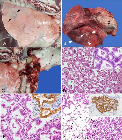
At postmortem examination, the sheep appeared emaciated and exhibited mark corneal opacity of the right eye due to traumatic injury to the periorbital tissue. Internally, there were some fibrous adhesions on the right caudal lobes between the visceral and parietal pleura. Overall, the lungs appeared pale and distended with rounded margins and were heavy and edematous (Figure 1A). There were two distinct types of tumoral infiltrations in the lung: the first types consisted of well-delineated firm tumoral masses (1-7 cm) protruding from the pleural surface (Figures 1A and 1B); the second type consisted of a locally extensive neoplastic infiltration grossly resembling broncho pneumonic consolidation (Figure 1B). The trachea and bronchi contained large quantities of frothy fluid (Figure 1C), the heart showed mark right-sided dilation, and the liver was congested. No other significant lesions were observed on postmortem examination. Tissues were fixed in 10 % in buffered formalin and routinely processed for histopathological examination.

A. Lateral view of the right lung showing partially distended and pale pulmonary parenchyma. The dorsal caudal lobe shows a focal, dark grey discoloration (arrows) with a firm texture on palpation. B. Ventral view of the lungs and the pericardial sac (p). Note a locally extensive area of dark consolidation in the left caudal lobe resembling bronchopneumonia (arrowheads) and a prominent raised tumoral nodule (arrow). C. Abundant foamy fluid (asterisk) oozing from the trachea (t). D. Microscopic view of tumor showing alveoli-like structures composed of thin bands of stromal tissue lined by cuboidal and columnar neoplastic cells. Hematoxylin-eosin. E. Alveolar-like structures are composed of a thin layer of connective tissue lined by cuboidal or columnar cells. Hematoxylin-eosin. Inset: Neoplastic cells show strong immunolabeling for JSRV-Env oncogenic protein in the cytoplasm and cell membrane. IHC; Monoclonal antibody. F. Microscopic view of the junction between the tumor and normal lung (dotted circle). Note lack of encapsulation or junctional fibrosis between tumor and adjacent lung tissue. Hematoxylin-eosin. Inset: Cluster of neoplastic cells showing strong immunolabeling for JSRV-Env oncogenic protein. IHC; Monoclonal antibody.
Figure 1 Ovine pulmonary adenocarcinoma; lung
Microscopically, the tumoral masses were poorly delineated, unencapsulated and composed of focal to coalescing clusters of epithelial cells growing in lepidic and papillary patterns on thin bands of connective tissue and forming alveoli-like structures (Figure 1D). Neoplastic cells were cuboidal or columnar, had abundant eosinophilic cytoplasm with round nuclei and subtle chromatin patterns containing one or two nucleoli (Figures 1D and 1E). There was mild anisokaryosis and 1-2 mitotic figures per high power field (40x). The surrounding alveoli were normal except for the presence of some edema and scattered alveolar macrophages, but without evidence of encapsulation (Figure 1F). Based on the gross and microscopic findings, a tentative diagnosis of OPA was made. Paraffin-embedded samples of the affected lung were shipped away for immune-detection of JSRV-Env oncogenic protein using monoclonal antibodies11,16, and the results were positive (Figures 1E and 1F).
Infection with JSRV is distributed globally in all continents, with the notable exception of Australia and New Zealand9,13,14. In some countries, JSRV infection is endemic and affects 80 % of the ovine population, although only 4-30 % of the infected sheep go onto developing OPA8,17. The first confirmed case of OPA reported in Mexico was in 2019 in a flock of sheep in the state of Tabasco in southern Mexico7. Until 2016 OPA was an official notifiable exotic disease in Mexico, but starting in 2018, the Official Mexican Sanitary Norm no longer recognizes OPA as a reportable disease18,19, perhaps conforming to the revised OIE recommendations. The true prevalence of JSRV infection and OPA incidence in Mexico is unknown mainly because the overall postmortem and laboratory investigations for farm animals are seldom performed in this country.
The clinical history, gross, microscopic and immunohistochemical findings for the 2-yr-old sheep reported here were classical of OPA. According to the veterinary literature, respiratory signs and weight loss usually manifest between 2-4 yr of age, but initial infections likely occur during colostrum ingestion and early life2,5. The abundant liquid secretion from the nostrils in a sheep with chronic cough and weight loss is a piece of relevant information that OPA needs to be investigated by the practicing veterinarian. Holding the affected sheep upside-down by the hind limbs (“wheelbarrow” test) results in clear whitish liquid's runoff from the nostrils8,12,17. Unfortunately, this hallmark sign was not adequately investigated by the local veterinarian. It should be noted that OPA's exuded nasal fluid is not edema or exudate "senso strictu", but rather excessive fluid oversecreted by neoplastic bronchiolar Club cells and alveolar type II pneumonocytes2,15. Secondary bacterial infections are common in OPA lungs, but this was not the case in these sheep.
OPA clinical signs are nonspecific and can be seen in other chronic respiratory infections of sheep12,15. Also caused by a retrovirus (genus lentivirus), Maedi induces a severe progressive lymphocytic interstitial pneumonia in sheep, and the clinical signs are indistinguishable from OPA3,15. Unfortunately, no routine laboratory assays are currently available to distinguish these two retroviral diseases in everyday ovine practice. Electron microscopy would be a rather unpractical and costly method to detect JRSV in OPA neoplastic cells8,13. Therefore, necropsy, histopathology and virus detection by immunolabelling or PCR are indispensable for conventional diagnostic work. Besides OPA and Maedi, other non-viral sheep diseases also manifest clinically with chronic cough and weight loss such, as chronic bacterial bronchopneumonia, verminous pneumonia, caseous lymphadenitis, and other lung neoplasms2,3,12. There was no evidence of any of these lung diseases on postmortem and microscopic examination in our sheep.
One unique feature of OPA is that tumors often have cranioventral lung distribution, mimicking bronchopneumonia's classical distribution. In some other cases like ours, the tumors involve all pulmonary lobes indistinctively, as it happens with most pulmonary neoplasia in domestic animals2,3,15. Tumor distribution in this case was dorsal, not cranioventral, as reported in the literature. The pulmonary changes in OPA can be grouped into two morphologic patterns, namely "classical" and "atypical"20. The classical OPA tends to be locally extensive, has a cranioventral distribution and on the cut-surface oozes clear fluid. In contrast, the atypical OPA tends to be focal and sometimes solitary with no fluid evidence on the cut-surface. It seems that this case deviated slightly from the classical presentation, also showing the atypical pattern. Regardless of the l type of presentation, the microscopic findings are the same12.
In this sheep, there was no evidence of metastasis to the mediastinal or bronchial lymph nodes or distant tissues, which was not surprising since only 10 % of OPA metastasize to these nodes3,17, and metastasis to other distant organs is extremely rare8. The OPA microscopic features are classic of a bronchioloalveolar carcinoma; however, this same type of pulmonary neoplasia can also develop spontaneously in sheep's lungs in the absence of JSRV infection3,12. The classification of lung carcinomas in human and veterinary pathology has changed continuously in the last three decades. Current classification distinguishes microscopically five main types of lung adenocarcinomas based on the predominant growth pattern of neoplastic cells: lepidic, papillary, acinar, squamous and adenosquamous3. Lepidic growth relates to proliferating neoplastic cells along the intact surface of alveolar walls without vascular stromal or vascular invasion12,15. In this case, the prevailing growth patterns were lepidic and papillary types, which are the most commonly seen in OPA3,20.
Retroviral-induced neoplasia of small ruminants such as OPA and also enzootic (ethmoidal) nasal carcinoma are of immense interest to researchers in human medicine worldwide. OPA has been used extensively as an animal model for human carcinogenesis studies9,10,16. There is still much to learn about sheep retroviral oncogenesis, and the current scientific advances in molecular pathology will likely lead to important discoveries soon.


 texto en


