SciELO - Scientific Electronic Library Online

 
vol.12 número3La coexistencia de Desmodus rotundus con la población humana en San Luis Potosí, MéxicoTratamiento de la laminitis crónica en equinos utilizando células troncales mesenquimales alogénicas de la médula ósea índice de autoresíndice de assuntospesquisa de artigos
Home Pagelista alfabética de periódicos  

Serviços Personalizados

Journal

Artigo

Indicadores

Links relacionados

  • Não possue artigos similaresSimilares em SciELO

Compartilhar


Revista mexicana de ciencias pecuarias

versão On-line ISSN 2448-6698versão impressa ISSN 2007-1124

Rev. mex. de cienc. pecuarias vol.12 no.3 Mérida Jul./Set. 2021  Epub 14-Mar-2022

https://doi.org/10.22319/rmcp.v12i3.5469 

Articles

Detection of Pasteurella multocida, Mannhemia haemolytica, Histophilus somni and Mycoplasma bovis in cattle lung

Seyda Cengiza  * 

M. Cemal Adıgüzela 

Gökçen Dinçb 

a Atatürk University Faculty of Veterinary Medicine Department of Microbiology 25240 Erzurum. Turkey.

b Erciyes University Faculty of Medicine Department of Microbiology Kayseri. Turkey.


Abstract

In this study, it was aimed to determine of P. multocida, M. haemolytica, H. somni and M. bovis in macroscopically healthy cattle lungs by PCR. The study was carried out on 82 macroscopically healthy cattle lung. DNA extraction was performed to the lung samples. PCR was then performed using all specific primers. By molecular evaluation, positive results were achieved for P. multocida, M. haemolytica, H. somni and M. bovis in 4 (4.8 %), 4 (4.8 %), 6 (7.3 %) and 3 (3.6 %) of the samples, respectively. Mix infections were detected in five samples. Of the samples, two were positive for both P. multocida and M. haemolytica, two were positive for both M. haemolytica and H. somni and one was positive for both P. multocida and H. somni. However, a positive sample, which carried all of pathogens, was not detected. In conclusion, P. multocida, M. haemolytica, H. somni and M. bovis are the important opportunistic pathogens of respiratory tract in cattle and these pathogens have a major role during infections. But multifactorial nature of bovine respiratory disease and immune system affected the formation of the disease. Hence, firstly cattle’s immunity should be strengthened and other conditions should be kept under control.

Key words Cattle; Molecular analysis; Lung; Respiratory disease

Resumen

En este estudio se tuvo como objetivo determinar P. multocida, M. haemolytica, H. somni y M. bovis en pulmones de bovinos macroscópicamente sanos mediante PCR. El estudio se llevó a cabo en 82 pulmones de bovinos macroscópicamente sanos. Se realizó la extracción de ADN de las muestras pulmonares. Después, se realizó la PCR utilizando todos los cebadores específicos. Mediante evaluación molecular, se lograron resultados positivos para P. multocida, M. haemolytica, H. somni y M. bovis en 4 (4.8 %), 4 (4.8 %), 6 (7.3 %) y 3 (3.6 %) de las muestras, respectivamente. Se detectaron infecciones mixtas en cinco muestras. De las muestras, dos fueron positivas tanto para P. multocida como para M. haemolytica, dos fueron positivas tanto para M. haemolytica como para H. somni y una fue positiva tanto para P. multocida como para H. somni. Sin embargo, no se detectó una muestra positiva que transportara todos los patógenos. En conclusión, P. multocida, M. haemolytica, H. somni y M. bovis son los patógenos oportunistas importantes del tracto respiratorio en bovinos y estos patógenos tienen un papel importante durante las infecciones. Pero la naturaleza multifactorial de la enfermedad respiratoria bovina y el sistema inmunológico afectaron la formación de la enfermedad. Por lo tanto, en primer lugar, se debe fortalecer la inmunidad de los bovinos y se deben mantener bajo control otras condiciones.

Palabras clave Bovinos; Análisis molecular; Pulmón; Enfermedades respiratorias

Introduction

Bovine respiratory disease (BRD) is one of the major health problems in calves and adult cattle, and has great economic impact on the cattle industry1-3. BRD in herds causes economic losses due to increased treatment costs, decreased production and culling4. BRD has a complex etiology, which involves bacterial and viral agents. Additionally, some predisposing factors such as management failures, environmental and host defense problems are influential on infection occurrence5.

The most frequent bacterial agents isolated from respiratory disease are Pasteurella multocida (P. multocida), Mannheimia haemolytica (M. haemolytica), Histophilus somni (H. somni) and Mycoplasma bovis (M. bovis)6,7. Pasteurella multocida is one of the primary bacterial pathogens and leads to clinical symptoms during BRD in neonatal calves and cattle. The bacterium, which is detected not only in infected but also healthy cattle, is isolated from lung, nasopharyngeal and nasal swabs and trans-tracheal washes. Therefore, diagnose of P. multocida becomes an issue, if clinical symptoms associated with pneumonia are detected in cattle8.

Similarly, M. haemolytica is normally presented in nasal pharyngeal mucosa in healthy cattle. However, the bacterium becomes a pathogen under inadequate conditions such as nutritional deficiency and overcrowded housing, and viral infections. Following to rapid proliferation of M. haemolytica within the infected lung, severe fibrinopurulent bronchopneumonia is presented. Additionally, it produces a potent leukotoxin that destroys the macrophages and neutrophils9,10. Because of these properties, this bacterium is accepted as the most harmful pathogen for lung.

H. somni is a bacterium which is normally presented not only in respiratory but also in reproductive tract. Similar with mentioned pathogens above, H. somni is also causing infections such as thrombotic meningoencephalitis (TME), pneumonia, septicemia, mastitis, arthritis, myocarditis and reproductive infections under inappropriate conditions with clinical symptoms5,11-13.

M. bovis is not only respiratory disease but also arthritis, mastitis, genital infections and abortus3. Moderate infections in cattle has the potential to cause an infection with severe clinical manifestations, as well as difficulty diagnosis; penicillin and its derivates are also an important problem in cattle breeding enterprises with the resistance mechanism of antibiotics14. At the same time, the rapid spread of bacteria in the cattle herd was a result of M. bovis makes it important15.

BRD is known as polymicrobial infection in cattle herds and mainly recorded in younger cows13. Thus, diagnosis of BRD is required to use different methods (conventional and molecular) to determine the bacteria that are effective in etiology. In particular, the use of different media, incubation conditions (temperature and O2 ratio), and differences between methods in conventional diagnostic methods require the use of faster methods. Molecular diagnosis of the pathogens based on Polymerase Chain Reaction (PCR) techniques can be used for identification and detailed evaluations. Molecular techniques, which are more sensitive than bacteriological methods, are preferred especially for direct identification of pathogens from tissue samples 8,9,15,16.

The aim of this study was to determine of P. multocida, M. haemolytica, H. somni and M. bovis of macroscopically healthy cattle lungs by PCR.

Material and methods

Sampling procedure

A total of 83 lung samples were collected from the different slaughterhouses. A piece of lung sample was taken from lungs without any lesions and placed in sterile tubes transported to the laboratory in cool chain.

Culture and DNA extraction

A piece of sample was taken and placed in eppendorf tube. Briefly, each sample was put into sterile petri dishes and then samples were break into parts using sterile bistouries and pens. Broth culture was only used for M. bovis. A piece of lung sample for M. bovis isolation inoculated in PPLO broth medium and incubated at 37 oC in %5 CO2 for 5 d. PPLO broth cultures were used for M. bovis DNA extraction, lyzed lung samples were used for other bacteria DNA extraction. DNA was extracted from the lung samples by using genomic DNA purification kit (Qiagen-DNeasy Blood and Tissue Kit-USA). Manufacturer instructions were followed.

Polymerase Chain Reaction

PCR procedures, which involved cycle conditions and reaction mixture, were performed according to previous reports17,18,19 (Table 1). The reaction mixture was prepared with a total volume of 50 μl contained 3 mM MgCl2, 200 μl dNTP, 0.5 μM each of primer and 1.25 units Taq DNA polymerase (Vivantis, MY) with minor revisions for pathogens. Extracted DNA (1 μL) was used as template. Amplification was carried by thermalcycler (The SuperCycler Trinity, Kyratech, AU). All samples of PCR amplification products (10 μL) were subjected to electrophoresis. DNA was visualized by UV fluorescence after staining with ethidium bromide.

Table 1 PCR conditions and oligonucleotids sequences 

Pathogen Cycle condition (°C/min) Oligonucleotids Base pair (bp) Reference
P.
multocida
94/1
69/1 30
cyc
72/1
F:GGCTGGGAAGCCAAATCAAAG R:CGAGGGACTACAATTACTGTAA 1432 Miflin and Blackall,
2001
M.
haemolytica
94/1
55/1 30
cyc
72/1
F:TGTGGATGCGTTTGAAGAAGG R:ACTTGCTTTGAGGTGATCCG 1145 Akan et al,
2006
H. somni 94/1
55/1 35
cyc
72/1
F:GAAGGCGATTAGTTTAAGAG R:TTCGGGCACCAAGTRTTCA 400 Angen et al,
1998
M. bovis 94/1
54/1 30
cyc
72/1
F: TATTGGATCAACTGCTGGAT R: AGATGCTCCACTTATCTTAG 447 Foddai et al,
2005

Results

In molecular evaluation, positive results were achieved for P. multocida, M. haemolytica, H. somni and M. bovis in 4 (4.8 %), 4 (4.8 %), 6 (7.3 %) and 3 (3.6 %) of the samples, respectively (Figure 1-3). Mix infections were detected in five samples. Of the samples, two were positive for both P. multocida and M. haemolytica, two were positive for both M. haemolytica and H. somni and one was positive for both P. multocida and H. somni. However, a positive sample, which carried all of the pathogens, was not detected.

M= Marker (100 bp DNA Ladder Plus), 1-3= P.multocida

Figure 1 Molecular evaluation of P.multocida 

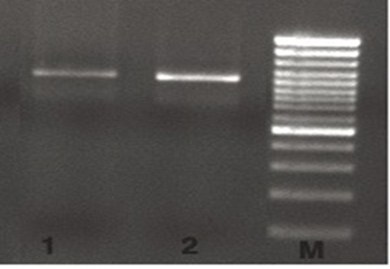
M= Marker (100 bp DNA Ladder Plus), 1-2= M. haemolytica

Figure 2 Molecular evaluation of M. haemolytica 

M= Marker (100 bp DNA Ladder Plus), 1-2= H. somni, 3= M.bovis

Figure 3 Molecular evaluation of H. somni and M.bovis 

Discussion

Bovine respiratory diseases, which cause economic losses due to production decrease and culling, have major importance in cattle herds. Although, cattle of all ages and sex are susceptible to the disease, it is more harmful for calves6,20,21. Bacteria that cause respiratory infections can be transmitted to sensitive animals from healed or immunologically strong animals (no clinical signs)2,22. In addition to pathogens, some predisposing factors such as overcrowded and bad-ventilated barns, inadequate feeding and other infectious diseases increased the infection risk. In these herds, transmission usually occurs horizontally7,9,23. Previous studies were usually focused on the detection of P. multocida, M. haemolytica, H. somni and M. bovis in the tonsils, nasopharynx and upper respiratory tract in carrier animals2,24,25. Various results about the presence of pathogens in cows were achieved in the reports. Positivity of P. multocida, M. haemolytica, H. somni and M. bovis in cattle varied between 0.4-57.4 %, 1.6-35 %, 2-45 % and 14-59 % in previous reports (Figure 4)9,19,26-30.

Figure 4 Proportional change of pathogens according to studies 9,20,24,27,29,30,31  

In the present study, the number of positive animals identified for P. multocida was found lower than Onat et al28 but was found higher than others24,27. Positivity of M. haemolytica was found lower than some other works2,27,29, but was found higher than Hajikolaei et al20. In terms of positivity H. somni and M. bovis was found different from other studies. In studies, in both bacteria was evaluated different diagnostic method in pneumonic cows15,30,31. Additionally, the variation among the results may be associated with difference in diagnostic methods, vaccination, and bacterial properties2,23,29. For instance, conventional culture methods can be inadequate in detection of the pathogens in healthy cows due to lower bacterial count in the samples. Likewise, vaccination can reduce bacteria carriage and lesions16,32,33. In addition, detection of some respiratory system pathogens as H. somni and M. bovis in culture media is difficult although the samples are collected from infected cows9,19. Thus, PCR tests, which involve specific primers for 16S rRNA, are suggested for identification of this bacterium during mix respiratory infections5,13. Therefore, genetic material basis molecular technics, which allow the detection of the pathogens even though lower bacterial count, would be preferable rather than culture methods in determining of carrier cows 16,34

The presence of these bacteria in the absence of clinical symptoms in animals or macroscopic lesions in the necropsy supports the opportunistic character of these bacteria. However, in these cases, histopathological examinations should be done and the animal's health status should be questioned. Additionally, because the occurrence of the disease has association with carriage22,24,27,35, detection of reservoir animals is important for reduction of the risk in herds. So that, detection of carrier cows, which have potential risk for contamination, is an approach for control22.

Conclusions and implications

In conclusion, P. multocida, M. haemolytica, H. somni and M. bovis, which cause economic losses and death in animals, are also important opportunistic pathogens. Therefore, the immune system should be developed by vaccination in animals. Moreover, housing conditions and management, awareness of the staff (owner) should be improved to establish an effective and sustainable control program for respiratory system diseases.

Literature cited

1. Headley SA, Alfieri AF, Oliveira VHS, Beuttemmüller EA, Alfieri AA. Histophilus somni is a potential threat to beef cattle feedlots in Brazil. Vet Rec 2014;175:249-250. [ Links ]

2. Jaramillo-Arango CJ, Hernandez-Castro R, Suarez-Guemes F, Martinez-Maya JJ, Aguilar-Romero F, Jaramillo-Meza L, Trigo FJ. Characterisation of Mannheimia spp. strains isolated from bovine nasal exudate and factors associated to isolates, in dairy farms in the Central Valley of Mexico. Res Vet Sci 2008;84:7-13. [ Links ]

3. Margineda GA, Zielinski GO, Jurado S, Alejandra F, Mozgovoj M, Alcaraz AC, López A. Mycoplasma bovis pneumonia in feedlot cattle and dairy calves in Argentina. Braz J Vet Pathol 2017;10(2):79 - 86. [ Links ]

4. Angen Ø, Thomsen J, Larsen LE, Larsen J, Kokotovic B, Heegaard PMH, Enemark JMD. Respiratory disease in calves: Microbiological investigations on trans-tracheally aspirated bronchoalveolar fluid and acute phase protein respons. Vet Microbiol 2009;137:165-171. [ Links ]

5. Tegtmeier C, Angen SNG, Riber U, Friis NF. Aerosol challenge of calves with Haemophilus somnus and Mycoplasma dispar. Vet Microbiol 2000;72:229-239. [ Links ]

6. Klima CL. Characterization of the genetic diversity and antimicrobial resistance in Mannheimia haemolytica from feedlot cattle [Thesis]. Alberta, Canada: University of Lethbridge; 2009. [ Links ]

7. Kurćubić VS, Đoković RD, Ilić ZŽ, Stojković JS, Petrović MP, Petrović VC. Modern approach to the enigma of bovine respiratory disease complex: A review. Pak Vet J 2014; 34:11-17. [ Links ]

8. Dabo SM, Taylor JD, Confer AW. Pasteurella multocida and bovine respiratory disease. Anim Health Res Rev 2008;8:129-150. [ Links ]

9. Tegtmeier C, Angen Ø, Ahrens P. Comparison of bacterial cultivation, PCR, in situ hybridisation and immunohistochemistry as tools for diagnosis of Haemophilus somnus pneumonia in cattle. Vet Microbiol 2000;76:385-394. [ Links ]

10. Bielsa JM. New solution for the control of the bovine respiratory complex. Congress of the Mediterranean Federation for Health and Production of Ruminants (FeMeSPrum) Zadar, Croatia, 2008;35-42. [ Links ]

11. Humphrey JD, Stephens LR. Haemophilus somnus: a review. Vet Bull 1983;53:987-1004. [ Links ]

12. Corbeil LB, Widders PR, Gogolewski RP, Arthur JE, Inzana TJ, Ward ASC. Haemophilus somnus: bovine reproductive and respiratory disease. Can Vet J 1986;27: 90-93. [ Links ]

13. Angen Q, Ahrens P, Tegtmeier C. Development of a PCR test for identification of Haemophilus somnus in pure and mixed cultures. Vet Microbiol 1998;63:39-48. [ Links ]

14. Nicholas RAJ. Recent developments in the diagnosis and control of mycoplasma infections in cattle. 23rd. World Buiatric Congress, Canada. 2004. [ Links ]

15. Petersen MB. Mycoplasma bovis in dairy cattle. [PhD Thesis] Denmark, Department of Veterinary and Animal Sciences Faculty of Health and Medical Sciences. University of Copenhagen; 2018. [ Links ]

16. Casademunt S. The role of Histophilus somni in bovine respiratory disease: An update. 2011. http://www.hipra.com . Accesed 30 May, 2018. [ Links ]

17. Miflin JK, Blackall P. Development of a 23S rRNA-based PCR assay for the identification of Pasteurella multocida. Lett App Microbiol 2001;33:216-221. [ Links ]

18. Akan M, Oncel T, Sareyyupoglu B, Hazıroglu R, Tel OY, Cantekin Z. Vaccination studies of lambs against experimental Mannheimia (Pasteurella) haemolytica infection. Small Ruminant Res 2006;65:44-50. [ Links ]

19. Foddai A, Idini G, Fusco M, Rosa N, Fe A, Zinellu S, Corona L, Tola S. Rapid differential diagnosis of Mycoplasma agalactiae and Mycoplasma bovis based on a multiplex-PCR and a PCR-RFLP. Mol Cell Prob 2005;19:207-212. [ Links ]

20. Hajikolaei HMR, Ghorbanpour M, Sayfi-Abadshapouri MR, Rasooli A, Ebrahimkhani D, Jabbari AR. Bacteriological and serological studies on Mannheimia haemolytica infection in cattle slaughtered at Ahvaz (southwestern Iran) abattoir. Iranian J Vet Res 2010;11:84-87. [ Links ]

21. Headley SA, Oliveira VH, Figueira GF, Bronkhorst DE, Alfieri AF, Okano W, Alfieri AA. Histophilus somni-induced infections in cattle from southern Brazil. Trop Anim Health Prod 2013;45:1579-1588. [ Links ]

22. Dziva F, Muhairwa A, Bisgaard M, Christensen H. Diagnostic and typing options for investigating diseases associated with Pasteurella multocida. Vet Microbiol 2007;128:1-22. [ Links ]

23. Taylor JD, Fulton RW, Mady DS, Lehenbauer TW, Confer AW. Comparison of genotypic and phenotypic characterization methods for Pasteurella multocida isolates from fatal cases of bovine respiratory disease. J Vet Diagn Invest 2010;22:366-375. [ Links ]

24. Hajikolaei HMR, Ghorbanpour M, Seyfi-Abadshapouri MR, Rasooli A, Moazeni Jula GR, Ebrahimkhani D. Study on the Prevalence of Pasteurella multocida carriers in slaughtered cattle and relationship with their immunity status at Ahvaz Abattoir. J Vet Res 2008;63:31-35. [ Links ]

25. Aebi M, Borne BHP, Raemy A, Steiner A, Pilo P, Bodmer M. Mycoplasma bovis infections in Swiss dairy cattle: a clinical investigation. Acta Vet Scan 2015;57:1-11. [ Links ]

26. Hajikolaei HMR, Ghorbanpour M, Sayfi-Abadshapouri MR, Rasooli A, Jahferian H. Occurrence of Pasteurella multocida in the nasopharynx of healthy buffaloes and their immunity status. Bull Vet Inst Pulawy 2006;50:435-438. [ Links ]

27. Barbour EK, Nabbut NH, Hamadeh SK, Al-Nakhli HM. Bacterial identity and characteristics in healthy and unhealthy respiratory tracts of sheep and calves. Vet Res Commun 1997;21:421- 430. [ Links ]

28. Onat K, Kahyaoğlu S, Çarlı KT. Frequency and antibiotic susceptibility of Pasteurella multocida and Mannheimia haemolytica isolates from nasal cavities of cattle. Turk J Vet Anim Sci 2010;34:91-94. [ Links ]

29. Alexander TW, Cook S, Klima CL, Topp E, McAllister TA. Susceptibility to tulathromycin in Mannheimia haemolytica isolated from feedlot cattle over a 3-year period. Front Microbio 2013;4:1-8. [ Links ]

30. Tenk M. Examination of Mycoplasma bovis infection in cattle [PhD Thesis]. Budapest: Szent Istvan University; 2005. [ Links ]

31. D’Amours GH, Ward TI, Mulvey MR, Read RR, Morck DW. Genetic diversity and tetracycline resistance genes of Histophilus somni. Vet Microbiol 2011;150:362-372. [ Links ]

32. Pérez DS, Pérez FA, Bretschneider G. Histophilus Somni: Pathogenicity in cattle. An update. An Vet (Murcia) 2010;26:5-21. [ Links ]

33. Lipsitch M. Vaccination against colonizing bacteria with multiple serotypes. Proc Natl Acad Sci 1997;94:6571-6576. [ Links ]

34. Deressa A, Asfaw Y, Lubke B, Kyule MW, Tefera G, Zessin KH. Molecular Detection of Pasteurella multocida and Mannheimia haemolytica in sheep respiratory infections in Ethiopia. Intern J Appl Res Vet Med 2010;8:101-108. [ Links ]

35. Derosa DC, Mechor GD, Staats JJ, Chengappa MM, Shryock TR. Comparison of Pasteurella spp. simultaneously isolated from nasal and transtracheal swabs from cattle with clinical signs of bovine respiratory disease. J Clin Microbiol 2000;38:327-332. [ Links ]

Received: August 07, 2019; Accepted: December 07, 2020

*Corresponding author: seydacengiz@atauni.edu.tr

Creative Commons License This is an open-access article distributed under the terms of the Creative Commons Attribution License