


Services on Demand
Journal
Article
 text in
  English (pdf)
 Article in xml format
 Article references
 Send this article by e-mail
Indicators
-
 Cited by SciELO
 -
 Access statistics
Related links
-
 Similars in
SciELO
Similars in
SciELO
Share

 Permalink
Revista mexicana de ciencias agrícolas
Print version ISSN 2007-0934
Rev. Mex. Cienc. Agríc vol.9 n.7 Texcoco Sep./Nov. 2018 Epub Sep 30, 2020
https://doi.org/10.29312/remexca.v9i7.472
Investigation note
Characterization CaCO3 and CaC2O4 with microphotographic analysis in Opuntia ficus-indica (L.) Miller
1Faculty of Agronomy-Autonomous University of Nuevo León. Av. Francisco Villa s/n, col. Ex-hacienda el Canadá, Nuevo León, Mexico. Tel. 01(81) 13404399.
2Postgraduate Studies-Campus Montecillos. Mexico-Texcoco Highway km 36.5, Montecillo, Texcoco, State of Mexico. Tel. 01(55) 58045900.
3Postgraduate Studies-Campus San Luis Potosí. Calle de Iturbide 73, San Agustín, Salinas de Hidalgo, San Luis Potosí, Mexico. CP. 78622. Tel. 01(444) 9630240.
The consumption of calcium (Ca++) is more frequent in the human diet due to its bioavailability and viability naturally in the cultivation of the nopal vegetable (Opuntia ficus-indica L. Miller.); however, its availability is limited by water stress in the plant by sequestering Ca++ in the form of calcium oxalate (CaC2O4). The study delves into sequential microphotographic analysis to produce high definition digital mosaics in undisturbed samples of the nopal cladodes with 60 days of age at water differentials 10 and 30% of available water (AD) and are processed in thin sections (SD) of 30 microns (μm) in thickness, besides being analyzed densitometrically in three different light phases by means of a petrographic microscope, the characterization of CaC2O4 and CaCO3 was carried out by signature in RGB formats characterizing the type of compound, its nucleus, size and texture. The results obtained by size of CaC2O4 of 10 and 30% A y D were: 37.63 μm and 71.39 μm in these compounds and the nuclei of these compounds reached 20.4 μm and 24.04 μm; with a texture of prismatic and cubic shape, while the CaCO3 for the same levels of available water: they reached size of 19.15μm and 20.86μm with a preponderant cubic texture. Concluding that the size of CaC2O4 and CaCO3 depends on the state of dehydration of the plants, as well as a correlation was observed between the size of the nucleus of CaC2O4, where for each CaC2O4 a CaCO3 is fixed, becoming unavailable.
Keywords: digital mosaics; nopal; oxalate; water stress
El consumo de calcio (Ca++) es más frecuente en la dieta humana por su biodisponibilidad y viabilidad de forma natural en el cultivo del nopal verdura (Opuntia ficus-indica L. Miller.); sin embargo, su disponibilidad es limitada por estrés hídrico en la planta al secuestrar Ca++ en forma de oxalato de calcio (CaC2O4). El estudio incursiona en análisis microfotográfico secuencial para elaborar mosaicos digitales de alta definición en muestras inalteradas del cladodios del nopal con 60 días de edad a diferenciales hídricas 10 y 30% de agua disponible (AD) y son procesadas en secciones delgadas (SD) de 30 micras (µm) de espesor, además de ser analizadas densitométricamente en tres diferentes fases de luz mediante un microscopio petrográfico, la caracterización de CaC2O4 y CaCO3 fue realizada por firma en formatos RGB caracterizando el tipo de compuesto, su núcleo, tamaño y textura. Los resultados obtenidos por tamaño de los CaC2O4 de 10 y 30% AD fueron: de 37.63µm y 71.39 µm en estos compuestos y los núcleos de dichos compuestos alcanzaron de 20.4 µm y 24.04 µm; con una textura de forma prismática y cubica, mientras que los CaCO3 para los mismos niveles de agua disponible: alcanzaron tamaño de 19.15µm y 20.86 µm con una textura cubica preponderante. Concluyendo que el tamaño de CaC2O4 y CaCO3 depende del estado de deshidratación de las plantas, así también se observó una correlación entre el tamaño del núcleo de CaC2O4, donde para cada CaC2O4 se fija un CaCO3, que no está disponible.
Palabras clave: estrés hídrico; mosaicos digitales; nopal; oxalato
Calcium carbonate (CaCO3) is an abundant compound due to inorganic substances originated by living organisms, an important aspect in the development of new materials and field of application (Shu-Chen et al., 2007). These present three anhydrous crystalline polymorphs such as calcite, aragonite and vaterite ordered by their thermodynamic stability (Payne et al., 2007). So calcite is present in nopal as well as in egg shells, mollusk shells, crustaceans and other biominerals (Shu-Chen et al., 2007). The carbonates have a discrete radical that depend on the conditions of pressure and temperature to give rise to calcium carbonate minerals monohydrate (Young et al., 2003) or for their specific cubic form in plant species (Franceschi and Horner, 1980).
In developing countries like Mexico, the consumption of calcium in dairy products is limited by its high cost, presenting as an alternative nopal vegetable (Heaney, 2001; Fairweather, 2007). For body absorption, calcium is available in nopal vegetables unless they have high concentrations of oxalic acid (C2H2O4) or dietary fiber (Kennefick and Cashman, 2000; Heaney, 2001; Charoenkiatkul et al., 2008) since they limit its formation when sequestered as oxalates, preventing its absorption by the metabolism, in addition calcium oxalate (CaC2O4) is limited by the type and shape of crystals which varies between 8 and 50% of the dry weight of the cladode (Gallaher, 1975; McConn and Nakata, 2004). Likewise, the consumption of nopalito varies depending on the state of maturation (between 400 and 500 g) that reports high contents of oxalate-free calcium (3.4 g in 100 g) (Rodríguez et al., 2007).
On the other hand, it has been reported that nopal by its physiology produces calcium oxalate crystals (Franceschi and Horner, 1980) classified into four types by their morphology (Jauregui et al., 2003) in: rafidia (acicular crystals in aggregates), drusen (spherical crystalline aggregates), styloid (acicular crystals) and prisms. Where the morphology of the crystals and distribution within the tissues of the plant are specific, which may be in the form of whewellite (calcium oxalate monohydrate) or weddellite (calcium oxalate dihydrate) (Horner, 1980).
Attending the concern to know the shape and structure of different crystals present in the nopal Jauregui et al. (2003) used scanning electron microscopy coupled to an energy of dispersive X-ray spectrometer (EDS), or using systematic infrared spectroscopy to characterize the chemical nature and deposits of crystals (Moje and Baran, 2003), or characterization of drusen. of calcium oxalate in vegetal tissue (Monje and Baran, 2009) or characterization of inclusion cells of minerals including the chemical composition and its morphology by means of the identification by X-ray diffraction (XRD) and petrographic microscope (Barcénas et al., 2015).
However, these methodologies for the characterization of oxalates and calcium carbonates require very specific instruments, so it is proposed to implement procedures for the analysis of minerals within the plant structures and their quantification on a microscopic scale with micro photomosaics in Giga images of thin sections, as a viable alternative to analysis using transmission microscopy. This methodological proposal was used to quantify and identify the geospatial distribution in bacteria and microorganisms within the soil components (Tarquini and Favalli, 2010; Gutiérrez et al., 2016).
Despite this, the technique has not been applied in the characterization of calcium oxalates and carbonates, so it can be adapted to plant tissues, allowing sequential photographs of continuous fields to be used to form composite or micro-photomosaic images. The capture of sequential images is very precise, and allows the generation of micromosaics with high spectral and spatial resolution (Gutiérrez et al., 2016), of high precision (Li et al., 2008).
However, the quantification and spatial distribution of oxalates and calcium carbonates within plant structures, as well as the location of these, remains limited due to the scarcity of information on how to quantify and characterize these minerals in nopal vegetables. The objective of this work was to describe an image analysis procedure that can be used to characterize calcium carbonates and oxalates present in the nopal vegetable (Opuntia ficus-indica L. Miller.) in composite images derived from thin sections of plant tissue; therefore, the assumption suggests that the size, quantity and form in calcium carbonates is related to water stress, where the different levels of dehydration of cladodes will influence their presence or absence.
Procedures
The present investigation was carried out in the Experimental Field Marin in the Faculty of Agronomy of the Autonomous University of Nuevo Leon during the years 2015-2017. The experimental units were established in containers of 36 cm high, 28.5 cm in diameter, under controlled conditions.
The substrate used reported differential constants to water stress during the development of the cladodes. The stress consisted of reducing the amount of available water to 10% and 30% based on volume, taking as reference the field capacity (CC) and permanent wilting point (WTP) of the substrate. The watering moments were determined based on the periodic weight of the pots until reaching the referred humidity levels.
Vegetable tissue samples were obtained from cladodes of 60 days, by transversal cuts of 4 cm2 and subjected to a state of dehydration with 100% acetone (CH3CH3) for 6 days and impregnated with 90% resin and a styrene monomer (C8H8) at 10%. The impregnation was carried out in a vacuum chamber for 45 min at 15 lbs of tension, drying for 15 days thereafter, once they were dried, they were placed on coverslips fixed with apoxic resin and polished up to 30 μm thick.
The micromorphological analysis began capturing sequential microimages with different light sources at a 10x objective using a digital camera mounted on a petrographic microscope (Olimpus BX51), later high-resolution micro-photomosaics were created. The image analysis was using ERDAS Imagine 2014v®. The measurement of calcium oxalates and carbonates was achieved by converting raster information into vector format using ArcGIS v.10.1. The processes were based on the methodology reported by Gutiérrez et al. (2016).
Discussions
The capture of sequential photographs with different light phases: flat (Figure 1A), polarized or crossed (Figure 1B) and compensated (Figure 1C) formed high quality micro-mosaics in spatial resolution in the treatment 10% and 30% of AD (Figure 2A, B, C).
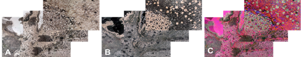
Figure 1 Treatment of 10% AD Construction of mosaics of high spatial resolution in different light sources and with different objectives. A) flat light; B) polarized or crossed light; and C) light compensated.
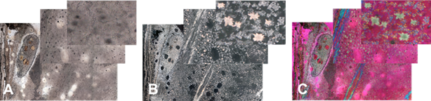
Figure 2 Treatment of 30% AD. Construction of mosaics of high spatial resolution in different light sources and with different objectives. A) flat light; B) polarized or crossed light; and C) light compensated.
The construction of micro-mosaics with Giga Images technique allowed observing the calcium oxalate crystals (drusen) (Figure 3) and carbonates (calcites) with polarized and compensated light (Figure 4).
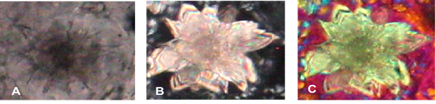
Figure 3 Visualization of calcium oxalate crystals with three light sources. A. flat light; B. Polarized or crossed light; and C. Light compensated.
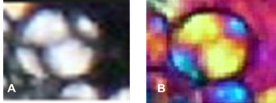
Figure 4 Calcium carbonates (calcite) with two light sources. A) polarized or crossed light; and B) compensated light.
Other advantages of the use of this methodology was the measurement of the minerals, which resulted in a significant difference between the calcium oxalates of the treatments of 10 and 30% of AD (Figure 5) with diameters of 5.9 and 11.11 μm respectively, also achieving the measurement of calcium carbonates (Figure 6).
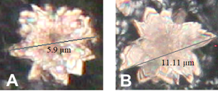
Figure 5 Measurement of calcium oxalates with a water stress of 10 and 30% of AD. A) calcium oxalate from the 10% treatment of AD with a diameter of 5.9 μm; and B) calcium oxalate from the treatment 30% available water with a diameter of 11.11 μm.
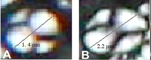
Figure 6 Measurement of calcium carbonates with water stress of 10 and 30% of AD. A) calcium carbonate of treatment 10% of AD with a measurement of 1.4 μm. B) calcium carbonate from treatment 30% of AD with a measurement of 2.2 μm.
In addition, it was possible to make a comparison of sizes between the oxalate nuclei and the calcium carbonates of some samples of the treatments of 10 and 30% of AD, demonstrating that for each oxalate a calcium carbonate is sequestered (Figure 7) when comparing the images of calcium oxalates and calcium carbonates in different light sources with petrographic microscopy and electronic scanning (Figure 8 and 9).
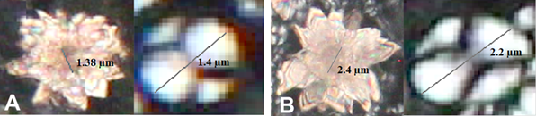
Figure 7 Comparison of measurements of the core of calcium oxalates and calcium carbonates. A) treatment of 10% of available water, shows the similarity in size of the oxalate nucleus 1.38 μm and the carbonate 1.4 μm; and B) treatment of 30% of available water, shows the similarity in size of the core of the oxalate 2.4 μm and the carbonate 2.2 μm.
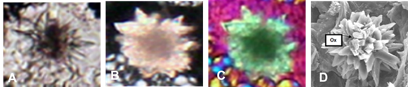
Figure 8 Comparison of photographs of calcium oxalate in different light sources with a petrographic and electronic microscope. A) flat light; B. polarized or crossed light; C) compensated light; and D) electronic scanning microscope by McCoonn and Nakata (2004).
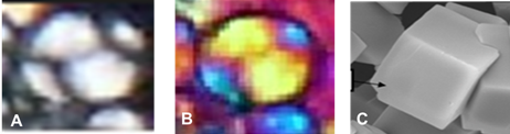
Figure 9 Comparison of photographs of calcium carbonate in different light sources with a petrographic and electronic microscope. A) polarized or crossed light; B) compensated light; and C) electronic scanning microscope by McCoonn and Nakata (2004).
A comparison of means of representative samples of treatments of 10 and 30% of AD (Table 1) where the diameter of the oxalates was significantly higher than that of the calcium carbonates with a diameter of 71.39 μm.
Conclusions
It is determined that water stress inhibits and promotes the size, quantity and shape of calcium oxalates and carbonates within vegetable structures of nopal vegetable. While the identification and characterization in undisturbed samples in cladodes with different stress was possible using multispectral analysis techniques in Giga Micro-photomosaic images determining the existing correlation between the nuclei that for each oxalate there is an unavailable calcium carbonate.
Acknowledgments
We thank CONACYT for the scholarship granted to the first author to carry out their doctoral studies in Agricultural Sciences. To the Laboratory of Soil Micromorphology, Postgraduate College, Montecillos. To the Faculty of Agronomy and to the PAICYT of the UANL.
REFERENCES
Bárcenas-Argüello, M. L.; Gutiérrez Castorena, M. C. and Terrazas, T. 2015. The polymorphic weddellita crystals in three species of Cephalocereus (Cactacea). J. Micron. 77:1-8 [ Links ]
Charoenkiatkul, S.; Kriengsinyos, W.; Tuntipopipat, S.; Suthutvoravut, U. and Weaver, C. M. 2008. Calcium absorption from commonly consumed vegetables in healthy Thai women. J. Food Sci. 73: H218-H221. [ Links ]
Fairweathert- Tait, S. M. 2007. Calcium bioavailability in relation to bone health. International J. Vitamin Nutr. Res. 72:13-18. [ Links ]
Franceschi, V. R. and Horner, H. T. 1980. Calcium oxalate crystals in plants. The Bot. Rev. 46:361-427. [ Links ]
Gallaher, R. N. 1975. The occurrence of calcium in plant tissue as crystals of calcium oxalate. Coom Soil Sci. Plant Anal. 6:315-321. [ Links ]
Gutiérrez, C. E. V.; Gutiérrez, C.; Ma. del C.: González- V.; T.; Cajuste, B. L.; Delgadillo, M. J.; Suástegui Méndez, E. y Ortiz- Solorio, C. A. 2016. Micromapping of microbial hotspots and biofilms from different crops using digital image mosaics of soil thin sections. Geoderma. 279:11-21. [ Links ]
Heaney, R. P. 2001. Meta-analysis of calcium bioavailability. Am. J. Therpeutics. 8:73-78. [ Links ]
Jáuregui-Zúñiga, D. J. P.; Reyes-Grajeda, J. D.; Sepúlveda-Sánchez, J.; Whitaker, R. and Moreno, A. 2003. Crystallochemical characterization of calcium oxalate crystals isolated from seed coats of Phaseolus vulgaris and leaves of Vitis vinifer. J. Plant Physiol. 160:239-245. [ Links ]
Kennefick, S. and Cashman, K. D. 2000. Inhibitory effect of wheat fibre extract on calcium absorption in Caco-2 cells: evidence for a role associated phytate rather than fibre per se. Eur. J. Nutr. 39:12-17. [ Links ]
Li, Y.: Onasch, C. M. and Guo, Y. 2008. GIS-based detection of grain bourdaries. J. Struct. Geol. 30:431-443. [ Links ]
McCoonn, M. M. and Nakata, P. A. 2004. Oxalate reduces calcium availability in the pads of the prickly pear cactus through formation of calcium oxalate crystals. J. Agric. Food Chem. 52:1371-1374. [ Links ]
Monje, P. V. and Baran, E. J. 2002a. Characterization of calcium oxalates generated as biominerals in cacti. Plant Physiol. 128:707-713. [ Links ]
Monje, P. V. and Baran, E. J. 2009b. Characterization of calcium oxalate biominerals in Pereskia species (Cactaceae). Z. Naturforsch. 64:763-766. [ Links ]
Payne, S. R.; Heppenstall-Butler, M. and Butler, M. F. 2007. Formation of thin calcium carbonate films on chitosan biopolymer substrates. Cryst Growth Design. 7:1262-1276. [ Links ]
Piochi, M.; Polacci, M.; De Astis, G.; Zanetti, A.; Mangiacapra, A.; Vannucci, R. and Giordano, D. 2008. Texture and composition of pumices and scoriae from the Campi Flegrei caldera (Italy): implications on the dynamic of explosive eruptions. Geochem. Geophys. Geosyst. doi:10.1029/ 2007GC001746 Q. [ Links ]
Rodríguez-García, M E.; de Lira, C.; Hernández Becerra, E.; Cornejo-Villegas, M. A.; Palacios-Fonseca, A. J.; Rojas-Molina, I. R.; Reynosa, L. C.; Quntero, A.; del Real, T.; Zepeda, A. and Muñoz- Torres. C. 2007. Physicochemical characterization of nopal pads (Opuntia ficus-indica) and dry Vacuum nopal powders as a function of the maturation. Plant Foods Human Nutr. 62:107-112. [ Links ]
Shu-Chen, H.; Kensuke, N. and Yoshiki, C. 2007. A carbonate controlled-addition method for amorphous calcium carbonate spheres satabilized by poly (acrylic acids). Langmuir. 23:12086-12095. [ Links ]
Tarquini, S. and Favalli, M. 2010. A microscopic information system (MIS) for petrographic analysis. Computers Geosc. 36:665-674. [ Links ]
Young, J.; Geisen, M.; Cros, L.; Kleijne, A.; Sprengel, C.; Probert, I. and Ostergaard, J. 2003. A guide to extant coccolithophores taxonomy. J. Nannoplankton Res. 1:125. [ Links ]
Received: September , 2018; Accepted: October , 2018
 Este es un artículo publicado en acceso abierto bajo una licencia
Creative Commons
Este es un artículo publicado en acceso abierto bajo una licencia
Creative Commons
