


Servicios Personalizados
Revista
Articulo
 texto en
  Inglés (pdf)
 Artículo en XML
 Referencias del artículo
 Enviar artículo por email
Indicadores
-
 Citado por SciELO
 -
 Accesos
Links relacionados
-
 Similares en
SciELO
Similares en
SciELO
Compartir

 Permalink
Revista mexicana de ciencias agrícolas
versión impresa ISSN 2007-0934
Rev. Mex. Cienc. Agríc vol.9 no.7 Texcoco sep./nov. 2018 Epub 30-Sep-2020
https://doi.org/10.29312/remexca.v9i7.683
Articles
First incidence of yellow vein virus of huasteco pepper in soybean in Mexico
1Valley of Culiacán Experimental Field-INIFAP. Highway Culiacán-Eldorado km 17.5, Culiacán, Sinaloa, Mexico. CP 80000. (sixjas@hotmail.com).
2Valley of Fuerte Experimental Field-INIFAP. Highway Mexico-Nogales International km 1609, Juan José Ríos, Sinaloa, Mexico. CP. 81110. (franklingrc2702@gmail.com; come60@yahoo.com; sauceda-raul@hotmail.com).
3Valley of Santo Domingo Experimental Site-INIFAP. Highway Transpeninsular km 208, Constitution City, Baja California Sur, Mexico. CP. 23600. (cmelgozavillagomez@gmail.com).
4General Terán Experimental Field-INIFAP. Road Montemorelos-China km 31, General Terán, Nuevo León, Mexico. CP. 67400.
In the state of Sinaloa, soybeans are one of the few crop alternatives during the summer; therefore, its reactivation is being reconsidered. The whitefly (Bemisia tabaci Genn, Biotype ‘B’) was one of the main causes of the reduction of soybean growing areas in Mexico as a result of direct damage to flower production and plant yield, as well as of the transmission of geminiviruses. During the summer of 2013-2014, a total of 11 soybean varieties were evaluated to determine the incidence of viral diseases in the Valley of Culiacán and Valle del Fuerte, Sinaloa, Mexico. Symptomatology, amplification of DNA by polymerase chain reaction (PCR) and sequence analysis of amplified fragments confirmed the incidence of yellow vein virus of huasteco pepper (PHYVV) in soybean grown in the west of Sinaloa, Mexico. Sequence analysis showed 100% of nucleotide and amino acid identities with respect to the PHYVV1GMES isolate from Mexico (GenBank accession number KT022087). This is the first report on the emergence of PHYVV in soybean in Mexico.
Keywords: begomovirus; enzymatic sequence; yellow vein virus of huasteco pepper
En el estado de Sinaloa, la soya es una de las pocas alternativas de cultivo durante el verano; por lo tanto, su reactivación está siendo reconsiderada. La mosca blanca (Bemisia tabaci Genn. Biotipo ‘B’) fue una de las principales causas de la reducción de las áreas de cultivo de soya en México como resultado de los daños directos en la producción de flores y rendimiento de la planta, así como de la transmisión de geminivirus. Durante el verano de 2013-2014, se evaluaron un total de 11 variedades de soya para determinar la incidencia de enfermedades virales en el Valle de Culiacán y Valle del Fuerte, Sinaloa, México. La sintomatología, la amplificación del ADN mediante la reacción en cadena de la polimerasa (PCR) y el análisis de la secuencia de los fragmentos amplificados confirmaron la incidencia del virus de la vena amarilla del chile huasteco (PHYVV) en la soya cultivada en el oeste de Sinaloa, México. El análisis de secuencia mostró 100% de identidades de nucleótidos y aminoácidos con respecto al aislado PHYVV1GMES de México (número de acceso de GenBank KT022087). Este es el primer informe sobre el surgimiento de PHYVV en la soya en México.
Palabras clave: begomovirus; secuencia enzimática; virus de la vena amarilla del chile huasteco
Introduction
Soy (Glycine max L.) Merr. also, known as ‘golden bean’ or ‘soy bean’ consists of more than 40% protein and 20% oil and for this reason has been recognized as a potential supplemental source of edible oil and nutritional food throughout the world. Mexico imports practically all the soybean that it consumes, from the year 2000 to 2008, the average annual production of soybean was only 119 924 t, while the imports were 3 905 596 t (http: //siap.sagarpa.gob.mx./ventana.php?idLiga =1592&type=0). In the state of Sinaloa, soybeans are one of the few crop alternatives during the summer (Martinez et al., 1998); therefore, its reactivation is being reconsidered. The whitefly (Bemisia tabaci Genn, Biotype ‘B’) was one of the main causes of the reduction of soybean growing areas in Mexico, as a result of direct damage to flower production and plant yield, as well as of geminiviruses transmission (Cortéz et al., 2005).
Geminiviruses are a diverse and widely distributed group of plant pathogens that cause great economic losses throughout the world. Its genome consists of one or two molecules of circular single-stranded DNA with a size of 2.5 to 3 Kb (Lazarowitz, 1992), known as DNA-A and DNA-B, which encodes all the functions of viruses required for DNA replication, control of gene expression, insect transmission and movement in the plant (Hanley et al., 1999). The single-stranded circular DNA of the genus Begomovirus, family Geminiviridae, are among the most important plant viruses (Brown and Bird, 1992, Moriones and Navas, 2000). This group of viruses is known as one of the economic factors that limit crops in many parts of the world, especially in tropical and subtropical regions (Zerbini et al., 2005). Begomoviruses only infect species of dicotyledonous plants and are transmitted exclusively by the whitefly Bemisia tabaci (Homoptera: Aleyrodidae) in a non-circulating way (King et al., 2012; Briddon, 2015).
These viruses are considered viruses of emerging plants, due to their increasing incidence and the severity of the diseases they cause (Polston and Anderson, 1997). It has also been reported that these viruses infect new hosts and have spread to more geographic locations (Mansoor et al., 2003; Varma and Malathi, 2003). Recombination and mutation may play an important role in the establishment and emergence of new begomovirus strains/species (Mansoor et al., 2006). Although only a small number of monopartite begomovirus have been discovered, it is known that all their essential functions are encoded by a single component in the DNA-A of the bipartite genome of begomovirus (Navot et al., 1991; Rojas et al., 2001).
Currently, the International Committee of Virus Taxonomy (ICTV) recognizes that four genera (mastrevirus, topocuvirus, curtovirus and begomovirus) belong to the geminiviridae family (Fauquet et al., 2008). There are several reasons why the genomes of begomovirus are the most diversified group, including: high global distribution, whitefly transmission, presence of a mono or bipartite genome and the ability to infect dicotyledonous plants. Components A and B of the bipartite begomoviruses share a common region, ranging from 160 to 230 base pairs (bp), which contain the cis elements necessary for viral replication (Argüello et al., 1994). Component A produces the proteins involved in the replication and encapsidation of the viral genome (Gutiérrez et al., 2004), while component B encodes two proteins that promote the movement of the virus through the plant (Gafni and Epel, 2002).
Currently, ICTV has noticed more than two hundred confirmed and tentative species of the genus Begomovirus (ICTV, King et al., 2012). These viruses are transmitted by insects of the homoptera (whiteflies and leafhoppers) to a variety of mono and dicotyledonous plants, producing symptoms such as: mosaics, specks, streaks, leaf deformation, dwarfism, yellowing and chlorosis. From an economic point of view, this transforms the Begomovirus group into one of the most important groups of plant pathogens, as a result of its high incidence, distribution and severity of symptoms.
In Mexico, this type of phytopathogen has caused great confusion about the etiology of different diseases, which were initially called ‘filling’, when their presence was detected in tomato crops (Solanum lycopersicum L.) in the state of Sinaloa (Gonzalez and Cervantes, 1973). In addition to this, infection by the virus has been called ‘tigers plant’ in Puebla (Garzón and Galindo, 1985) due to the symptoms of yellowing caused in pepper crops (Capsicum spp.).
Finally, in the pepper crops in the south of Tamaulipas, it is known as the ‘curly yellow’ disease (Garzón et al., 1993). It has been determined that these diseases are of geminiviral etiology and can be caused by the Huasteco pepper virus (PHV) (Brown et al., 1989; Brown and Bird, 1992; Garzon et al., 1993; Torres et al., 1996; Garzón et al., 2002). Currently, it is believed that the PHV is restricted to species of the Solanaceae family, especially tomato and pepper crops. Recently, it was detected and identified that the huasteco pepper yellow vein virus (PHYVV) affects tomato (Lugo et al., 2011) and pepper in Culiacan, Sinaloa (Retes et al., 2016; Melendrez et al., 2016).
Materials and methods
Surveys and sample collection
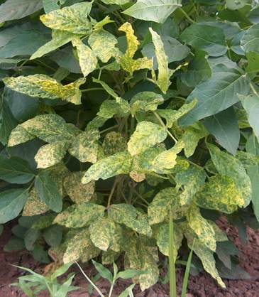
During the summer of 2013-2014, a total of 11 varieties of soybeans were evaluated for the incidence of viral diseases in experimental fields operated by the National Institute of Agricultural and Livestock Forestry Research (INIFAP), in the Valley of Culiacán and the Valle del Fuerte, Sinaloa, Mexico. The incidence of the disease in these areas varies from 25.2 to 39.8%. The collected leaves were immediately placed in plastic bags and transported to the laboratory and kept at 4 °C. For long-term storage, the leaf samples were dried using silica gel or a lyophilizer and stored at -20 °C. Of these, three varieties (Cajeme, Nainari and Esperanza) showed distortion of the leaf (Figure 1).
Extraction of DNA
The total DNA was extracted from 0.05 g of dry foliar tissue using the previously described method (Velarde et al., 2015). The tissue was macerated in a sterile porcelain mortar and pestle and finally cooled to -70 °C. Three mL of extraction buffer was added containing: 30 mM sodium chloride (NaCl), 30 mM ethylenedinitrile tetracytic acid (EDTA) (pH 8.5) and 250 mM tris base (pH 8.5). The maceration product was placed in sterile 1.5 mL Eppendorf tubes. Subsequently, a volume of 100 μL of 10% ammonium cetyltrimethylbromide (CTAB) was added and incubated at 95 °C for 10 min.
Then 250 μl of 5 M NaCl was added and incubated at -20 °C for 5 min and then centrifuged. The aqueous solution was added with a volume of cold absolute chloroform (v/v) and stirred with a vortex machine for a few seconds, and subsequently centrifuged at 10 000 rpm for 5 min, after this time, by pipetting, the aqueous solution was transferred to a new tube, adding a volume of cold absolute isopropanol (v/v) and homogenized manually.
Subsequently, the samples were stored in a refrigeration unit at -20 °C for one hour to allow the precipitation of the DNA. After this time, the samples in tubes were centrifuged at 13 000 rpm for 10 min to obtain the DNA pellet, for which it was waited two hours for the DNA to dehydrate and finally the DNA obtained from the different isolates were resuspended in 50 μL of nuclease-free water (Promega) and stored at 4 ºC for its preservation.
Polymerase chain reaction (PCR)
The DNA extracted from the tissues was analyzed by PCR using the degenerate oligonucleotides AV494 (direct; 5’- GCC(C/T)AT(G/A)TA(T/C)AG(A/G)AAGCC(A/C)AG-3’) and AC1048 (reverse) 5-‘GG(A/G)TT(A/G/T)GA(G/A)GCATG(T/A/C)GTACATG-3’ (Wyatt and Brown, 1996), corresponding to the 3’and 5’ ends of the capsid protein (PC) gene, respectively The cyclic profile consisted of 10 min of fusion at 95 °C, followed by 30 cycles of fusion, hybridization and polymerization of DNA for 1 min at 95 °C, 1 min at 55 °C and 1 min at 72 °C, respectively and a final extension of 10 min at 72 °C A thermal cycler was used (NyxTechnik Amplitronyx Series 6 A6 (ATC401 The final reaction mixture (15 μL) contained 100 nanograms of DNA, an equimolar mixture of dATP, dCTP, dGTP and dTTP, magnesium chloride (25 mM), PCR buffer (1X), DNA Taq polymerase (5U) (Promega® PCR Master Mix, catalog No. M7502) and 40 picomoles of each oligonucleotide (Sigma®).
The amplified products were analyzed in 1% agarose gels, stained with a Gel Red solution (Biotium catalog No. 41003), for which 15 μL of the amplified DNA were deposited in each well of the gel, adding 3 μL of the blue dye of bromophenol 6X (Sambrook and Rusell, 2001) and as reference the molecular weight markers 1Kb HyperLadder and HyperLadder 1kb Plus (Bioline). The run buffer and preparation of the agarose gels was 1X NaOH-borate (Brody and Scott, 2004). Run time was 30 min at 250 V (volts) and to visualize the DNA, the gel was exposed to a UV light transilluminator. The images were captured by a camera and scanned with the Launch Doc-ItLS (UVP) program. The sequence of oligonucleotides for all geminivirus strains and PCR conditions are given in Table 1.
Table 1 Alignment temperature conditions and sequences of degenerate primers AV494/AC1048 for the analysis of begomovirus PCR.
| Initiator | Sequence (5’ → 3’) | Alignment °C/virus cycles |
|---|---|---|
| F: AV494 | GCC(C/T)AT(G/A)TA(T/C)AG(A/G)AAGCC(A/C)AG | |
| R: AC1048 | GG(A/G)TT(A/G/T)GA(G/A)GCATG(T/A/C)GTACATG 55/30 | all geminivirus |
F= direct initiator; R= reverse initiator.
Enzymatic sequencing
Three DNA fragments of Geminivirus amplified by PCR on agarose gel based on silica columns (EZ-10 Spin Column BS354 DNA Gel Extraction Kit, Bio Basic Inc.), were purified, samples were sent for enzymatic sequencing using the method based on ddNTPs (Sanger et al., 1977) to the National Laboratory of Genomics for Biodiversity (LANGEBIO) of the Center for Research and Advanced Studies of the National Polytechnic Institute (CINVESTAV-IPN), Irapuato, Guanajuato, Mexico. A 3730 XL DNA sequencer (Applied Biosystems, Foster City, CA) and a Big Dye Terminator 3.1 kit (Applied Biosystems, Foster City, CA) were used for this purpose. The search for DNA sequence similarity was made using the BLAST program, comparing the values that identify the homology with the information found in the databases of the National Biotechnology Information Center (NCBI) (http: //www.ncbi.nlm nih.gov/BLAST/).
Phylogenetic analysis
The nucleotide sequences of the CP amplicon represent the region of 668 bp coordinated in the PHYVV, were compared with the golden mosaic virus Rhynchosia (RhGMV) and the Chinese tomato virus (CdTV), using the Neighbor-Joining phylogenetic dendrogram method (Saitou and Nei, 1987), based on the alignment of partial nucleotide sequences. Evolutionary distances were calculated using the Tajima-Nei method (Tajima and Nei, 1984). Evolutionary analyzes were performed on MEGA7 (Kumar et al., 2016).
Results and discussion
Identification of geminivirus
The genetic analysis showed amplicons of 668 bp of the CP gene obtained with a PCR of three randomly generated geminivirus isolates (Figure 2). Once purified, the amplified product was sent (Figure 3) for sequencing and subsequent search of databases using the BLASTn software: the resulting sequence of 668 bp had a 100% identity match with the sequence of a corresponding gene of the virus of the yellow vein of huasteco pepper ‘HE967672’ in the deposit of the Bank of Genes (GenBank) carried out by Rodelo et al. (2013). The sequence obtained was deposited in the GenBank of the National Biotechnology Information Center (NCBI), with the accession number KT022087. PHYVV was isolated from the variety Esperanza de soya.

Figure 2 Fragments amplified by PCR from DNA extracted from different soybean plants with symptoms and without symptoms of begomovirus infection, using primers AV494 and AC1048. Lane 1= hyperLadder 1Kb (Bioline); Lane 2= DNA isolated from begomovirus in symptomatic soy plants (positive control); lanes 3-5= DNA isolated from geminivirus in symptomatic soy plants; lane 6= negative control (without DNA).

Phylogenetic analysis
The phylogenetic relationships of the Begomovirus group were investigated with the Neighbor-Joining method, using the CP gene sequences of RhGMV and CdTV. This phylogenetic analysis is shown in Figure 4. The sequence analysis of KT022087 (PHYVV 1GMES) was similar to the begomovirus genes and was clearly differentiated from the other Begomovirus strains found in pepper and several hosts (Figure 4). Multiple sequence alignments using the CLUSTAL W program showed that the 1GMES nucleotide sequence was 100% identical to the Mexican isolates of the virus. Within this region, PHYVV1GMES isolated from soybeans in Sinaloa exhibited 100% identity with PHYVV isolates in pepper in different states of Mexico. Soy is a temporary refuge for a virus that affects horticultural species that have great economic importance for Sinaloa. Therefore, it is necessary to use resistant varieties (such as Guayparime S-10), as well as to comply with an adequate program for the management of insect vectors. This document is the first report on the appearance of PHYVV in soybean in Mexico.

Figure 4 Phylogenetic dendrogram generated by the Neighbor-Joining method based on the partial alignment of nucleotide sequences of the capsid protein gene of PHYVV, with which the PHYVV, RhGMV and CdTV were selected and isolated. The values in the nodes represent the percentage of boot cores (1 000 replicas), only ˃50 values are shown.
Conclusions
Based on the analysis of the nucleotide sequence of the fragments amplified by means of the Polymerase Chain Reaction, the yellow vein virus of huasteco pepper was identified for the first time, infecting soybean plants in the state of Sinaloa, Mexico. This new knowledge regarding the PHYVV could provide information on its ability to infect plants of the Solanaceae family.
Literatura citada
Argüello, A. G. R.; Guevara, G. R. G.; Herrera, E. L. R. and Rivera, B. R. F. 1994. Geminivirus replication origins have a group-specific organization of iterative elements: a model for replication. Virology. 203:90-100. [ Links ]
Briddon, R. W. 2015. Geminiviridae. eLS Online Publication. doi: 10.1002/ 9780470015902.a0000750.pub3. [ Links ]
Brody, J. R. and Scott, E. K. 2004. Sodium boric acid: a tris-free, cooler conductive medium for DNA electrophoresis. Biotechniques. 36(2):2-4. [ Links ]
Brown, J. K. and Bird, J. 1992. Whitefly-transmittedgeminiviruses and associated disorders in the Americas andthe Caribbean Basin: past and present. Plant Dis. 76:220-225. [ Links ]
Brown, J. K.; Pozo, C. O. and Nelson, M. R. 1989. A white fly transmitted geminivirus from peppers with tigré disease. Plant Dis . 73:610. [ Links ]
Cortez, M. E.; Rodríguez, C. F. G.; Martínez, C. J. L. y Macías, C. J. 2005. Tecnología de producción y manejo de la mosca blanca de la hoja plateada en el cultivo de soya en el Norte de Sinaloa. INIFAP-CIRNO. Campo Experimental Valle del Fuerte. Los Mochis, Sinaloa, México. Folleto técnico Núm. 25. 52 p. [ Links ]
Fauquet, C. M.; Briddon, R. W.; Brown, J. K.; Moriones, E.; Stanley, J.; Zerbini, M. and Zhou, X. 2008. Geminivirus strain demarcation and nomenclature. Arch. Virol. 153:783-821. [ Links ]
Gafni, Y. and Epel, B. L. 2002. The role of host and viral proteins in intraand inter-cellular trafficking of geminiviruses. Physiol. Mol. Plant. Pathol. 60:231-241. [ Links ]
Garzón, T. J. A.; Acosta, G. G.; Torres, P. I.; González, C. M.; Rivera, B. R. F.; Maya, H. V. y Guevara, G. R. G. 2002. Presencia de los geminivirus, huasteco del chile (PHV), texano del chile variante Tamaulipas (TPV-T), y chino del tomate (VCdT), en los estados de Guanajuato, Jalisco y San Luis Potosí, México. Rev. Mex. Fitopatol. 20:45-52. [ Links ]
Garzón, T. J. A. y Galindo, A. J. 1985. La planta ‘atigrada’ del chile (Capsicumannuum) en la región de Valsequillo, Puebla. Rev. Mex. Fitopatol. 3:10-13. [ Links ]
Garzón, T. J. A.; Torres, P. I.; Ascencio, I. J. T.; Herrera, E. L. and Rivera, B. R. F. 1993. Inoculation of peppers with infectious clones of a new geminivirus by a biolistic procedure. Phytopathology. 83:514-521. [ Links ]
González, G. R. y Cervantes, R. J. 1973. Enchinamiento de las plantas del tomate, enfermedad en observación y estudio en el Valle de Culiacán. In: Primera reunión sobre plagas y enfermedades de las hortalizas en Sinaloa. SAG, Culiacán, Sinaloa, México. 51-56 pp. [ Links ]
Gutiérrez, C.; Ramírez, P. E.; Mar, C. M.; Sanz, B. A. P.; Luque, A. and Missich, R. 2004. Geminivirus DNA replication and cell cycle interactions. Veterinary Microbiol. 98:111-119. [ Links ]
Hanley, B. L.; Settlage, S. B.; Orozco, B. M.; Nagar, S. and Robertson, D. 1999. Geminiviruses: models for plantDNA replication, transcription, and cell cycle regulation. Crit. Rev. Plant Sci. 18:71-106. [ Links ]
King, A. M. Q.; Adams, M. J.; Carstens, E. B. and Lefkowitz, E. J. 2012. Virus taxonomy: ninth report of the international committee on the taxonomy of viruses. Elsevier AcademicPress, Amsterdam, The Netherlands. [ Links ]
Kumar S.; Stecher, G. and Tamura, K. 2016. MEGA7: molecular evolutionary genetics analysis version 7.0 for bigger datasets. Mol. Biol. Evol. 33:1870-1874. [ Links ]
Lazarowitz, S. G. 1992. Geminiviruses: genome structure and gene function. Critical Reviews in Plant Sciences. 11:327-349. [ Links ]
Lugo, M. O. Y.; Guzmán, U. R.; García, E. R. S. y León, F. J. 2011. Geminivirus transmitidos por mosca blanca (Bemisiatabaci) en tomate del Valle Agrícola de Culiacán, Sinaloa. Rev. Mex. Fitopatol. 29:109-118. [ Links ]
Mansoor, S.; Briddon, R. W.; Zafar, Y. and Stanley, J. 2003. Geminivirus disease complexes: an emerging threat. Trends Plant Sci. 8:128-134. [ Links ]
Mansoor, S.; Zafar, Y. and Briddon, R. W. 2006.Geminivirusdisease complexes: the threat is spreading. Trends Plant Sci. 11:209-212. [ Links ]
Martínez, C. J. L.; Rodríguez, C. F. G.; Navarro, S. F. J. y López, A. B. 1998. Establecimiento de un manejo integrado para la mosquita blanca en soya en el norte de Sinaloa. Campo Experimental Valle del Fuerte. Juan José Ríos, Sinaloa, México. Folleto técnico núm. 14. 16 p. [ Links ]
Melendrez, B. N.; Magallanes. T. M. A.; Armenta, A. C.; Camacho B. E.; Rodríguez, N. E. A.; Leyva, L. N. E and Méndez, L. J. 2016. Pepper huasteco yellow vein virus associated to sweet pepper disease in Sinaloa, Mexico. Plant Dis . 100:23-38. [ Links ]
Moriones, E. and Navas, C. J. 2000. Tomato yellow leaf curl virus, an emerging virus complex causing epidemics worldwide. Virus Res. 71:123-134. [ Links ]
Navot, N.; Pichersky, E.; Zeidan, M.; Zamir, D. and Czosnek, H. 1991. Tomato yellow leaf curl virus-a whitefly-transmitted geminivirus with a single genomic component. Virology. 185:151-161. [ Links ]
Polston, J. E. and Anderson, P. K. 1997.The emergence of whitefly-transmitted geminiviruses in tomato in the Western hemisphere. Plant Dis . 81:1358-1369. [ Links ]
Retes, M. J. E.; Hernández, V. S.; Pariaud, B.; Melgoza, V. C. M.; Pacheco, O. A.; Parra, T. S. y Garzón, T. J. A. 2016. Detección de resistencia al virus huasteco vena amarilla del chile y su heredabilidad en genotipos silvestres de Capsicum annuum L. Interciencia. 41:541-547. [ Links ]
Rodelo, U. M.; Pagán, I.; González, J. P.; Betancourt, M.; Moreno, L. A.; Ayllón, M. A.; Fraile, A.; Piñero, D. and García, A. F. 2013. Landscape heterogeneity shapes host-parasite interactions and results in apparent plant-virus codivergence. Mol. Ecol. 22:2325-2340. [ Links ]
Rojas, M. R.; Jiang, H.; Salati, R.; Xoconostle, C. B.; Sudarshana, M. R.; Lucas, W. J. and Gilbertson, R. L. 2001. Functional analysis of proteins involved in movement of the monopartite begomovirus, tomato yellow leaf curl virus. Virology. 291:110-125. [ Links ]
Saitou, N. and Nei, M. 1987. The neighbor-joining method: a new method for reconstructing phylogenetic trees. Mol. Biol. Evol. 4:406-425. [ Links ]
Sambrook, J. and Rusell, D. 2001. Molecular cloning: a laboratory manual. Cold Spring Harbor Press, Thirth Edition, New York, USA. [ Links ]
Sanger, F.; Nicklen, S. and Chase, A. R. 1977. DNA sequencing with chain terminating inhibitors. PNAS. 74:5463-5468. [ Links ]
Tajima, F. and Nei, M. 1984. Estimation of evolutionary distance between nucleotide sequences. Mol. Biol. Evol. 1:269-285. [ Links ]
Torres, P. I.; Garzón, T. A.; Brown, J. K.; Bercerra, F. A. and Rivera, B. F. R. 1996. Detection and distribution of geminiviruses in Mexico and the Southern United States. Phytopathology. 86:1186-1192. [ Links ]
Varma, A. and Malathi, V. G. 2003. Emerging geminivirus problems: a serious threat to crop production. Ann. Appl. Biol. 142:145-164. [ Links ]
Velarde, F. S.; Ortega, M. P. F.; Fierros, L. G. A.; Padilla, V. I.; Gutiérres, P.E.; Rodríguez, C. F. G.; López, V. J. A.; Acosta, G. J. A. y Garzón, T. J. A. 2015. Identificación molecular y biológica de las razas 0 y 5 de Fusarium oxysporum Schlechtend.: Fr f. sp. Ciceris (Padwick) Matuo & K. Sato del garbanzo en el noroeste de México. Rev. Mex. Cienc. Agríc. 6:735-748. [ Links ]
Wyatt, S. D. and Brown, J. K. 1996. Detection of subgroup III geminivirus isolates in leaf extracts by degenerate primers and polymerase chain reaction. Phytopathology. 86:1288-1293. [ Links ]
Zerbini, F. M.; Andrade, E. C.; Barros, D. R.; Ferreira, S.S.; Lima, A. T. M.; Alfenas, P. F. and Mello, R. N. 2005. Traditional and novel strategies for geminivirus management in Brazil. Austral. Plant Pathol. 34:475-480. [ Links ]
Received: August , 2018; Accepted: October , 2018
 Este es un artículo publicado en acceso abierto bajo una licencia
Creative Commons
Este es un artículo publicado en acceso abierto bajo una licencia
Creative Commons
