


 text new page (beta)
 English (pdf)
 Article in xml format
 Article references
 Send this article by e-mail
 Cited by SciELO
  Similars in
SciELO
Similars in
SciELO 
 Permalink
Introduction
The coastline of Yucatán, in the southeastern part of Mexico, is composed of a variety of ecosystems such as coastal lagoons, coastal seas, mangroves, seagrass meadows, and sandy intertidal patches (Herrera-Silveira et al., 2013), where thousands of species of marine fauna live. Several groups of invertebrates such as mollusks, crustaceans, nematodes, and annelids are among the most studied benthic fauna in the region (Pech et al., 2007; Pech & Ardisson, 2010), with a main interest in those animals with economic importance at the local and regional level, such as shrimps (May-Kú & Ordóñez-López, 2006; May-Kú et al., 2014; Wakida-Kusunoki et al., 2016), lobsters (Ríos-Lara et al., 2010), or sea cucumbers (Hernández-Flores et al., 2015, 2017), among others. In addition, some inventories of these groups have been carried out as valuable steps to study their diversity and their ecological role in the ecosystems (Kuk-Dzul et al., 2019; Palomino-Álvarez et al., 2019; Pech & Ardisson, 2010). However, other invertebrate groups, such as echinoderms, sponges, tunicates, and cnidarians, have been less studied or even overlooked in most faunal studies or biodiversity assessments of the shallow water environments along the coast of Yucatán (Pech & Ardisson, 2010).
Sea anemones (Cnidaria: Anthozoa: Actiniaria) are among the benthic marine cnidarian groups that are commonly found in a wide range of coastal environments, including seagrass meadows, rocky bottoms, coral reefs, sandy patches, mangroves, and artificial substrates (González-Muñoz et al., 2016). They are recognized as valuable elements of the benthic fauna due to their role in the bidirectional exchange of energy between the benthic-pelagic coupling, as well as their propensity to establish close mutualistic relationships with other invertebrates (Daly et al., 2008). Some species of sea anemones have been reported in the coral reef formations off the coast of Yucatán, such as Bajo de Diez, Madagascar, Serpientes, and Alacranes reefs (González-Muñoz et al., 2013), but no previous studies have been conducted to document the species inhabiting other coastal environments along the shoreline.
In this study, we document 8 species of sea anemones from 5 coastal localities along the coast of Yucatán: Chelem, Yucalpetén, Telchac, Chabihau, and Dzilam de Bravo, and provide diagnoses and images of the external and internal anatomy and types of cnidae for 7 of them. Five species, Actinostella flosculifera (Le Sueur, 1817), Bunodosoma granuliferum (Le Sueur, 1817), Anemonia sargassensis Hargitt, 1908, Exaiptasia diaphana (Rapp, 1829), and Calliactis tricolor (Le Sueur, 1817), have been previously reported from the coral reef areas of the region, but Bunodosoma cavernatum (Bosc, 1802), Anthopleura dalyae González-Muñoz, Garese and Acuña, 2018, and Anthopleura krebsi (Duchassaing & Michelotti, 1860) are recorded for the first time in Yucatán.
Materials and methods
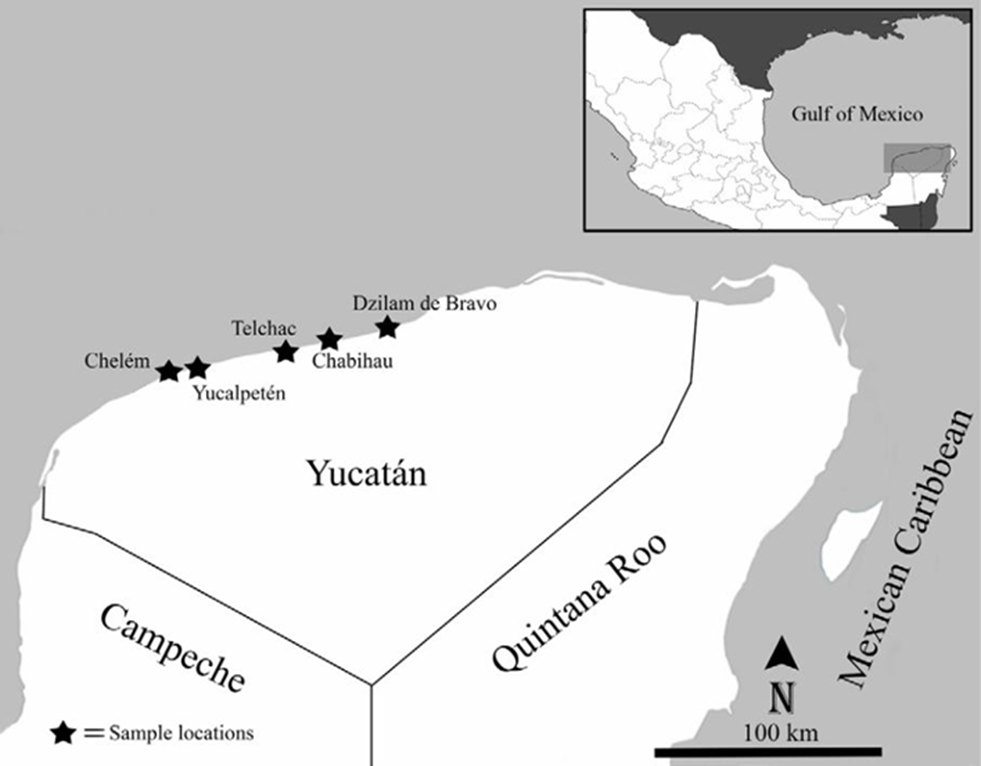
Specimens of sea anemones were collected from 5 beach locations along the Yucatán coastline, Mexico: Chelem (21°16’ N, 89°43’ W), Yucalpetén (21°15’ N, 89°39’ W), Telchac Puerto (21°16’ N, 89°13’ W), Chabihau (21°18’ N, 89°09’ W), and Dzilam de Bravo (21°20’ N, 88°24’ W) (Fig. 1). Specimens were collected by hand and snorkeling from 0-2 m depth, using a hammer and a chisel. Collected specimens were transferred to the laboratory and maintained in an aquarium to photograph their color while alive. Specimens were relaxed in 5% MgCl2 seawater solution and fixed in 10% seawater formalin. Measurements of specimens (i.e., column, oral, and pedal disc diameters, maximum length and width of the body, and number of tentacles) were obtained from fixed specimens. Histological sections 5-10 µm thick were made from 1-2 specimens from each species (except for C. tricolor due to poor preservation), and stained with hematoxylin and eosin (Estrada-Flores et al., 1982). Squash preparations of small amounts of tissue from tentacles, actinopharynx, mesenterial filaments, column, marginal projections, and acontia (if present) were made from at least 1 specimen from each species to observe their cnidae types. Cnidae were examined using a Nikon Eclipse E200 light microscope, and cnidae terminology follows Östman (2000). Specimen identifications are based on Carlgren (1949, 1952), Carlgren and Hedgpeth (1952), González-Muñoz et al. (2012, 2013, 2019), and Daly and den Hartog (2004). We followed the taxonomic classification implemented in Carlgren (1949) with modifications from Rodríguez et al. (2014). Taxa were organized according to their suborder and family and listed in alphabetical order. Voucher specimens of each species were deposited in the zoological collection (YUC-CC) of the Departamento de Zoología, of the Universidad Autónoma de Yucatán (UADY). This study was conducted under SEMARNAT collecting permit SGPA/DGSV/03407/16.
Results
Class Anthozoa
Subclass Hexacorallia
Order Actiniaria Hertwig, 1882
Suborder Enthemonae Rodríguez & Daly, 2014 in Rodríguez et al. (2014)
Superfamily Actinioidea Rafinesque, 1815 Family Actiniidae Rafinesque, 1815 Actinostella flosculifera (Le Sueur, 1817)
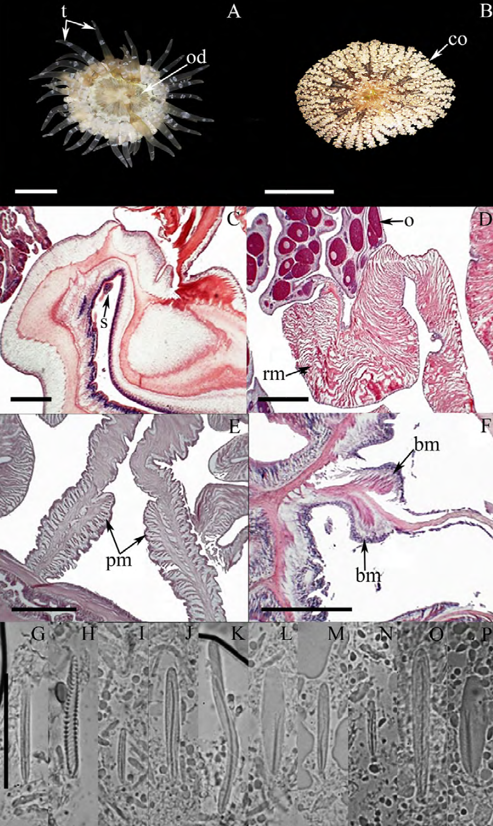
Figure 2. Actinostella flosculifera. A) Oral view, tentacles fully expanded; B) oral view, tentacles retracted and collar fully expanded; C) cross section through column, detail of siphonoglyph; D) cross section through column, detail of a mesentery and oocysts; E) cross section through column, detail of a parietobasilar muscle; F) longitudinal section through proximal column, detail of the basilar muscles. Cnidom: tentacles: G) basitrich, H) spirocyst; actinopharynx: I) small basitrich, J) basitrich, K) long curve basitrich, L) microbasic p-mastigophore; column: M) basitrich; Filaments: N) small basitrich, O) microbasic b-mastigophore, P) microbasic p-mastigophore. Abbreviations, bm: basilar muscles, co: collar, o: oocysts, od: oral disc, pm: parietobasilar muscles, rm: retractor muscles, s: siphonoglyph, t: tentacles. Scale bars, A, B: 10 mm; C-F: 200 μm; G-P: 10 μm.
Actinia flosculifera Le Sueur (1817)
Metridium praetextum: Couthouy in Dana, 1846
Actinostella Formosa [sic]: Duchassaing, 1850
Oulactis flosculifera: Milne-Edwards, 1857
Oulactis conquilega: Duchassaing & Michelotti, 1860
Oulactis Flosculifera [sic]: Duchassaing, 1870
Evactis flosculifera: Andres, 1883
Oulactis foliosa: Andres, 1883
Oulactis fasciculata: McMurrich, 1889a
Asteractis n. sp.: Duerden, 1897
Asteractis expansa: Duerden in McMurrich, 1898
Cradactis fasciculata: Haddon, 1898
Asteractis flosculifera: Verrill, 1899
Actinactis flosculifera: Verrill, 1900
Actinostella flosculifera: McMurrich, 1905
Actinostella conchilega: McMurrich, 1905
Phyllactis flosculifera: Stephenson, 1922
External anatomy. Oral disc flat, 21-31 mm in diameter, beige to dark-brown or light gray, sometimes with white spots; mouth whitish or beige (Fig. 2A). Margin with a collar (also called marginal ruff) formed by 48 rows of small frond-like fused papillae, located in the distal end of the column and surrounding the oral disc (Fig. 2B; collar green to brown or beige, sometimes with dark-red or pink spots. Tentacles smooth, conical, tapering distally, moderately long, contractile, hexamerously arranged in 4 cycles (between 44-48 in number), with the internal cycles longer than outer ones, white, translucent, and with small circular spots on its oral face (Fig. 2A). Column cylindrical, smooth, 20-27 mm in height and 21-34 mm in diameter, with longitudinal rows of small verrucae distally. Pedal disc well-developed, circular. Pedal disc and column beige to light-yellow, translucent when fully expanded.
Internal anatomy. Mesenteries hexamerously arranged in 3 cycles (24 pairs in the specimen examined), first and second cycles perfect, third imperfect; same number of mesenteries distally and proximally. Gametogenic tissue (i.e., oocytes) observed the strongest mesenteries of the first and second cycle, except the directives. Two pairs of directive mesenteries each attached to a well-developed siphonoglyph (Fig. 2C). Retractor muscles strong and restricted (Fig. 2D); parietobasilar muscles well-developed with a free mesogleal pennon (Fig. 2E). Basilar muscles well-developed (Fig. 2F). Marginal sphincter circumscribed. Longitudinal muscles of tentacles ectodermal. Zooxanthellate.
Cnidom. Basitrichs, microbasic b- and p-mastigophores, and spirocysts (Fig. 2G-P). For further information on the size-ranges of cnidae, see González-Muñoz et al. (2012).
Taxonomic summary
Material examined: 26 specimens (YUC-CC-250-9055-19056, 19104-19111, 19232-19246).
Distribution. This species is mainly distributed in the Western Atlantic, from Bermuda to Brazil, and along the Gulf of Mexico and Caribbean Sea (González-Muñoz et al., 2012), but has also been reported from the Canary Islands (Ocaña & den Hartog, 2002) and the Gulf of Guinea (Wirtz, 2003). Actinostella flosculifera has been previously recorded in the rocky intertidal shore and coral reefs from Veracruz (Vassallo et al., 2014; De la Cruz-Francisco & González-Muñoz, 2019), the Campeche Bank (González-Muñoz et al., 2013) and the Mexican Caribbean (González-Muñoz et al., 2012). Here we document for the first time its presence at the coastal localities of Chelem, Yucalpetén, Telchac, Chabihau, and Dzilam de Bravo.
Remarks
The taxonomic characteristics observed in the specimens examined of A. flosculifera agree well with those reported for the species (González-Muñoz et al., 2012; Schlenz & Belém, 1992). In addition, we note the presence of microbasic b-mastigophores (Fig. 2O) in the mesenteric filaments (as is usually observed in other Actiniidae) as well as large curved basitrichs in the actinopharynx (Fig. 2K), that were not previously reported for this species.
Anemonia sargassensis Hargitt, 1908 (Fig. 3A-S)

Figure 3. Anemonia sargassensis. A) Oral view, tentacles fully expanded; B) lateral view, tentacles expanded; C) cross section through column, detail of siphonoglyphs (black arrows); D) cross section through column, detail of a siphonoglyph and retractor muscles; E) longitudinal section through proximal column, detail of basilar muscles; F) longitudinal section through distal column, detail of the marginal sphincter muscle. Cnidom: tentacles: G) spirocyst, H) small basitrich, I) basitrich; actinopharynx: J) basitrich, K) small basitrich, L) microbasic p-mastigophore; column: M) basitrich; acrorhagi: N) small basitrich, O) basitrich, P) holotrich; filaments: Q) small basitrich, R) microbasic b-mastigophore, S) microbasic p-mastigophore. Abbreviations, bm: basilar muscles, c: column, e: epidermis, la: larvae, od: oral disc, rm: retractor muscles, s: siphonoglyph, sp: sphincter, t: tentacles. Scale bars, A, B: 10 mm; C-F: 200 μm; G-S: 10 μm.
Anemonia sargassensis Hargitt, 1908
Anemonia antilliensis: Pax, 1924
Anemonia sargassiensis [sic]: Carlgren, 1949
Anemonia melanaster: den Hartog, 1995
External anatomy. Oral disc flat, wide, 6-14 mm in diameter in specimens examined, dark-orange, dark-red, light-yellow, dark-brown or beige, with white or yellowish endocoelic radial stripes from the base of the tentacles to the mouth (Fig. 3A); mouth yellowish, red, brown or purple. A ring of soft, yellowish or whitish marginal projections with acrorhagi (containing holotrichs and basitrichs) surrounding the oral disc. A shallow fosse is present between the oral disc and the margin of the column. Tentacles smooth, thin, slender, tapering distally, contractile, irregularly arranged in 4-5 cycles (47-111 in number), inner cycles longer than outer ones, dark-red to light-yellow or dark-brown, sometimes with the tips with purple or pink flashes. Column cylindrical, short, cup-shaped, smooth, 3-6 mm in height and 5-11 mm in diameter, bright-red, light-yellow or dark-brown (Fig. 3B). Pedal disc well-developed, circular, wider than column, yellowish to beige.
Internal anatomy. Mesenteries irregularly arranged in 4 cycles (45-52 pairs). Directive mesenteries absent; 5-8 siphonoglyphs observed, each one attached to a regular pair of mesenteries (Fig. 3C). Gametogenic tissue not observed; some larvae observed in the coelenteron. Retractor muscles diffuse to restricted (Fig. 3D); parietobasilar muscles well-developed with a short mesogleal lamella. Basilar muscles well-developed (Fig. 3E). Marginal sphincter muscle endodermal, weak and diffuse (Fig. 3F). Longitudinal muscles of tentacles ectodermal. Zooxanthellate.
Cnidom. Basitrichs, microbasic b- and p-mastigophores, holotrichs, and spirocysts (Fig. 3G-S). For further information on the size-ranges of cnidae see González-Muñoz et al. (2013).
Taxonomic summary
Material examined: 30 specimens (YUC-CC-250-9061-19068, 19112-19121, 19220-19231).
Distribution. Anemonia sargassensis is distributed in the Western Atlantic, from the northern coast of United States to Brazil, and along the Gulf of Mexico and Caribbean Sea (González-Muñoz et al., 2012). This species has been documented in the Mexican Atlantic from coral reefs of Veracruz (De la Cruz-Francisco & González-Muñoz, 2019), the Campeche Bank (González-Muñoz et al., 2013), and the Mexican Caribbean (González-Muñoz, Simões et al., 2015), but this is its first report from Yucatán, at the localities of Chelem and Chabihau.
Remarks
All the taxonomic characteristics observed in A. sargassensis fit well with those previously described, including the presence of more than 2 siphonoglyphs and the absence of directive mesenteries (González-Muñoz et al., 2013). The irregularities of the hexameral pattern are due to asexual reproduction by longitudinal fission, which are commonly reported for this species (Carlgren & Hedgpeth, 1952; González-Muñoz et al., 2013).
Anthopleura dalyae González-Muñoz, Garese & Acuña, 2018

Figure 4. Anthopleura dalyae. A) Oral view, tentacles fully expanded; B) lateral view; C) longitudinal section through column, detail of verrucae; D) cross section through column, detail of a siphonoglyph, retractor and parietobasilar muscles; E) longitudinal section through proximal column, detail of basilar muscles; F) longitudinal section through distal column, detail of the marginal sphincter muscle. Cnidom: tentacles: G) basitrich, H) spirocyst; actinopharynx: I) small basitrich, J) microbasic p-mastigophore, K) microbasic b-mastigophore; column: L) basitrich, M) holotrich; Acrorhagi: N) holotrich, O) basitrich; filaments: P) microbasic p-mastigophore, Q) basitrich, R) microbasic b-mastigophore. Abbreviations, ac: acrorhagi, bm: basilar muscles, c: column, od: oral disc, pm: parietobasilar muscles, rm: retractor muscles, s: siphonoglyph, sp: sphincter, t: tentacles, ve: verrucae. Scale bars, A, B: 10 mm; C-F: 200 μm; G-R: 10 μm.
non Anthopleura varioarmata: Watzl, 1922
Anthopleura vario-armata [sic]: Carlgren, 1952Anthopleura varioarmata: Belém & Monteiro, 1981 Anthopleura variarmata [sic]: Zamponi et al., 1998 Anthopleura texaensis: González-Muñoz, Tello-Musi & Simões, 2015
External anatomy. Oral disc smooth and wide, 4-6 mm in diameter, olive-green, light-green to dark-green, or yellowish with white radial stripes (Fig. 4A); mouth dark-green or dark-brown. A ring of rounded marginal projections with acrorhagi in the fosse (containing holotrichs and basitrichs) surrounding the oral disc. A deep fosse is present between the oral disc and the margin of the column. Tentacles smooth, slender, conical, relatively short, tapering distally, contractile, irregularly arranged in 4-5 cycles (41-76 in number), inner cycles longer than outer ones, gray, light-yellow, olive-green to dark-green, with white spots on the oral side and along their entire length (Fig. 4A). Column cylindrical, stout, 6-17 mm in height and 5-12 mm in width, with longitudinal rows of verrucae from margin to limbus but more pronounced distally, olive-green to dark-green, yellowish, pale-orange or whitish (Fig. 4B-C). Pedal disc well-developed, slightly wider than column, beige, and yellowish to white.
Internal anatomy. Mesenteries irregularly arranged in 3-4 cycles (19-31 pairs): first, second, and some of the third cycle perfect, others imperfect; same number of mesenteries distally and proximally. Gametogenic tissue not observed. 2 pairs of directive mesenteries each attached to a well-developed siphonoglyph (Fig. 4D). Retractor muscles strong and restricted; parietobasilar muscles well-developed with a short mesogleal pennon (Fig. 4D). Basilar muscles well-developed (Fig. 4E). Marginal sphincter muscle ectodermal, strong and circumscribed (Fig. 4F). Longitudinal muscles of tentacles ectodermic.
Cnidom. Basitrichs, microbasic b- and p-mastigophores, holotrichs, and spirocysts (Fig. 4G-R). For further information on cnidae size-ranges see González-Muñoz et al. (2019).
Taxonomic summary
Material examined: 18 specimens (YUC-CC-250-19069-19071, 19137-19140, 19247-19257).
Distribution. This species has been reported in Brazil and from the coast of Veracruz, Mexico (Belém & Monteiro, 1981; González-Muñoz et al., 2019), but this is the first record of A. dalyae from Yucatán, at the locality of Chabihau.
Remarks
Taxonomic characteristics observed fit well with those previously described for A. dalyae (González-Muñoz et al., 2019) including the number of tentacles and restricted rather than diffused retractor muscles as in the species Anthopleura texaensis (Carlgren & Hedgpeth, 1952) (Daly & den Hartog, 2004). In addition, we add the presence of microbasic b-mastigophores in the actinopharynx (Fig. 4K) to the cnidom of this species.
Anthopleura krebsi (Duchassaing & Michelotti, 1860)

Figure 5. Anthopleura krebsi. A) Oral view, tentacles fully expanded; B) lateral view; C) cross section through column, detail of a mesentery and retractor muscles; D) cross section through column, detail of a mesentery, retractor and parietobasilar muscles; E) longitudinal section through proximal column, detail of basilar muscles; F) longitudinal section through distal column, detail of the marginal sphincter muscle and acrorhagi. Cnidom: tentacles: G) spirocyst, H) basitrich; actinopharynx: I) microbasic b-mastigophore, J) microbasic p-mastigophore, K) small basitrich, L) basitrich; column: M) basitrich, N) holotrich; acrorhagi: O) holotrich, P) basitrich; filaments: Q) microbasic b-mastigophore, R) microbasic p-mastigophore, S) small basitrich, T) long curve basitrich. Abbreviations, ac: acrorhagi, bm: basilar muscles, c: column, fm: filaments, fo: fosse, od: oral disc, pm: parietobasilar muscles, rm: retractor muscles, sp: sphincter, t: tentacles. Scale bars, A, B: 10 mm; C-F: 200 μm; G-T: 10 μm.
Anthopleura Krebsi [sic] Duchassaing & Michelotti, 1860 Anthopleura Krebsii [sic]: Duchassaing & Michelotti, 1864
Bunodes Krebsi [sic]: Duerden, 1897
Bunodactis stelloides carneola: Verrill, 1907
Anthopleura varioarmata: Watzl, 1922
Anthopleura kerbsi: Bigger, 1980
Anthopleura carneola: McCommas, 1983
External anatomy. Oral disc smooth, narrow, 5-7 mm in diameter, beige to dark-brown, dark-gray, or olive-green, sometimes with white flashes; mouth whitish or beige (Fig. 5A). A ring of rounded endocoelic marginal projections with acrorhagi in the fosse (containing holotrichs and basitrichs) surrounding the oral disc. A deep fosse is present between the oral disc and the margin of the column. Tentacles smooth, slender, conical, relatively short, tapering distally, contractile, irregularly arranged in 3-4 cycles (19-45 in number), inner cycles longer than outer ones, gray, yellowish to white color, translucent, with oral white spots along their entire length (Fig. 5A-B). Column cylindrical, sometimes trumped-shape, 3-10 mm in height and 3-7 mm in diameter, with longitudinal rows of verrucae from margin to limbus but more pronounced distally, pale-pink with a distinctive bright-red to bright-pink dot on each verrucae (Fig. 5B). Pedal disc well-developed, slightly wider than column, pale-pink to beige.
Internal anatomy. Mesenteries irregularly arranged in 2-3 cycles: first and part of the second cycle perfect, others imperfect. Gametogenic tissue not observed. Directive mesenteries and siphonoglyphs variable in number, 1-4 in specimens examined. Retractor muscles diffuse to restricted (Fig. 5C-D); parietobasilar muscles well-developed with a short mesogleal lamella (Fig. 5C-D). Basilar muscles well-developed (Fig. 5E). Marginal sphincter muscle strong and circumscribed (Fig. 5F). Longitudinal muscles of tentacles ectodermal. Zooxanthellate.
Cnidom. Basitrichs, microbasic b- and p-mastigophores, holotrichs, and spirocysts (Fig. 5G-T). For further information on cnidae size-ranges see Daly and den Hartog (2004).
Taxonomic summary
Material examined: 27 specimens (YUC-CC-250-19072-19082, 19127-19136, 19258-19263).
Distribution. Anthopleura krebsi is distributed in the Western Atlantic, from the Gulf of Mexico and the Caribbean Sea to Brazil (González-Muñoz et al., 2012). In Mexico this species has been previously reported from Veracruz (De la Cruz-Francisco & González-Muñoz, 2019; González-Muñoz, Tello-Musi et al., 2015), and the Mexican Caribbean (INE, 1998), but this is its first record from Yucatán, at the locality of Chabihau.
Remarks
The taxonomic characteristics agree well with those reported for A. krebsi, including the pattern of coloration of column and verrucae, number of tentacles, and cnidom (Belém & Pinto, 1990; Daly & den Hartog, 2004). In addition, we report the presence of microbasic b-mastigophores in the actinopharynx (Fig. 5I); however, we do not observe the small microbasic b-mastigophores in the mesenteric filaments as documented by Daly and den Hartog (2004).
Bunodosoma cavernatum (Bosc, 1802)

Figure 6. Bunodosoma cavernatum. A) Oral view, tentacles fully expanded; B) lateral view; C) cross section through column, detail of a siphonoglyph and retractor muscles; D) cross section through column, detail of retractor and parietobasilar muscles; E) longitudinal section through proximal column, detail of basilar muscles; F) longitudinal section through distal column, detail of the marginal sphincter muscle and vesicles. Cnidom: tentacles: G) basitrich, H) spirocyst; actinopharynx: I) small basitrich, J) basitrich, K) microbasic p-mastigophore; column: L) basitrich; acrorhagi: M) holotrich, N) basitrich; filaments: O) microbasic b-mastigophore, P) microbasic p-mastigophore, Q) basitrich. Abbreviations: bm: basilar muscles, c: column, od: oral disc, rm: retractor muscles, pm: parietobasilar muscles, s: siphonoglyph, sp: sphincter, t: tentacles, vs: vesicles. Scale bars: A, B: 10 mm; C-F: 200 μm; G-Q: 10 μm.
Actinia cavernataBosc, 1802
Actinia (Monostephanus) cavernata: Brandt, 1835
Urticina cavernata: Duchassaing, 1850
Bunodes cavernata: Verrill, 1864
Phymactis cavernata: Andres, 1883
Bunodosoma cavernata: Verrill, 1899
Anthopleura cavernata: Cary, 1906
Bunodosma [sic] cavernata: Daly, 2003
External anatomy. Oral disc flat, wide and smooth, 8-28 mm in diameter, olive-green, beige or pale-red, with whitish or yellowish radial stripes from tentacles to mid oral disc on endocoelic spaces of the first and second cycles; mouth reddish (Fig. 6A). A ring of rounded marginal projections with acrorhagi (containing holotrichs and basitrichs) surrounds the oral disc. A deep fosse is present between the oral disc and the margin of the column.
Tentacles smooth, conical, tapering distally, moderately long, contractile, irregularly arranged in 5 cycles (71-98 in number), internal cycles longer than outer ones, grey, greyish-red or beige, with a red stripe in the aboral side on the tentacles of the inner cycles (never observed in the last cycle of tentacles) (Fig. 6A, B). Column cylindrical, 16-44 mm in length and 14-31 mm in width, densely covered by rounded vesicles arranged in longitudinal rows alternating between large and small vesicles, which are beige or greyish with dark-brown or black in the center each vesicle (Fig. 6B).
Internal anatomy. Mesenteries hexamerously arranged in 4 cycles (48 pairs): first, second, and part of the third cycle perfect, others imperfect; same number of mesenteries distally and proximally. Gametogenic tissue (i.e., spermatic cysts) observed in the strongest mesenteries of the first, second, and third cycle (except the directives). 2 pairs of directive mesenteries each attached to a well-developed siphonoglyph (Fig. 6C). Retractor muscles strong and restricted; parietobasilar muscles well-developed with a free mesogleal pennon (Fig. 6D). Basilar muscles well-developed (Fig. 6E). Marginal sphincter muscle endodermic, strong and circumscribed (Fig. 6F). Longitudinal muscles of tentacles ectodermal. Zooxanthellate.
Cnidom. Basitrichs, microbasic b- and p-mastigophores, holotrichs, and spirocysts (Fig. 6G-Q). For further information on the size-ranges of cnidae, see González-Muñoz et al. (2013).
Taxonomic summary
Material examined: 9 specimens (YUC-CC-250-19188 -19205).
Distribution. Bunodosoma cavernatum is distributed in the Western Atlantic, from the northern coast of United States to the Gulf of Mexico and Caribbean Sea (González-Muñoz et al., 2012, 2013, 2016), and in the Caroline Islands, Micronesia (Bosc, 1802). This species has been reported in coral reefs of Veracruz (De la Cruz-Francisco & González-Muñoz, 2019; González-Muñoz et al., 2013), but this is its first report from Yucatán, at the localities of Chabihau and Dzilam de Bravo.
Remarks
All taxonomic features agree well with those previously reported for B. cavernatum, except for the presence of 2 cnidae size categories of basitrichs in the column, while only one category is clearly distinguishable in the specimens examined. In addition, we found 2 size categories of basitrichs in the actinopharynx (Fig. 6I-J), which were not observed by González-Muñoz et al. (2013).
Bunodosoma granuliferum (Le Sueur, 1817)

Figure 7. Bunodosoma granuliferum. A) Oral view, tentacles fully expanded; B) lateral view; C) longitudinal section through column, detail of vesicles; D) cross section through column, detail of a pair of mesenteries and retractor muscles; E) longitudinal section through proximal column, detail of basilar muscles; F) longitudinal section through distal column, detail of the marginal sphincter muscle. Cnidom: tentacles: G) basitrich, H) spirocyst; actinopharynx: I) microbasic p-mastigophore, J) basitrich, K) microbasic b-mastigophore; column: L) basitrich; acrorhagi: M) basitrich, N) holotrich; filaments: O) microbasic p-mastigophore, P) basitrich, Q) microbasic b-mastigophore, R) small basitrich. Abbreviations: bm: basilar muscles, c: column, od: oral disc, rm: retractor muscles, sp: sphincter, t: tentacles, vs: vesicles. Scale bars: A, B: 10 mm; C-F: 200 μm; G-R: 10 μm.
Actinia granulifera Le Sueur, 1817
Urticina Lessoni [sic]: Duchassaing, 1850
Oulactis granulifera: Milne-Edwards, 1857
Urticina granulifera: Duchassaing & Michelotti, 1860
Cereus Lessoni [sic]: Duchassaing & Michelotti, 1860
Anthopleura granulifera: Duchassaing & Michelotti, 1864
Anthopleura Granulifera [sic]: Duchassaing, 1870
Aulactinia granulifera: Andres, 1883
Bunodes taeniatus: McMurrich, 1889b
Bunodes granulifera: Duerden, 1897
Bunodosoma granulifera: Verrill, 1899
Bunodosoma granuliferum: Pax, 1910
Phymactis granulifera: Stephenson, 1922
External anatomy. Oral disc flat, wide and smooth, 13-43 mm in diameter, variable in coloration, from white with yellow or greenish to dark-brown, sometimes with whitish of yellowish radial stripes marking the endocoelic spaces; mouth dark-brown, reddish, orange or yellow (Fig. 7A). A ring of rounded marginal projections with acrorhagi (containing holotrichs and basitrichs) surrounds the oral disc. A deep fosse is present between the oral disc and the margin of the column. Tentacles smooth, conical, tapering distally, moderately long, contractile, mostly hexamerously arranged in 5 cycles (83-101 in number), internal cycles longer than outer ones, olive-green, gray, white and translucent, with oral white spots and purple flashes at the tips (Fig. 7A-B). Column cylindrical, 12-24 mm in length and 19-33 mm in width in specimens examined, densely covered by rows of rounded vesicles arranged in 24 alternating dark and light longitudinal bands, which are orange, green, gray or light-pink (Fig. 7B-C). Pedal disc well-developed, cylindrical, orange, olive-green or gray.
Internal anatomy. Mesenteries hexamerously arranged in 4 cycles (48 pairs): first, second, and part of the third cycle perfect, others imperfect; same number of mesenteries distally and proximally. Gametogenic tissue (i.e., spermatic cysts) observed in the strongest mesenteries of the last 2 cycles. 2 pairs of directive mesenteries each one attached to a well-developed siphonoglyph. Retractor muscles strong and restricted; parietobasilar muscles well-developed with a free mesogleal pennon (Fig. 7D). Basilar muscles well-developed (Fig. 7E). Marginal sphincter muscle endodermic, strong and circumscribed (Fig. 7F). Longitudinal muscles of tentacles ectodermal. Zooxanthellate.
Cnidom. Basitrichs, microbasic b- and p-mastigophores, holotrichs, and spirocysts (Fig. 7G-R). For further information on the size-ranges of cnidae see González-Muñoz et al. (2012).
Taxonomic summary
Material examined: 15 specimens (YUC-CC-250-19052, 19122-19126, 19179-19187).
Distribution. This species is distributed in the Western Atlantic, from Bermuda to Brazil, and along the Gulf of Mexico and Caribbean Sea (González-Muñoz et al., 2012). In Mexico, B. granuliferum has been reported from Veracruz (De la Cruz-Francisco & González-Muñoz, 2019; González-Muñoz, Tello-Musi et al., 2015; Vassallo et al., 2014), the Campeche Bank (González-Muñoz et al., 2013), and the Mexican Caribbean (González-Muñoz et al., 2012), but this is its first report from Yucatán, at the localities of Chelem and Chabihau.
Remarks
All the taxonomic characteristics of the specimens examined of B. granuliferum agree well with those reported for the species, including the pattern of column coloration and the number of tentacles, except that we observe only 1 size category of basitrichs in the actinopharynx and column, instead of 2 as reported by González-Muñoz et al. (2012). In addition, we added the presence of microbasic b-mastigophores in the actinopharynx and filaments (Fig. 7K), as well as small basitrichs in filaments (Fig. 7R), to the cnidom of the species.
Superfamily Metridioidea Carlgren, 1893 Family Aiptasiidae Carlgren, 1924 Exaiptasia diaphana (Rapp, 1829)

Figure 8. Exaiptasia diaphana. A) Oral view, tentacles fully expanded; B) lateral view; C) longitudinal section through column, detail of a siphonoglyph and retractor muscles; D) cross section through column, detail of retractor muscles and spermatic cysts; E) cross section through column, detail of oocysts; F) longitudinal section through proximal column, detail of basilar muscles. Cnidom: tentacles: G) microbasic p-amastigophore, H) basitrich, I) spirocyst; actinopharynx: J) basitrich, K) microbasic p-amastigophore; column: L) basitrich, M) microbasic p-amastigophore, N) microbasic b-mastigophore; acontia: O) small microbasic p-amastigophore, P) microbasic p-amastigophore, Q) basitrich; filaments: R) microbasic p-amastigophore, S) small basitrich, T) basitrich. Abbreviations: a: acontia, bm: basilar muscles, c: column, od: oral disc, oo: oocysts, rm: retractor muscles, s: siphonoglyph, sc: spermatic cysts, t: tentacles. Scale bars, A, B: 10 mm; C-F: 200 μm; G-T: 10 μm.
Actinia diaphana Rapp, 1829
Cribina diaphana: Deshayes & Milne-Edwards, 1840
Actinia elongata Delle-Chiaje, 1841
Adamsia diaphana: Milne-Edwards, 1857
Dysactis pallida: Agassiz in Verrill, 1864
Bartholomea tagetes: Duchassaing & Michelotti, 1864
Bartholomea inula: Duchassaing & Michelotti, 1864
Dysactis mimosa: Duchassaing & Michelotti, 1864
Dysactis minuta: Verrill, 1867
Paranthea minuta: Verrill, 1868
Paranthea pallida: Verrill, 1868
Disactis mimosa [sic]: Duchassaing, 1870
Aiptasia saxicola: Andres, 1881
Aiptasia diaphana: Andres, 1883
Aiptasia Agassizii [sic]: Andres, 1883
Aiptasia inula: Andres, 1883
Aiptasia minuta: Andres, 1883
Aiptasia mimosa: Andres, 1883
Aiptasia tagetes: Andres, 1883
Aiptasia pallida: McMurrich, 1887
Aiptasia leiodactyla: Pax, 1910
Aiptasioides pallida: Stephenson, 1918
Aiptasiomorpha diaphana: Stephenson, 1920
Aiptasiomorpha leiodactyla: Stephenson, 1920
Aiptasia insignis: Carlgren, 1941
Aiptasia pulchella: Carlgren, 1943
Aiptasia californica: Carlgren, 1952
Aiptasiomorpha minuta: Uchida & Soyama, 2001
Aipstasia pulchella [sic]: Reimer et al., 2007
Exaiptasia pallida: Grajales & Rodríguez, 2014 External anatomy. Oral disc smooth, 1-18 mm in
diameter, olive-green to beige, more or less translucent with whitish or yellowish scattered dots; mouth slit-like, white, yellowish or beige (Fig. 8A). Column cylindrical, trumped-shaped when fully expanded, smooth, 2-19 mm in height and 1-20 mm in diameter, divided in capitulum and scapus: capitulum short, beige to dark-brown with whitish, yellowish, or pale-orange spots; scapus long, beige, translucent, with the mesenterial insertions visible (Fig. 8B). 2-3 rings of cinclides around the mid part of scapus. Tentacles smooth, slender, relatively short, irregularly arranged in 3-5 cycles (23-113 in number), inner cycles longer than outer ones, olive-green, yellowish, whitish, dark-brown, beige, or black (Fig. 8A-B). Pedal disc well-developed, beige, translucent. White acontia (with microbasic p-mastigophores and basitrichs).
Internal anatomy. Mesenteries irregularly arranged in 2-4 cycles (about 12-48 pairs in specimens examined): first cycle perfect, others imperfect. Gametogenic tissue (oocysts and spermatic cysts) was observed in the strongest mesenteries of the first, second, and third cycle, except the directives (Fig. 8D-E). 2 pairs of directive mesenteries each one attached to a well-developed siphonoglyph (Fig. 8C). Retractor muscles diffuse to restricted (Fig. 8E); parietobasilar muscles poorly-developed. Basilar muscles well-developed (Fig. 8F). Marginal sphincter muscles mesogleal, short and diffuse. Longitudinal muscles of tentacles ectodermal. Zooxanthellate.
Cnidom. Basitrichs, microbasic b-mastigophores and p-amastigophores, and spirocysts (Fig. 8G-T). For further information on cnidae size-ranges see Carlgren (1952), González-Muñoz et al. (2012), and Grajales and Rodríguez (2014).
Taxonomic summary
Material examined: 27 specimens (YUC-CC-250-19057-19060, 19095-19103, 19206-19219).
Distribution. This species is distributed mainly in the Western Atlantic, from the northeast coast of the United States to Brazil, along the Gulf of Mexico and Caribbean Sea, but it has also been reported from the east and west coasts of the Pacific, Japan, Hawaii, and the islands of Santa Helena (Grajales & Rodríguez, 2014). Exaiptasia diaphana has been previously reported from Veracruz (González-Muñoz, Tello-Musi et al., 2015), the Campeche Bank (González-Muñoz et al., 2013), and the Mexican Caribbean (González-Muñoz et al., 2012), but this is its first report from the coast of Yucatán, at the localities of Chabihau, Yucalpetén, Telchac, and Chelem.
Remarks
All taxonomic characteristics of the examined specimens of E. diaphana are consistent with those reported for the species (González-Muñoz et al., 2012; Grajales & Rodríguez, 2014), including the pattern of cinclides in the column, the pattern of coloration, and cnidae. In addition, we note that the cnidocysts of the column identified as basitrich by González-Muñoz et al. (2012: Fig. 9M) corresponds to a microbasic b-mastigophore (Fig. 8N).

Figure 9. Calliactis tricolor. A) Oral view, tentacles fully expanded; B) lateral view. Abbreviations: c: column, ci: cinclides, od: oral disc, t: tentacles. Scale bars: A, B: 10 mm.
Family Hormathiidae Carlgren, 1932 Calliactis tricolor (Le Sueur, 1817) (Fig. 9A-B)
Actinia tricolor: Le Sueur, 1817
Actinia bicolor: Le Sueur, 1817
Cereus bicolor: Milne-Edwards, 1857
Adamsia tricolor: Milne-Edwards, 1857
Adamsia Egletes [sic]: Duchassaing & Michelotti, 1864
Adamsia egletes: Duchassaing & Michelotti, 1866
Calliactis bicolor: Verrill, 1869
Adamsia sol: McMurrich, 1893
Adamsia bicolor: Andres, 1883
Adamsia tricolor: Andres, 1883
Calliactis tricolor: Haddon, 1898
External anatomy. Oral disc smooth, wide, 5-30 mm in diameter in specimens examined, white to gray with purple flashes; mouth slit-like in shape, with bright-yellow or bright-orange lips, and with a purple or bright-pink ring around the mouth (Fig. 9A). Tentacles smooth, conical, relatively short, contractile, tapering distally, hexamerously arranged in 4-6 cycles (48-192 in number), translucent, beige, with oral white spots along their entire length, sometimes with pink rings distally (Fig. 9A-B). Column cylindrical or trumped-shaped, divided in capitulum and scapus: capitulum very short, smooth, beige to dark-brown; scapus stout, wrinkled in appearance, pale-red to dark-red, beige to dark-brown, or pale-orange. One or 2 rings of dark-red or brown cinclides present in the proximal part of the column, near to the limbus (Fig. 9B). Pedal disc well-developed, wider than column, beige to dark-brown, translucent with the mesenterial insertions and acontia visible.
Internal anatomy. See González-Muñoz et al. (2013).
Cnidom. See González-Muñoz et al. (2013).
Taxonomic summary
Material examined: 5 specimens (YUC-CC-250-19090-19094).
Distribution. Calliactis tricolor is distributed in the Western Atlantic, from North Carolina to the north coast of Brazil, and along the Gulf of Mexico and Caribbean Sea (Tello-Musi et al., 2018). This species has been previously reported in the Mexican Atlantic, from Veracruz (Tello-Musi et al., 2018), and the Campeche Bank (González-Muñoz et al., 2013), but this is the first report from Yucatán, at the locality of Dzilam de Bravo.
Remarks
All the taxonomic characteristics of the external anatomy of the specimens examined agree well with those reported for the species (González-Muñoz et al., 2013), including the pattern of coloration of the column, cinclides and acontia, and the number of tentacles.
Discussion
Among the species found in the sampled locations, A. flosculifera and E. diaphana are the most widely distributed, being both found in almost all the places visited. Moreover, the nearest previous record of these 2 species in the region, as well as for C. tricolor, is in Madagascar reef (González-Muñoz et al., 2013), located about 20 km from Chelem beach. Actinostella flosculifera is often found in habitats associated with shallow seagrasses and sandy bottoms, and E. diaphana is frequently found attached to small rocks and submerged wood, or even on sponges, between patches of sand and seagrass (González-Muñoz et al., 2012), which are common environments in much of the Yucatán coastal area (Herrera-Silveira et al., 2013). The species C. tricolor is usually found attached to gastropod mollusk shells occupied by hermit crabs (Tello-Musi et al., 2018). However, this species was found attached to buried hard substrate in the intertidal zone of Dzilam de Bravo.
The species A. dalyae, A. krebsi, A. sargassensis, B. cavernatum, and B. granuliferum were found attached to rocks or submerged remains of old constructions that serve these animals as hard substrates. This artificial substrate, such as bricks or concrete remains of the breakwater constructions, as well as the rubble of old summer houses that were affected by the loss of beaches and hurricane events, facilitate the settlement of the sea anemones and other benthic invertebrates in the area. The nearest report of A. sargassensis and B. granuliferum is from Alacranes reef, located approximately 130 km north from the coast of Yucatán (González-Muñoz et al., 2013). The species A. dalyae, A. krebsi, and B. cavernatum were previously recorded in Mexico at the Veracruz Reef System (González-Muñoz, Tello-Musi et al., 2015), but these are their first records in Yucatán.
González-Muñoz et al. (2013) documented the presence of 13 species of sea anemones in coral reefs of Yucatán, 5 of which are also reported here as inhabitants in the coastal area. The other 8 species are documented so far only in coral reef environments in the region (Table 1). The new records of A. dalyae, A. krebsi, and B. cavernatum increase to 16 the number of known species of sea anemones in the region. These species do not represent all species found in Yucatán, but they are those for which there are no unresolved taxonomic problems, and that are more abundant and conspicuous. In addition, we observed and collected some individuals identified so far as Diadumene sp., but their complete taxonomic identification is still in progress.
Table 1. List of sea anemones species reported from the coast and coral reefs of Yucatán. Citation: 1) González-Muñoz et al. (2013). The symbol “*” indicates a new record for the locality, and “†” indicates new records for Yucatán.
| Species | ||||||||||
| Chelém | Yucalpetén | Telchac | Chabihau | Dzilam de Bravo | Bajo de Diez | Madagascar | Serpientes | Alacranes | ||
| 1 | Actinostella flosculifera (Le Sueur, 1817) | * | * | * | * | * | 1 | 1 | ||
| 2 | Anemonia sargassensis Hargitt, 1908 | * | * | 1 | ||||||
| 3 | Anthopleura dalyae González-Muñoz, Garese & Acuña, 2018 † | * | ||||||||
| 4 | Anthopleura krebsi (Duchassaing & Michelotti, 1860) † | * | ||||||||
| 5 | Anthopleura pallida Duchassaing & Michelotti, 1864 | 1 | ||||||||
| 6 | Bartholomea annulata (Le Sueur, 1817) | 1 | 1 | 1 | 1 | |||||
| 7 | Bunodeopsis antilliensis Duerden, 1897 | 1 | ||||||||
| 8 | Bunodosoma cavernatum (Bosc, 1802) † | * | * | |||||||
| 9 | Bunodosoma granuliferum (Le Sueur, 1817) | * | * | 1 | ||||||
| 10 | Calliactis tricolor (Le Sueur, 1817) | * | 1 | 1 | ||||||
| 11 | Condylactis gigantea (Weinland, 1860) | 1 | 1 | 1 | ||||||
| 12 | Exaiptasia diaphana (Rapp, 1829) | * | * | * | * | 1 | 1 | 1 | ||
| 13 | Laviactis lucida (Duchassaing & Michelotti, 1860) | 1 | ||||||||
| 14 | Lebrunia neglecta (Duchassaing & Michelotti, 1860) | 1 | ||||||||
| 15 | Phymanthus crucifer (Le Sueur, 1817) | 1 | 1 | |||||||
| 16 | Stichodactyla helianthus (Ellis, 1768) | 1 |
The documentation of the sea anemones species in the coastal areas of Yucatán is an important step to inventory the actinofauna in Mexico, as well as to study the biological and ecological role of these animals in coastal ecosystems.

