


 nueva página del texto (beta)
 Inglés (pdf)
 Artículo en XML
 Referencias del artículo
 Enviar artículo por email
 Citado por SciELO
  Similares en
SciELO
Similares en
SciELO 
 Permalink
Pseudogynoxis benthamii Cabrera (= P. cabrerae H.Rob. & Cuatrec.) is an evergreen, suffrutescent, climbing, ornamental Asteraceae native to Argentina, Brazil, and Paraguay. Its stout peduncles hold capitula with bright orange female ray-florets, and reddish hermaphrodite disk florets (Hind, 1992; Robinson & Cuatrecasas, 1977). In March 2016, 10 of 40 plants of P. benthamii in breeding programs in Hurlingham (Buenos Aires, Argentina) suddenly wilted after infrequent hot conditions (Fig. 1a, b). The lower foliage turned yellow and brown, and plants died within a week. White mycelial mats and roundish sclerotia appeared at the basal portion of the stems (Fig. 1c, d), and the lesions spread rapidly to girdle and rot the base of the stems and roots (Fig. 1e). The aim of this study was to identify the causal agent of the disease.
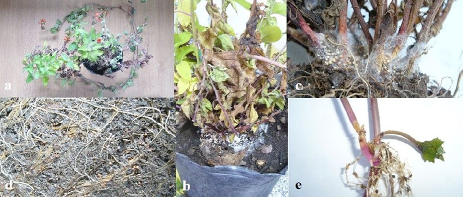
Figure 1 Wilt of Pseudogynoxis benthamii caused by Sclerotium rolfsii. a, Wilting plant, with the lower foliage turned brown, and hanging on the plant; b, advanced stage of wilt, showing the growth of the pathogen at the base of the plant; c, white mycelial mats colonizing the lower stems, and roundish sclerotia at different stages of development; d, plant eased out of the pot showing sclerotia formed in the substrate and near rooted roots; e, intense rot of the lower stem and roots.
Sclerotia that had developed at the base of 2 wilting plants were gently picked with a tong, disinfected by immersion in a NaOCl: water solution (20% Cl) for 30 s, washed in sterilized distilled water for 1 min, and incubated on potato dextrose agar (PDA, Merck) at 27 °C. The growing fungal colonies were kept at 8 °C in darkness, and one of the 5 phenotypically identical isolates that developed from the sclerotia was chosen for further studies.
Vigorous shoots with 2 leaf buds were cut from young non-lignified branches of mother plants of P. benthamii and dipped into a talcum powder mixture containing 250 ppm of indolbutiric acid (Hartman et al., 2011). The cuttings were planted in 25-alveole plastic molded plug trays filled with a mixture of Grow Mix Tabaco S2 (Terrafertil) and expanded perlite (1:1), and placed on a bench in a greenhouse equipped with 80% shadow netting and mist system, at 23-26 °C. After 15 days, each rooted plantlet was transplanted into a 12 cm diameter plastic pot filled with the same substrate, watered and fertilized routinely for 3 months prior to inoculation.
The fungal isolate was cultivated on PDA at 25 oC and pieces of 1.7 cm2 were cut from the edge of 5-days colonies with a scalpel. Pathogenicity was tested in a climatic chamber on twelve potted plants inoculated by placing 2 plugs of inoculum on the soil near the stem bases. For the control, 5 plants were treated with sterile PDA plugs. Each plant was enclosed in a transparent polyethylene bag, and incubated at 27 °C for 72 h. The plants remained in the chamber with natural daylight conditions. The test was repeated twice. To satisfy Koch’s postulates, the fungus was re-isolated from symptomatic tissues. Following DNA extraction (Stenglein & Balatti, 2006), a PCR was carried out in an XP thermal cycler (Bioer Technology) to amplify the nuclear ribosomal DNA internal transcribed spacer (ITS) region using primer pairs ITS1 (5 ́-TCC GTA GGT GAA CCT GCG G-3 ́) / ITS4 (5 ́-TCC TCC GCT TAT TGA TAT GC-3 ́) (White et al. 1990). The sequence fragment was compared with BLASTn (Altschul et al., 1990) to publicly-available sequences deposited in GenBank. The sequence was subsequently submitted to GenBank (accession KY216142).
Fungal colonies consistently developed on PDA, and were preliminarily placed in the genus Sclerotium. They grew fast on PDA, covering the whole area of the 10 cm diameter plates in 4 days, and even growing outwards in 7 days, while the aerial mycelium was observed to reach the lid of the plates. The mycelium was white, with strands showing a fan shaped expansion. Almost spherical sclerotia 0.7 to 1.5 mm diameter with a shiny surface developed from the mycelium. They were white at first, and turned tan to brown with age (Fig. 2a). The hyphae formed typical clamp connections (Fig. 2b).
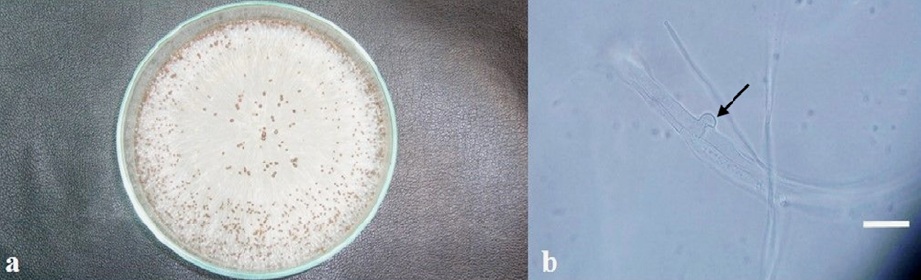
Figure 2 Cultural and morphological characters of Sclerotium rolfsii. a, 7-day-old colony on PDA; b, the arrow indicates a typical clamp connection structure formed in a hypha (bar = 25 μm).
Two days after inoculation, hyphal strands emerged from the plugs and grew towards the substrate and the base of the plants (Fig. 3a), which gradually withered (Fig. 3b). The lower stems appeared surrounded by pathogen’s hyphae (Fig. 3c), and showed water-soaked lesions 5 days from inoculation. All the inoculated plants died within 2 weeks, while the controls remained healthy.

Figure 3 Results of target inoculations of Pseudogynoxis benthamii with Sclerotium rolfsii. a, Hyphal strands emerging from the inoculum plugs, and developing towards the substrate and the lower stem; b, from left to right: Control plant, and wilting infected plants 7 days from inoculation; c, hyphal growth around the stem base.
On the basis of colony characteristics, symptoms, and pathogenicity to the host, the isolate was identified as S. rolfsii Sacc. (Mordue, 1974). It was deposited in the fungal collection the Instituto de Floricultura INTA (entry coded INTA-IF-501). The resulting 684 bp of ITS region sequence revealed that it was 99-100% identical to S. rolfsii (ex. HQ420816, KX186998, KU760984).
Sclerotium rolfsii usually infects the lower stem near the soil surface of many crops causing the disease known as Southern blight. Infections are favored by high temperature and moisture (Punja, 1985), which correspond to the environmental conditions before the disease was detected in this case. This is a widely distributed soil-borne species, but P. benthamii is not included in the 2,579 fungus-host records for Sclerotium rolfsii (Farr & Rossman, 2017). The pathogen can overwinter as mycelium in infected plants, plant debris, or as sclerotia. These data should to be taken into account when planning control measures. To the best of our knowledge, this is the first report of S. rolfsii on P. benthamii and the first pathogen anywhere reported on this plant species.

