


 text new page (beta)
 English (pdf)
 Article in xml format
 Article references
 Send this article by e-mail
 Cited by SciELO
  Similars in
SciELO
Similars in
SciELO 
 Permalink
Introduction
Current estimates of fungal diversity based on the plant:fungi ratio found in countries where both populations are sufficiently well studied suggest the existence of 1.5 million species (Hawksworth, 1991, 2001; Mueller & Schmit, 2007). The study of fungal diversity is important because fungi are decomposers of organic matter and comprise a major proportion of microbial biomass. Recently, global climate change and the better known role of fungi in biogeochemical cycles have enforced the importance of studying fungal diversity (Chapin et al., 2000; Wardle & Giller, 1996).
Studies of fungal diversity have been limited by the lack of appropriate microbiological methods (Kimura, 2006; Torsvik & Ovreas, 2002). The application of molecular approaches such as extracting, cloning and amplifying DNA from environmental samples currently allows us to explore biodiversity without the need of culturing. In this regard, 18S ribosomal DNA (rDNA) sequences have been extensively used to explore fungal diversity (Hunt, Boddy, Randerson, & Rogers, 2004; Le Calvez, Burgaud, Mahé, Barbier, & Vandenkoornhuyse, 2009; Monchy et al., 2011; Piquet, Bolhuis, Meredith, & Buma, 2011) and many specific primers have been designed for this purpose (Borneman & Hartin, 2000; Moon-van der Staay, De Wachter, & Vaulot, 2001; Vainio & Hantula, 2000). The capacity of these primers to reveal fungal diversity in environmental samples is based on their specificity in preferentially priming fungal sequences and also their ability to represent all fungal phyla at the same time (Anderson, Campbell, & Prosser, 2003; Hunt et al., 2004). Molecular tools, including 18S rDNA sequence analysis, have been used recently to re-define fungal taxonomy based on multi-locus phylogenetic analyses (Hibbett et al., 2007; James et al., 2006). As a consequence, our view of traditional fungal groups has changed drastically.
If terrestrial fungi are still largely under-described, aquatic fungi are even less well known. Most of the cultivated aquatic species belong to the Chytridiomycota and Ascomycota phyla (Mueller & Schmit, 2007; Mueller, Bills, & Foster, 2004; Shearer et al., 2007) and many fungal-related microbes belonging to the straminipiles (oomycetes and hyphochytriomycetes in particular) have been described (Mueller et al., 2004; Van der Auwera et al., 1995). In Mexico, an important effort to explore fungal diversity has been made (Guzmán, 1998). Exploration of aquatic fungal diversity has also been conducted by traditional methods isolating fungi from freshwater and marine environments (González & Chavarría, 2005; González, Hanlin, Herrera, & Ulloa, 2000; González, Hanlin, & Ulloa, 2001; Heredia, Reyes, Arias, Mena-Portales, & Mercado Sierra, 2004). In marine environments, ascomycetes and mitosporic fungi were mainly found, although one basidiomycete was reported (González et al., 2001).
To explore the diversity of aquatic fungi in Mexico and to demonstrate the potential of a classification system based on a single molecular marker, we present an investigation of different water bodies (marine, brackish and fresh water) using a fragment of 18S rDNA sequences and the results of our phylogenetic clustering approach.
Materials and methods
Description of the sampled locations
Zempoala, Morelos (fresh water, 19°01′20″ N, 99°16′20″ W). The Zempoala Lagoons comprises 7 pristine water bodies located in a protected park in the state of Morelos at 2670-3686 masl. The area is surrounded by a temperate forest of pines, firs and oaks. Samples were obtained from one of the permanent lagoons.
Carboneras, Tamaulipas (brackish water, 24°37′41.88″ N, 97°42′59.19″ W). Fishery and leisure town located at 58 km from San Fernando in the state of Tamaulipas. It belongs to the central section of Laguna Madre.
Mezquital, Tamaulipas (sea water, 25°14′55.70″ N, 97°31′05.54″ W). Located in the eastern side of the wider part of Laguna Madre, it is connected to the Gulf of Mexico through an artificial navigation channel.
Media Luna, Tamaulipas (brackish water, 25°09′47.64″ N, 97°40′16.35″ W). Located at the western side of the wider part of Laguna Madre, and due to poor road conditions and to the absence of large settlements, this is one of the less spoiled areas. The distance between the Mezquital and Media Luna sampling places is approximately 12 km.
El Rabón, Tamaulipas (hypersaline sea water, 25°26′23.68″ N, 97°24′34.79″ W). At the northern end of Laguna Madre, this area has suffered serious transformations due to human activity and had become dry. Recently, the wetlands have been restored by pumping in sea water.
Carpintero. Tamaulipas (fresh water, 22°14′01.12″ N, 97°51′20.67″ W). Belonging to the Pánuco River basin and located in a protected natural park covering 7 ha, Carpintero Lagoon is currently used for fish and crocodile breeding grounds. In spite of the urban location of the site in the City of Tampico it is relatively unspoiled.
Vicente Guerrero, Tamaulipas (fresh water, 24°03′43.70″ N, 98°44′13.44″ W). This water body is a dam built in 1971 in the Soto La Marina River basin. It has a surface of 22.1 km2 at 134 m above sea level. With an average volume of 500 million m3 it is used for aquatic sports and sport fishing.
Bahía de Banderas, Jalisco (sea water, 20°38′58.93″ N, 105°24′51.94″ W). Located in the border between the states of Nayarit and Jalisco in the Pacific Coast, it is the largest bay in Mexico with a surface of 773 km2. The Ameca River mouth divides the bay, which has a high population density based on Puerto Vallarta.
Cruz de Huanacaxtle, Nayarit (sea water, 20°44′12.96″ N, 105°23′17.53″ W). A coastal site with low anthropogenic impact located in the north end of Bahía de Banderas in the state of Nayarit. The distance between the Bahía de Banderas and Cruz de Huanacaxtle sampled sites is approximately 15 km.
Zacapulco, Chiapas (mangrove swamp water, 15°04′07″ N, 92°45′20″ W). Unspoiled mangrove area located 200 km northwest of the border of the state of Chiapas with Guatemala on the Pacific Ocean coastline. At 14 masl, the estuarine salinity oscillates with the tides. The perennial vegetation provides coverage for the habitat of many aquatic birds. There is no seasonal daytime change and temperatures vary between 25 and 35 °C.
Santa Catarina, Querétaro (fresh water, 20°47′30.6″ N, 100°27′01.66″ W). An artificial water reservoir located 25 km northwest of the city of Querétaro in the state of Querétaro at 2035 masl.
Sample collection
We decided to collect samples from water instead from organic matter to recover a wider selection of the fungal population in each site, not only of those directly involved in decay. Samples were collected from 0.5 m below the water surface using a clean and surface sterilized container. Typical sample volumes were 20 L. The whole sample was passed through 3 layers of sterile cheesecloth and afterwards filtered through 5 μm PVDF membranes (Millipore). The biomass layer was scrapped with a spatula from the membranes, washed in 3-5 ml of the same water and centrifuged in 3 different Eppendorf tubes for replica analysis. Final biomass volumes were of 0.1-0.3 ml. Pellets were frozen at −20 °C until extraction.
DNA extraction
The whole pellet was processed and total DNA extracted using the Ultra Clean Soil DNA Kit (MoBio, Carlsbad, USA) in accordance to the manufacturer's instructions. Total DNA was analyzed for integrity by agarose gel electrophoresis.
Ribosomal DNA amplification
Total DNA aliquots were amplified using primers nu-SSU-0817 and nu-SSU-1536 (Borneman & Hartin, 2000), yielding a primary product of approximately 750 bp. Reaction mixtures contained 2.5 mM MgCl2, 1 mM dNTPs mix, 5 pM of each primer, 50 ng of total DNA as template, 1X reaction buffer and 5 U of Taq polymerase (Altaenzymes, Alberta, Canada). Reaction mixtures were subjected to an initial denaturation step of 5 min at 95 °C followed by 30 amplification cycles (30 s at 95 °C, 30 s at 55 °C, 45 s at 72 °C) and a final extension step of 2 min at 72 °C. Amplifications were performed in a PCR sprint thermal cycler (Thermo Electron Corporation, USA). The resultant fragments were separated by agarose gel electrophoresis, the amplified fragment was visualized by ethidium bromide staining, excised and purified with the QIAquick gel extraction kit (QIAGEN, Hilden, Germany).
Library construction
Amplified fragments were directly cloned in the pGemT-easy vector (Promega, Madison, USA) and transformed by electroporation into Escherichia coli strain DH5α. Insert-containing clones were detected by the lack of coloration in the presence of X-gal and IPTG and picked in fresh plates. Plasmids from 6 randomly selected clones from each library were extracted by alkaline lysis and sequenced for insert verification. In those cases where the amplification product could not be detected in at least 4 out of the 6 clones or when the sequenced inserts were not of fungal ribosomal genes, the procedure was repeated to obtain a better yield or quality.
DNA sequencing
A group of 384 randomly selected clones from each library was sequenced in sets of 4 96-well plates. Colonies were picked and plasmid DNA extracted by alkaline lysis. The concentration and integrity of isolated DNA were verified by agarose gel electrophoresis and sequenced with the T7 primer in the Sequencing Unit of the Centro de Ciencias Genómicas (UNAM).
Control experiments
Genomic DNA of different organisms was used as control before sample amplification. From prokaryotic sources: E. coli, Rhizobium meliloti and Spirulina maxima. From fungal sources: Aspergillus nidulans, Debaryomyces hansenii, Yarrowia lipolytica, Saccharomyces cerevisiae, Schizophyllum commune and Bjerkandera adusta. For an arthropod source we used DNA from Centruroides limpidus and from plant sources, Arabidopsis thaliana and Phaseolus vulgaris. In all cases the samples were kindly provided by colleagues at the Instituto de Biotecnología (UNAM) and the Centro de Investigación en Biotecnología (UAEM).
Sequence analysis
Sequenced DNA files were provided in FASTA format and the name of each file was manually edited in order to allow clear identification of each sequence in the future. An initial depuration was performed for each set of 384 sequences and very short or ambiguous sequences were removed. The rest of the sequences were submitted to individual identification against GenBank using the BLAST tool (Altschul et al., 1997). While most of the sequences matched entries from fungal origins those that clearly corresponded to other phyla were removed. In samples from marine sources, the cutoff was less clear based on the poor number of well-characterized marine fungi so we decided to leave all ambiguous sequences in the set for further analysis. Approved sets were automatically aligned using the Clustal W algorithm (Larkin et al., 2007) contained in Mega version 4 (Tamura, Dudley, Nei, & Kumar, 2007). Most of the sequence aligned in the first round, although optimization of the surroundings of variable regions required manual alignment. Vector borne fragments were removed after alignment. A set of ribosomal sequences from well-identified organisms (Table 1) was added to each alignment for internal reference and the group was re-aligned and manually optimized with Clustal W. We sometimes detected groups of experimental sequences that did not cluster with reference sequences but within themselves. In these cases we identified each one of the sequences by looking for the closest match in the databases using the BLAST tool. For some sequences, the closest match resulted to be an entry from a characterized species but in other cases we recovered entries from environmental surveys. Phylogenies were reinforced by including sequences from characterized species to the reference sequence list and, sometimes, sequences from uncultured sources (Table 2). Sequence clustering was performed using the Neighbor Joining algorithm (Saitou & Nei, 1987) contained in Mega version 4.
Table 1 Taxonomic classification of reference sequences used in this work.
| Phylum | Class/Subphylum | Order | Family | Species | GI |
|---|---|---|---|---|---|
| Ascomycota | Saccharomycetes | Saccharomycetales | Mitosporic Saccharomycetales | Candida sake strain JCM 8894 | 4586748 |
| Candida fluviatilis | 4586709 | ||||
| Candida membranifaciens strain W14-3 | 124494629 | ||||
| Saccharomycetaceae | Kazachstania sinensis | 114050511 | |||
| Kluyveromyces hubeiensis | 33114591 | ||||
| Saccharomyces cerevisiae | 270308944 | ||||
| Eurotiomycetes | Onygenales | Onygenaceae | Castanedomyces australiensis | 21732245 | |
| Mycocaliciales | Mycocaliciaceae | Chaenothecopsis savonica | 2804615 | ||
| Chaetothyriales | Incertae sedis | Coniosporium sp. MA 4597 | 66990818 | ||
| Eurotiales | Trichocomaceae | Penicillium sp. Enrichment culture clone NJ-F4 | 270311611 | ||
| Aspergillus sp. Z3b | 151384867 | ||||
| Aspergillus unguis strain F3000054 | 120431388 | ||||
| Leotiomycetes | Helotiales | Bulgariaceae | Bulgaria inquinans islote AFTOL-ID 916 | 91841147 | |
| Dermateaceae | Pezicula carpinea isolate AFTOL-ID 938 | 91841226 | |||
| Dothideomycetes | Capnodiales | Davidiellaceae | Mycosphaerella tassiana strain TS01 | 238734423 | |
| Capnodiaceae | Leptoxyphium sp. MUCL 43740 | 50726934 | |||
| Pleosporales | Pleosporaceae | Alternaria sp. enrichment culture clone NJ-F7 | 270311614 | ||
| Glyphium elatum | 17104830 | ||||
| Phoma sp. CCF3818 | 213876689 | ||||
| Incertae sedis | Norrlinia peltigericola | 56555555 | |||
| Sordariomycetes | Xylariales | Mitosporic Xylariales | Dicyma olivacea | 13661088 | |
| Hypocreales | Mitosporic Hypocreales | Fusarium oxysporum | 291482357 | ||
| Ophiocordycipitaceae | Hirsutella citriformis | 11125693 | |||
| Incertae sedis | Putative Paecilomyces sp. 080834 | 89112992 | |||
| Diaporthales | Valsaceae | Valsella salicis isolate AFTOL-ID 2132 | 112785209 | ||
| Cryphonectriaceae | Chrysoporthe cubensis isolate AFTOL-ID 2122 | 112785199 | |||
| Basidiomycota | Ustilaginomycetes | Ustilaginales | Ustilaginaceae | Pseudozyma sp. JCC207 18S | 77167276 |
| Agaromycetes | Polyporales | Polyporaceae | Coriolopsis byrsina | 288557592 | |
| Agaricales | Cyphellaceae | Radulomyces hiemalis isolate 5444a | 116687716 | ||
| Auriculariales | Auriculariaceae | Auriculariaceae clone Amb_18S_699 | 134022019 | ||
| Russulales | Peniophoraceae | Peniophora nuda | 2576440 | ||
| Tremellomycetes | Tremellales | Tremellaceae | Cryptococcus vishniacii | 7262452 | |
| Filobasidiales | Filobasidiaceae | Filobasidium globisporum | 21326776 | ||
| Cystofilobasidiales | Cystofilobasidiaceae | Cystofilobasidium infirmominiatum isolate AFTOL-ID 1888 | 109289344 | ||
| Microbotrymycetes | Leucosporidiales | Non identified | Leucosporidium scotti isolate AFTOL-ID 718 | 51859977 | |
| Sporidiobolales | Mitosporic Sporidiobolales | Rhodotorula glutinis AFTOL-ID 720 | 111283841 | ||
| Blastocladiomycota | Blastocladiomycetes | Blastocladiales | Catenariaceae | Catenomyces sp. JEL342 isolate AFTOL-ID 47 | 49066429 |
| Chytridiomycota | Chytridiomycetes | Chytridiales | Chytridiaceae | Blyttiomyces helicus isolate AFTOL-ID 2006 | 108744678 |
| Chytriomyces sp. JEL378 isolate AFTOL-ID 1532 | 108744670 | ||||
| Chytriomycetaceae | Entophlyctis helioformis isolate AFTOL-ID 40 | 49066425 | |||
| Entophlyctis sp. JEL174 isolate AFTOL-ID 38 | 49066423 | ||||
| Cladochytriaceae | Polychitrium aggregatum strain JEL109 | 47132215 | |||
| Rhizophlyctidiales | Rhizophlyctidaceae | Rhizoplhyctis rosea isolate AFTOL-ID 43 | 490664428 | ||
| Rhizophydium elyense isolate AFTOL-ID 693 | 108744666 | ||||
| Triparticalcar arcticum isolate AFTOL-ID 696 | 108744667 | ||||
| Fungi incertae sedis | Mucoromycotina | Mucorales | Mucoraceae | Mucor plumbeus strain UPSC 1492 | 33334392 |
| Thamnidiaceae | Backusella ctenidia | 11078007 | |||
| Fungi/metazoan incertae sedis | Protozoa | Eccrinales | Eccrinaceae | Eccrinidus flexilis isolate SPA11C45 | 50083273 |
| Rozellida | Rozelliidae | Rozella allomycis isolate AFTOL-ID 297 | 49066437 | ||
| Rozella sp. JEL347 isolate AFTOL-ID 16 | 47132211 | ||||
| Ichthyosporeae | Non identified | Non identified | Ichthyoponida sp. LKM51 | 3894141 | |
| Ichtyophonida | Non identified | Anurofeca richardsi | 4322029 | ||
| Non identified | Choanoflagellida | Acanthoecidae | Stephanoeca diplocostata | 37359232 | |
| Salpingoecidae | Lagenoeca sp. antarctica | 120407515 | |||
| Alveolata | Dinophyceae | Non identified | Unclassified Dynophyceae | Gymnodinium simplex strain ccmp 419 | 88659160 |
Table 2 Non-cultured reference sequences used in this work.
| Definition | Clone ID | GI number | Closest match by BLAST |
|---|---|---|---|
| Uncultured fungi | Clone BAQA254 | 20377933 | Phaeopleospora eugeniicola (Ascomycota) |
| Uncultured basidiomycete clone H18E12_8 | 149786618 | Filobasidium globisporum (Basidiomycota) | |
| Uncultured basidiomycete clone MV2E_89 | 149786738 | Cryptococcus vishniacci (Basidiomycota) | |
| Uncultured basidiomycete clone MV5E_EF18 | 149786752 | Pseudozyma sp. (Basidiomycota) | |
| Uncultured basidiomycete clone LC23_5EP_14 | 95115857 | Pseudozyma sp. (Basidiomycota) | |
| Uncultured ascomycete | 27530772 | Davidiella tassiana (Ascomycota) | |
| Uncultured ascomycete clone LC23_4EP_18 | 95115853 | Bulgaria inquinans (Ascomycota) | |
| Uncultured ascomycete isolate | 21902393 | Phoma sp. (Ascomycota) | |
| Uncultured rhizosphere clone | 23504803 | Mucor plumbeus (Mucoromycotina) | |
| Uncultured Chytridiomycota clone MV5E2_91 | 149786790 | Rhizophydium elyensis (Chytridiomycota) | |
| Clone CCW24 | 29423782 | Triparticalcar arcticum (Chytridiomycota) | |
| Clone CCW48 | 27802600 | Entophlyctis confervae-glomeratae (Chytridiomycota) | |
| Clone control46 | 151413777 | Rozella sp. JEL347 (Chytridiomycota) | |
| Clone RBfung138 | 90904231 | Candida sp. Y6 EG-2010 (Ascomycota) | |
| Clone WIM48 | 113926798 | Basidiobolus haptosporus (Fungi incertae sedis) | |
| Clone F47 (S2) | 86604435 | Preussia lignicola (Ascomycota) | |
| Clone NAMAKO-37 | 114217391 | Basidiobolus haptosporus (Fungi incertae sedis) | |
| Clone SSRPD64 | 126033366 | Phaeophleospora eugeniicola (Ascomycota) | |
| Clone Zeuk2 | 59709949 | Phaeophleospora eugeniicola (Ascomycota) | |
| Uncultured eukaryotes | Clone 051025_T2S4_W_T_SDP12_094 | 223030789 | Cryothecomonas longipes (Cryomonadida) |
| Clone 18BR20 | 124541005 | Paracalanus aculeatus (Copepoda) | |
| Clone SCM15C21 | 56182170 | Pentapharsodinum tyrrhenicum (Dinoflagellate) | |
| Clone SCM27C27 | 50541716 | Acartia longiremis (Copepoda) | |
| Clone SCM28C135 | 56182295 | Diaphanoeca grandis (Choanoflagellidae) | |
| Clone SCM37C13 | 50541727 | Pantachogon haeckeli (Cnidaria) | |
| Clone SCM38C38 | 50541719 | Paracalanus parvus (Copepoda) | |
| Clone SCM38C41 | 56182194 | Ichthyodinium chabelardi (Alveolata) | |
| Clone SCM38C62 | 56182178 | Dinophyceae sp. CCMP1878 (Dinoflagellate) | |
| Clone SCM38C9 | 50541718 | Paracalanus parvus (Copepoda) | |
| Clone SSRPB26 | 126033229 | Gymnodinium aureolum (Alveolata) | |
| Clone TAGIRI-8 | 67624905 | Gymnodinium beii (Alveolata) | |
| Clone CYSGM-24 | 133778655 | Amastigomonas mutabilis (Apusozoa) | |
| Isolate E3 | 30144455 | Duboscquella sp. Hamana/2003 (Alveolata) | |
| Clone MB04.31 | 146157556 | Oithona similis (Copepoda) | |
| Clone T37A2 | 58531834 | Allas sp. JJP-2003 (Cercozoa) | |
Results
Amplification of fungal 18S ribosomal DNA from reference isolates
Control genomic DNA from different sources was tested for amplification with primers nu-SSU-0817 and nu-SSU-1536. Templates from non-fungal sources were unable to support amplification while fungal sources specifically amplified the internal fragment of 18S rDNA (data not shown). In 5 out of 6 fungal samples (A. nidulans, D. hansenii, Y. lipolytica, S. cerevisiae, and B. adusta) the size of the amplified product matched the expected 762 bp (data not shown). Sequence data quality was assessed by sequencing a control library generated by amplification of 18S rDNA from S. cerevisiae.
Identification and phylogenetic analysis of nu-SSU-0817 and nu-SSU-1536 amplification libraries
Total DNA from water samples from 11 different sites (see Section ‘Materials and methods’) was isolated, a fragment of the 18S rDNA amplified and cloned in genetic libraries for individual clone sequencing. In those rare cases where rDNA sequences from phylogenetic groups other than fungi were recovered, these were removed from the collection before clustering. The only exception to this behavior was the sample from Carpintero lagoon, where the 146 sequences recovered were more similar to reference sequences from dinoflagellates than from fungi. This set of sequences was removed from our analysis.
Clustering analysis of the libraries
We tested the ability of our molecular marker, a fragment of the 18 rDNA gene containing regions V4-V8, to reconstruct a clustering profile consistent with the taxonomic classification using our ensemble of reference sequences (Fig. 1). Once the robustness of the clustering method was demonstrated, the environmental sequences from 10 of our 11 libraries (after removing the samples from Carpintero Lagoon for the reasons described above) were organized by site along with the fungal reference sequences as described in Section ‘Materials and methods’. Serial reconstructions of the 10 libraries were performed using the manually optimized multiple alignment from each site, which included the recovered reference sequences, until a robust and informative phylogram was obtained. Based on these phylograms, we identified the most probable taxonomic classification of the environmental sequences by their clustering with reference sequences.
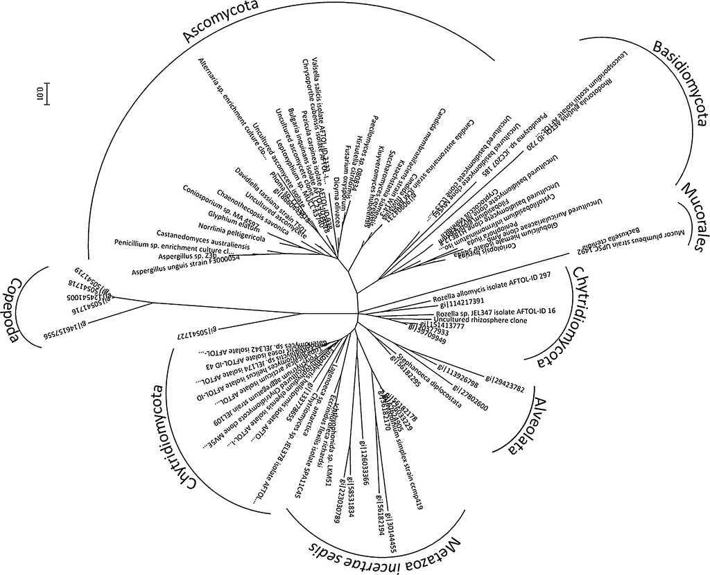
Figure 1 Reconstructed phylogram of reference sequences including supporting environmental sequences and other metazoa (Alveolata, Eccrinales, Ichtryosporeae and Choanoflagellida) indicating the taxonomic classification of the organisms. Sequences from Arthropoda and Cnidaria were added to root the phylogram.
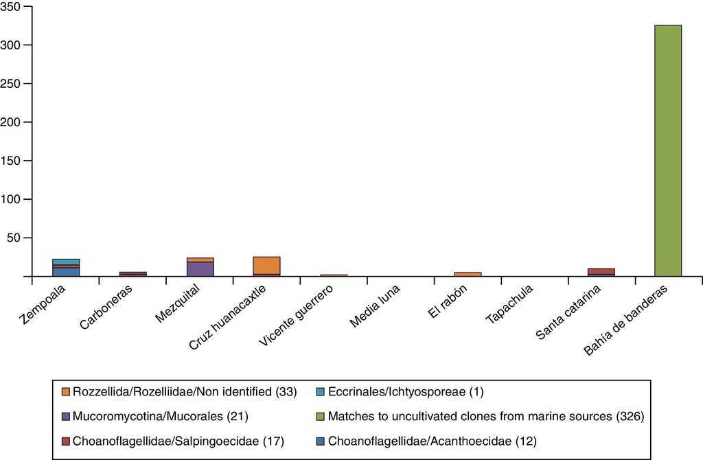
By the methodology described above, we were able to identify 529 of the environmental sequences at the family level, 1077 at the order level and 288 at the Class/Subphylum level. Only 326 sequences, all from the same site, did not group with any reference sequence but to uncultivated clones from marine sources (Table 3). The most abundant phylum found was Ascomycota (1458 sequences) (Fig. 2), followed by Chytridiomycota (133 sequences) (Fig. 3). Sequences belonging to phylum Basidiomycota (45 sequences) (Fig. 4) and to subphylum Mucoromycotina (21 sequences) were seldom recovered as well as non-fungal sequences belonging to Choanoflagellidae and Eccrinales (29 sequences) (Fig. 5).
Table 3 Identification of environmental sequences.
| Sampled sites | |||||||||||||||
|---|---|---|---|---|---|---|---|---|---|---|---|---|---|---|---|
| Phylum | Class/Subphylum | Order | Family | Genus | Zempoala | Carboneras | Mezquital | Cruz Huanacaxtle | Vicente Guerrero | Media Luna | El rabón | Zacapulco | Santa Catarina | Bahía Banderas | Total by genus |
| Saccharomycetes (363) | Saccharomycetales | Mitosporic Saccharomycetal | Candida | 63 | 6 | 69 | |||||||||
| Non identified | Non identified | 9 | 213 | 71 | 293 | ||||||||||
| Non identified | Non identified | Non identified | 1 | 1 | |||||||||||
| Non identified (260) | Non identified | Non identified | Non identified | 7 | 253 | 260 | |||||||||
| Leotiomycetes (3) | Heliotiales | Dermateaceae | Pezicula | 3 | 3 | ||||||||||
| Ascomycota (1279) | Eurotiomycetes (18) | Eurotiales | Trichocomaceae | Aspergillus | 1 | 1 | 5 | 7 | |||||||
| Chaetothyriales | Non identified | Non identified | 6 | 6 | |||||||||||
| Verrucarriales | Verrucariaceae | Norrlinia | 1 | 1 | |||||||||||
| Non identified | Non identified | Non identified | 4 | 4 | |||||||||||
| Sordariomycetes (630) | Hypocreales | Non identified | Non identified | 1 | 10 | 109 | 120 | ||||||||
| Diaporthales | Cryphonectriaceae | Chysoporthe | 8 | 1 | 9 | ||||||||||
| Hypocreales | Mitosporic Clavicipitaceae | Paecilomyces | 24 | 12 | 61 | 11 | 234 | 26 | 368 | ||||||
| Non identified | Non identified | 133 | 133 | ||||||||||||
| Euascomycetes (1) | Pleosporales | Pleosporaceae | Phoma | 1 | 1 | ||||||||||
| Dotyideomycetes (1) | Capnodiales | Davidiellaceae | Davidiella | 1 | 1 | ||||||||||
| Non identified (3) | Non identified | Non identified | Non identified | 2 | 1 | 3 | |||||||||
| Mucorales (27) | Non identified (27) | Non identified | Non identified | Non identified | 4 | 4 | 19 | 27 | |||||||
| Chytridiomycota (299) | Non identified | Non identified | Non identified | 3 | 3 | ||||||||||
| Non identified | Non identified | 35 | 35 | ||||||||||||
| Spizellomycetales | Spizellomyceteaceae | Tripalcalcar | 6 | 6 | |||||||||||
| Spizellomycetales incertae sedis | Rozella | 13 | 179 | 4 | 23 | 1 | 5 | 225 | |||||||
| Chytridiomycetes (299) | Chytridiales | Endochytriaceae | Non identified | 1 | 1 | ||||||||||
| Non identified | Non identified | 4 | 4 | ||||||||||||
| Chytridiaceae | Blyttiomyces | 10 | 10 | ||||||||||||
| Chytriomyces | 1 | 3 | 4 | ||||||||||||
| Cladochytriaceae | Polychytrium | 4 | 4 | ||||||||||||
| Rhizophydiales | Rhizophydiaceae | Rhizophydium | 4 | 2 | 1 | 7 | |||||||||
| Basidiomycota (40) | Tremellomycetes (9) | Filobasidiales | Non identified | Non identified | 3 | 3 | |||||||||
| Filobasidieaceae | Non identified | 2 | 1 | 3 | |||||||||||
| Cystofilobasidales | Cystofilobasidiaceae | Non identified | 3 | 3 | |||||||||||
| Agaromycetes (14) | Polyporales | Non identified | Non identified | 1 | 13 | 14 | |||||||||
| Pucciniomycotina (2) | Microbotrimycetes | Leucosporidiales | Leucosporidium | 2 | 2 | ||||||||||
| Atractielomycetes (2) | Soporidiobolales | Non identified | Non identified | 2 | 2 | ||||||||||
| Ustilagomycetes (2) | Non identified | Non identified | Non identified | 2 | 6 | 8 | |||||||||
| Non identified (5) | Non identified | Non identified | Non identified | 1 | 4 | 5 | |||||||||
| Marine clones (326) | Non identified (326) | Non identified | Non identified | Non identified | 326 | 326 | |||||||||
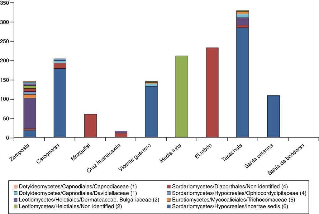
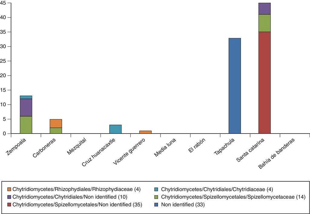
Figure 3 Chytridiomycota diversity in the sampled sites identified by Class (sub-phyllum)/order/family.
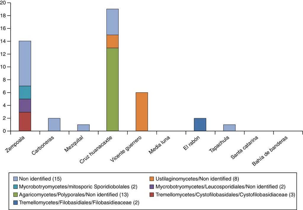
Figure 4 Basidiomycota diversity in the sampled sites identified by Class (sub-phyllum)/order/family.
Diversity estimation
Molecular Operational Taxonomical Units (MOTUS) diversity was estimated using the Shannon Index, which gives a measure of both species numbers and the evenness of their abundance as presented in Table 4 (Shannon, 1948). The value of the index ranges from low values (reduced species richness and evenness) to high values (extended species evenness and richness). In this work, the lowest diversity value was obtained for Media Luna and Bahía de Banderas, samples with a single MOTU identified in each (H′ = 0). In contrast, the highest diversity value was obtained for the Zempoala lagoon (H′ = 2.285), slightly higher than the total diversity by family (H′ = 2.130).
Table 4 Shannon diversity values calculated for the sampled site.
| Location | Environment | N per site | S per site | H′ per site |
|---|---|---|---|---|
| Zempoala | Freshwater temperate lagoon | 171 | 25 | 2.285 |
| Carboneras | Brackish coastal lagoon | 215 | 9 | 0.743 |
| Mezquital | Marine coastal lagoon | 85 | 4 | 0.769 |
| Cruz de Huanacaxtle | Pacific Ocean coastline | 64 | 8 | 1.749 |
| Vicente Guerrero | Dam | 153 | 7 | 0.583 |
| Media Luna | Brackish coastal lagoon | 213 | 1 | 0 |
| El rabón | Hypersaline coastal lagoon | 241 | 3 | 0.148 |
| Zacapulcp | Mangrove swamp | 361 | 8 | 0.906 |
| Santa Catarina | Artificial water reservoir | 164 | 6 | 1.003 |
| Bahía de Banderas | Pacific Ocean coastline | 326 | 1 | 0 |
| Total by family | 1,993 | 39 | 2.130 |
To determine if the different groups were randomly present in the various sample sites or if there was a predominance of certain groups in a given site, Friedman's test for the 2-way classification was performed. The entire population was distributed using the 10 different sites (blocks) and 18 taxonomic groups classified at the order level (treatments). Application of Friedman's procedure to the data resulted in a X2 value of 35.71 and 17 degrees of freedom.
Discussion
In aquatic environments organic matter decomposition occurs through a complex but well defined fungal succession (Barlocher & Kendrick, 1974; Gessner, Thomas, Jean-Louis, & Chauvet, 1993). The most common approach used for aquatic fungi diversity surveys involves the collection of organic material from natural sources such as plant debris or from artificial baits and the microscope-based estimation of species richness. Using this approach, richness depends on the ability of the species in the community to sporulate. Alternatively, molecular methods may be applied for the identification of fungal species, independently of their metabolic status or life cycle stage. Comparative studies performed on decaying leaves indicate that both approaches are complementary in the elucidation of population composition and dynamics (Nikolcheva, Cockshutt, & Barlocher, 2003).
Unfortunately, little attention has been paid to aquatic fungi, so it is difficult to compare our results with those of other groups. Available studies focus mainly on fungi adhered to organic matter (mainly wood), which limit the study to wood-decomposing fungi or to specific water bodies, mainly lakes or rivers. It is also true that most of these studies use traditional cultivation techniques to explore biodiversity, and this is also a limitation. Many of these studies refer only to certain groups of fungi (yeasts, for example).
Small subunit ribosomal DNA sequences (18S rDNA) have been used as molecular markers for reconstructing fungal taxonomy (Bruns et al., 1992; Hibbett et al., 2007; James et al., 2006) and for the description of fungal diversity in soils and water bodies (Anderson et al., 2003; Hunt et al., 2004; Monchy et al., 2011; Piquet et al., 2011). Small subunit rDNA sequences have been used to explore biological diversity and specialized software has been developed to discriminate among prokaryotic and eukaryotic sequences (Bengtsson et al., 2011). 18S rDNA sequences are still widely used to explore environmental samples. The set of primers used here was designed for the amplification of fungal ribosomal DNA, in particular an internal fragment of the 18S ribosomal particle gene between variable regions V4 and V8 (Borneman & Hartin, 2000). In the original paper these primers were unable to amplify DNA isolated from organisms other than fungi and operational specificity was demonstrated by the targeted amplification of fungal 18S ribosomal sequences from soil samples (Anderson et al., 2003).
Species designation of non-cultured individuals based on molecular markers presents the intrinsic weakness of lacking a statistically sound method. In some cases the identification is based on the overall similarity of query sequences to reference sequences in public databases in paired alignments using arbitrarily designated limits. In order to enforce the taxonomical robustness of our work, we adapted the identity interval rank concept originally devised for the classification of plant-nodulating bacterial species (Lloret et al., 2007; Martínez-Romero, Ormeño-Orrillo, Rogel, López-López, & Martínez-Romero, 2010).
Our approach is based on clustering of experimental sequences and a set of taxonomically classified reference sequences encompassing known fungal phyla and basal lineages. This gives enough information to reconstruct the taxonomic classification of the organisms. Sometimes clustering patterns were sensitive to the distribution of experimental sequences unless more reference sequences were included. In these cases, we identified clusters of experimental entries lacking reference sequences and used them to retrieve their best match in the databases. Inclusion of these new reference sequences in the alignments settled tree topology. Each cluster was taxonomically identified using the classification of the reference sequences located within, at different levels, from phylum to family.
The sequence variability of the 18S rDNA fragments supports the clustering reconstruction of our reference sequences assembled with taxonomic consistency. This includes the identification of a subgroup of Chytridiomycetes belonging to the Rozella genus, which is known to cluster separately (Hibbett et al., 2007). It is important to note that we were able to recover sequences from all fungal phyla, indicating little bias for the collection procedure or the amplification primers used. From the entire collection, 23.8% of the sequences could be identified at the family level, 48.5% at the order level and 13% at the class/subphylum level.
The important role of wood and leaf litter degradation has been ascribed to aquatic ascomycetes since basidiomycetes are scarce in water habitats and other organisms such as bacteria rarely have the ability to completely mineralize lignin (Simonis, Raja, & Shearer, 2008). Thus, it was not surprising that the Ascomycota was the most frequently recovered phylum and Sordariomycetes the most frequently recovered class. The occurrence of non-identified ascomycetes was documented in only 3 of the sampled sites supporting that idea that this phylum is the best characterized even in aquatic habitats. The second most abundant phylum described in aquatic environments is Chytridiomycota and, accordingly, it was the second most frequently found group for our sequence with 41% of the sequences identified at the family level.
The large number of clones analyzed allowed us to assess diversity using the Shannon index obtaining values ranging from 0 to 2.363. With a single exception, these values are below those described for soil fungal populations (1.87-2.82) (Klaubauf et al., 2010), suggesting that aquatic fungi are less diverse than their soil counterparts. The only site with high diversity was Zempoala, a pristine environment located in a temperate zone. The less diverse sites were Media Luna and Bahía de Banderas, both coastal sites with high human impact. The combination of organisms identified was specific for each site as demonstrated by the direct application of the Friedman test for the 2-way classification. This result emphasizes organism specialization and therefore the need for regular evaluation of the local biodiversity in aquatic environments.

