


Serviços Personalizados
Journal
Artigo
 Inglês (pdf)
 Artigo em XML
 Referências do artigo
 Enviar este artigo por email
Indicadores
-
 Citado por SciELO
 -
 Acessos
Links relacionados
-
 Similares em
SciELO
Similares em
SciELO
Compartilhar

 Permalink
Revista mexicana de biodiversidad
versão On-line ISSN 2007-8706versão impressa ISSN 1870-3453
Rev. Mex. Biodiv. vol.84 no.4 México Dez. 2013
https://doi.org/10.7550/rmb.32149
Anatomía
Sclerophylly in mangrove tree species from South Brazil
Esclerofilia de las especies de manglares del sur de Brasil
Carolina Sereneski-de Lima1, María Regina Torres-Boeger2*, Letícia Larcher-de Carvalho2, Andressa Pelozzo1 and Patricia Soffiatti1
1 Universidade Federal do Paraná, Programa de Pós Graduação em Botânica, Departamento de Botânica, Caixa Postal 19031, 81531-990 Curitiba, PR, Brasil.
2 Universidade Federal do Paraná, Programa de Pós-Graduação em Ecologia e Conservação, Setor de Ciências Biológicas, Caixa Postal 19031, 81531-990 Curitiba, PR, Brasil. *rboeger@ufpr.br
Recibido: 17 junio 2012
Aaceptado: 27 junio 2013
Abstract
Sclerophylly, a morphological trait that defines coriaceous and hard leaves, is presently accepted as a non-specific response to environments with acting multiple stresses. In mangroves, features such as flooded and unconsolidated soil, low availability of oxygen, and high salinity characterize this stressful environment. From 2 mangroves areas in the coast of Paraná state, leaves of 3 species (Rhizophora mangle, Laguncularia racemosa and Avicennia schaueriana) were collected and analyzed nutritionally and morphologically. Sclerophylly indices (Rizzini index and specific leaf area) indicated that all species are sclerophyllous. Considering nutritional and morphological traits, only some of them suggest sclerophylly, such as total leaf thickness in all species, the presence of a sub-epidermal layer in Rhizophora mangle and Avicennia schaueriana and sclereids in Rhizophora mangle. Comparatively, leaves presented different degrees of sclerophylly, in the following order: R. mangle > L. racemosa > A. schaueriana, considering all characteristics analyzed. This gradient of sclerophylly appears to be consequence of different strategies developed by each species in response to the stressful abiotic conditions of mangroves, especially the mechanisms for salinity tolerance.
Key words: leaf morphology, nutrient concentration, salinity, sclerophylly index, specific leaf area.
Resumen
La esclerofilia es un rasgo morfológico que define hojas coriáceas y duras. Actualmente se acepta como una respuesta no específica a los ambientes con múltiples factores estresantes. Los manglares se consideran un ambiente estresante debido a sus suelos inundados y no consolidados, una baja disponibilidad de oxígeno y la alta salinidad. En 2 áreas de manglar en la costa del estado de Paraná, se recolectaron hojas de 3 especies de mangle (Rhizophora mangle, Laguncularia racemosa y Avicennia schaueriana) y se realizaron análisis nutricionales y morfológicas. Los índices de esclerofilia (índice de Rizzini y área específica foliar) indicaron que todas las especies son esclerófilas. Considerando los rasgos nutricionales y morfológicos, sólo unos pocos indican esclerofilia, como la espesura total de la hoja para todas las especies, la presencia de un estrato subepidérmico en Rhizophora mangle y en Avicennia schaueriana, y esclereidas en Rhizophora mangle. Comparando todos los caracteres analizados, las hojas presentaron diferente grados de esclerofilia, en el siguiente orden: R. mangle > L. racemosa > A. schaueriana. Este gradiente de esclerofilia parece ser consecuencia de las diferentes estrategias desarrolladas por cada especie en respuesta a las condiciones abióticas estresantes de los manglares, especialmente, como mecanismos para tolerar la salinidad.
Palabras clave: morfología foliar, concentración de nutrientes, salinidad, índice de esclerofilia, área específica foliar.
Introduction
Sclerophylly was initially defined by Schimper (1903) to distinguish xeromorphic plants with leathery leaves of the Mediterranean region from those exhibiting succulence or leaflessness (Edwards et al., 2000). Later, sclerophylly was interpreted as a morpho-physiological adaptation of plants as a response to low soil fertility of tropical forests, mainly nitrogen and phosphorous (Loveless, 1962), and as a defense against herbivory (Choong et al., 1992).
Nowadays, sclerophylly is accepted as a non-specific response to a wide range of environmental stresses (Read et al., 2006). It can be adaptive, enhancing leaf longevity and photosynthetic efficiency across a range of stressful environments (Turner, 1994), or non adaptive, resulting from the combination of morphological and physiological traits that result in more stress resistant leaves (Read et al., 2006).
Leathery leaves characterize sclerophylly and are a consequence of the presence of mechanical tissues (Shimper, 1903). Therefore, sclerophylly can be measured by indices that utilize leaf dry mass and leaf area. Specific leaf area (SLA), which represents the investment of photosynthetic mass by area unit, has been extensively used as a sclerophylly index (Camerik and Werger, 1981; Marin and Medina, 1981; Bongers and Popma, 1990; Witkowski and Lamont, 1991; Perez, 1994; Groom and Lamont, 1999; Boeger and Wisniewski, 2003), due to the ease of data collection (Witkowski and Lamont, 1991). Besides that, SLA is a measure of relative growth of species (Vendramini et al., 2002). Leaves with low mean values of SLA have more mechanical tissues, making leaves more leathery and hard, with lower growth rates. Another sclerophylly index is the Rizzini index (Rizzini, 1976), that relates leaf dry mass and leaf surface and classifies leaves as sclerophyllous when those values are higher than 0.6.
Although leaf hardness of sclerophyllous plants can be determined mainly by the amount of mechanical tissue, the combination of morpho-anatomical traits such as leaf thickness, thick cuticle and epidermis, presence of hypodermis, palisade/spongy parenchyma ratio > 1, compacted mesophyll and high crude fibers/crude proteins ratio can also generate rigidity in leaves (Turner, 1994; Read and Sanson, 2003). According to Turner (1994), the degree of sclerophylly can be evaluated by the morphological characteristics mentioned above, besides the values of AEF and foliar Nitrogen and Phosphorous concentrations.
Mangroves are located in protected coastal areas, in the tropics and sub-tropics (Shaeffer-Novelli, 1995), and are characterized by muddy soils rich in nutrients. Mangrove forests are constantly submitted to limiting environmental conditions such as high salinity, unconsolidated substrate, low oxygenation and frequent submersion by tides (Schaeffer-Novelli et al., 2000; Paraguassu and Silva, 2007).
Although mangrove trees present thick and leathery leaves, mangroves are not classified as a sclerophyllous environment. The few studies about sclerophylly in mangroves evaluated species separately and only in experimental conditions of different salinities and nutrient concentrations (Feller, 1996; Sobrado, 2005). Species such as Rhizophora mangle are classified as sclerophyllous only in high salinity concentration (Feller, 1996). However, under uncontrolled conditions, the occurrence of sclerophylly in mangrove species can be considered a strategy of the plants to enhance the efficiency of water use (Naidoo, 2010).
This study evaluated if the 3 most representative tree species from South Brazilian mangroves are sclerophyllous according to a) sclerophyllous indices (SLA and Rizzini index), b) leaf Nitrogen and Phosphorous concentrations, and c) presence of morphological traits that indicate sclerophylly (sensu Turner, 1994). Our hypothesis is that all studied mangrove species are sclerophyllous, caused by stressful environmental conditions, such as high salinity.
Materials and methods
This study was conducted in the Antonina Bay, Paraná, Brazil (25°29'57" S, 48°42'44" W). The Antonina Bay is a large water body, with irregular shape, that occupies 3 882 km2 (Fávaro et al., 2007). The climate is classified as Cfa type, according to Koppen's classification, i.e. subtropical, mesothermic, with hot summers, without a dry season. The average temperature is 20.5° C, with 16.7° C and 26.4° C minimum and maximum values, respectively. The annual precipitation is 2 773 mm. The soil type is Histosol thiomorphic sapric salic/sodic (EMBRAPA, 2009). Measurements of salinity of the interstitial water varied from 13.2 to 19% and the potential redox varied from -294.8 to -347.8 mV.
In the studied area, 3 tree species were identified: Rhizophora mangle L. (Rhizophoraceae), Avicennia schaueriana Stapf and Leachman (Acanthaceae) and Laguncularia racemosa (L.) Gaertn (Combretaceae). Fifteen individuals of each species were selected, each at least, 8 m high. From each individual, 30 totally expanded and mature leaves were collected between the third and the sixth nodes, from 5 to 7 branches directly exposed to sunlight. Senescent and juvenile leaves were discarded due the potential presence of chlorose or injuries, or to avoid using leaves not fully developed. Of a total of 450 leaves collected for each species, 150 were used for analyses of morphology, 30 were used for anatomy, and 270 for chemistry.
For each leaf, we measured the following parameters: 1) leaf thickness of the median region of the lamina, with a digital caliper; 2) dry mass, estimated with a digital analytical balance, from previously dehydrated leaves; 3) leaf area (cm2), measured from images obtained with a flatbed scanner calibrated with Sigma Scan PRO software (version 5.0, SPSS Inc., Chicago, IL, USA), and 4) specific leaf area (SLA)= leaf area (cm2)/leaf dry mass (g) and sclerophylly index (IE)= leaf dry mass (g)/2 x leaf area (dm2), according to Rizzini (1976). This latter index defines sclerophylly as IE> 0.6 and mesophylly as IE< 0.6. Leaf density (DF, g.cm-3) was estimated by the following equation: specific leaf mass (leaf dry mass (g)/leaf area (cm2))*1/thickness.
Salt secretion gland density (mm2) was estimated from clear nail polish imprints from the median region of the epidermal surface of leaves, for 30 leaves, using a light microscope coupled with a camera lucida.
For anatomical analysis, leaves were fixed in FAA 70 and conserved in ethanol 70%. Previously fixed plant material was sectioned transversally on the median region of the leaf laminas, with a razor blade then cleared with sodium hypochlorite 10%, stained with Toluidine Blue 1% and mounted in glycerin. In the transverse sections, adaxial and abaxial epidermis, sub-epidermal layers, palisade and spongy parenchyma thickness and total thickness were measured. The ratio palisade/spongy parenchyma was calculated. All
The nitric-perchloric digestion (Martins and Reissmann, 2007) was used for phosphorus (P); potassium (K) and sodium (Na) analysis. For nitrogen (N), the determination was made by the Kjeldahl method (Souza 1999). All elements determinations were made by coupled plasma optical emission spectrometry (OES) with argon source.
For every quantitative variable, mean and respective standard deviations were calculated. To test for morphological differences among species, we used Anova, with 5% significance. Post-hoc Tukey's test was performed to test for differences between pairs of means, using the software Statistica version 7.0, (Statsoft, Inc., Tulsa, OK, USA). Also, we tested the correlation (Pearson's Correlation) with morphological traits and leaf nutrients (nitrogen, phosphorus, potassium and sodium). In all cases, we tested the homogeneity of the variances using the test of Levene (Zar 1999), when necessary, data were log-transformed.
Results
All 3 species studied presented quantitative differences related to leaf morphology. The leaves of Rhizophora mangle showed the higher values of leaf area and leaf dry mass and lower values for total thickness of the lamina, when compared to Avicennia schaueriana and Laguncularia racemosa (Table 1).
Avicennia schaueriana presented lower mean values of leaf area, dry mass and total leaf thickness. However, this species presented the highest mean values of specific leaf area (Table 1), followed by R. mangle and L. racemosa. Considering the sclerophylly index (sensu Rizzini), the highest mean values were observed in L. racemosa, followed by R. mangle and A. schaueriana. In spite of the significant differences among species, all of them were classified as sclerophyllous (Table 1). Leaf density was the only variable similar among all 3 species (Table 1).
Avicennia schaueriana presented the highest density of salt glands /mm2, in both epidermis surfaces, followed by L. racemosa and R. mangle (Table 1). Avicennia schaueriana showed the highest density of salt glands on the adaxial surface, while L. racemosa presented higher density on the abaxial surface. Rhizophora mangle presented salt secretion structures only on the abaxial surface.
The leaf lamina of the 3 studied species has a uniseriate epidermis, covered by the cuticle (Figs. 1a, c, e). The leaf is hypostomatic in A. schaueriana (Fig. 1a) and R. mangle (Fig. 1c) and amphistomatic in L. racemosa (Fig. 1e). Salt glands (Fig. 1d) occur on both leaf surfaces in A. schaueriana and L. racemosa and on the adaxial surface of R. mangle. In A. schaueriana, besides salt glands, glandular trichomes are present on the abaxial surface (Fig. 1b) in large quantities widely covering the surface forming an indumentum.
Internally to the epidermis, a sub-epidermal layer occurs in A. schaueriana leaves, formed by 3 or 4 layers of cells on the adaxial surface (Fig. 1a). In R. mangle, the sub-epidermal layer is composed by 5 to 7 layers (Fig. 1e).
The mesophyll is heterogeneous in all species. The palisade parenchyma presented 3 to 5 layers in A. schaueriana (Fig. 1a), 2 to 3 in L. racemosa (Fig. 1c), and 1 to 2 layers in R. mangle (Fig. 1e). The spongy parenchyma varied between 6 to 8 layers in A. schaueriana (Fig. 1a), 12 to 14 in L. racemosa (Fig. 1c), and 9 to 10 layers in R. mangle (Fig. 1e). Collateral vascular bundles are scattered in the mesophyll. In R. mangle, H-shaped sclereids were observed immersed in the mesophyll (Fig. 1f).
All 3 species showed significantly distinct values of nutrient concentrations (Table 2). Avicennia schaueriana showed higher concentrations of N, K, P and Na, when compared to L. racemosa and R. mangle. Lower concentrations of N, K and Na were found in L. racemosa. The mean values of nutrient concentration followed this order: A. schaueriana: N>K>Mg>Na>Ca>P; L. racemosa: Ca>N>K>Na>Mg>P and R. mangle: N>Ca>K>Na>Mg>P (Table 2). In the 3 species studied, P presented the lower concentration related to other nutrients, while Na maintained higher values. Other nutrients varied in concentration values among the 3 species.
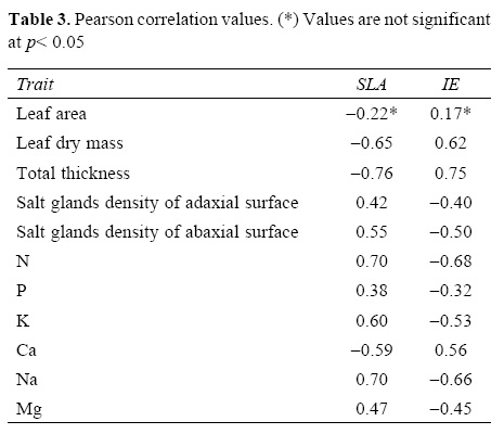
Specific leaf area was directly correlated with N, K and Na concentration and inversely correlated with leaf dry mass and total leaf thickness (Table 3). The sclerophylly index (sensu Rizzini) showed the same correlations but inversely to SLA, since this index (leaf dry mass/leaf area) is proportionally inverse to SLA (leaf area/ leaf dry mass) (Table 3).
Discussion
The leaves from the 3 species studied are sclerophyllous according to both indices used, although these values were significantly different between species. Laguncularia racemosa was the most sclerophyllous species among them. Values similar to those observed in the present study for L. racemosa were reported for: 1) two perennial species from the Venezuelan savannas (Montes and Medina, 1977); 2) species from the upper region of Rio Negro, Amazonia (Medina et al., 1990); 3) tree species of initial stages of succession in the coastal forest of South Brazil (Boeger and Wisniewski, 2003), and 4) species of a very dry Venezuelan forest (Marin and Medina, 1981). All these forest types are considered sclerophyllous. Lower values of SLA were found for A. germinans in different mangroves in the Maranhäo state (Goncalves-Alvim et al., 2001), compared to the values obtained in this study.
The studies that have used SLA as the sclerophylly index related this condition with different types of stress such as low soil fertility and water deficit (Sobrado and Medina, 1980; Medina et al., 1990; Boeger and Wisniewski, 2003) and high light intensities (Groom and Lamont, 1997; Mendes et al., 2001). The low values of SLA suggest that leaves invest more in dry mass per area unit, especially in mechanical tissues. According to Wilson et al. (1999), leaves with low values of SLA occur in environments with low availability of resources, where the retention of these resources is of high priority.
One of the difficulties in the use of SLA as an index of sclerophylly is the lack of parameters for comparison. Although many studies show that low values of SLA indicate sclerophylly, there are no values establishing the limit between sclerophylly and mesophylly, such as those defined for the Rizzini index. Furthermore, the use of SLA as an index of sclerophylly has been criticized since it ignores the fact that plant tissues are made of distinct components that generate distinct mechanical properties, which interfere with the calculation of SLA (Edwards et al., 2000). Sclerophylly could be a result of different processes such as lignification, cutinization, and/or silification, which can occur isolated or combined, influencing the SLA values (Beadle, 1966; Balsamo et al., 2003).
SLA is strongly and inversely correlated with total leaf thickness (Table 3), indicating that small differences on leaf thickness can influence SLA values (Witkoswski and Lamont, 1991). This is evident when the mean values of leaf density of the studied species were compared with each other, because the leaf density equation takes leaf thickness into consideration. On the other hand, while SLA is different among species, the leaf density average is similar among them (Table 2).
The leaf thickness of the studied species results mainly from the combination of several layers of sub-epidermal and clorophyll parenchyma cells. Both sub-epidermical layers and spongy parenchyma in L. racemosa are formed by large cells, which probably perform a water storage function.
Regarding the morphological traits used as sclerophylly indicators (sensu Turner, 1994), the studied species presented only some of the characteristics (Table 4): thick leaves (> 450 urn) in all species, presence of a hypodermis in R. mangle and A. schaueriana and sclereids in R. mangle. Considering this set of traits, only A. schaueriana and R. mangle can be considered sclerophyllous (Table 4), while L. racemosa can be considered sclerophyllous only according to the sclerophylly indices (SLA and Rizzini).
The mean concentrations of nutrients were either higher or similar to values of other mangrove species (Cuzzuol and Campos, 2001; Bernini et al., 2010). The N and P mean values obtained in this study were higher than values for sclerophyllous leaves (Montes and Medina, 1977; Medina et al., 1990), indicating that the sclerophylly observed in our study is not determined by soil oligotrophy. Soil N and P appear not to be limited in the mangrove system.
One of the few studies that considered R. mangle as sclerophyllous is Feller's study (1996) of dwarf trees of R. mangle from Twin Cays (Belize), due to soil oligotrophy caused by P deficiency. In that study, experiments with soil P enrichment demonstrated a reduction on the degree of sclerophylly of the species, through the reduction of hypodermis thickness and, consequently, reduction of the lamina thickness, corroborating with the sclerophylly hypothesis by P soil deficiency (Loveless, 1962; Feller, 1996). However, leaves from dwarf R. mangle trees that were 1100 urn thick were considered sclerophyllous, while leaves 600 μm thick were not (Feller, 1996). The leaves of the dwarf red mangrove trees are thicker than the leaves from trees evaluated in the present study (450 μm).
The mean concentration of leaf Na of the studied species was similar to mangrove species from Rio de Janeiro state (Bernini et al., 2010). This concentration can be considered within the expected limits (0.5 to 30 g.kg-1; Finck, 1969), and is probably due to the presence of several adaptations of these species to high salinity through the dilution of Na in water storage tissue, salt secretion, and excretion mechanisms, as well as the low values of salinity of interstitial water, when compared to other mangroves (Feller, 1996; Naidoo, 2010).
Associated to water storage tissues, all species presented salt glands, with variations in density and position on the lamina surface. This variation of salt glands among species appears to be influenced by the degree of tolerance of each species to salinity and by the different mechanisms of elimination of Na (Tomlinson, 1986; Parida and Jha, 2010). According to different authors, species of Laguncularia are salt secretors, Rhizophora species are salt excluders and salt accumulators, andAvicennia species are concomitantly secretors, "excluders" and accumulators (Parida and Jha, 2010).
Although SLA and the Rizzini index indicated that the leaves studied are sclerophyllous, the morphological and nutritional traits did not. The only leaf trait that indicates sclerophylly for all species is leaf thickness, due to the presence of several layers of parenchyma tissue or the combination of palisade parenchyma and sub-epidermal tissue (hypodermis).
In this study, we were able to classify leaves according to different degrees of sclerophylly, considering all analyzed characteristics, in the following decreasing order: R. mangle > L. racemosa > A. schaueriana. This sclerophyllous gradient appears to be a consequence of the different strategies these mangrove species developed to survive in an environment with multiple stresses, especially mechanisms for salt tolerance.
However, it is important to point out that the evaluated morphological traits, such as leaf thickness and dry mass, are highly plastic and depend on environmental conditions, which may result in populations with distinct levels of sclerophylly depending on the site conditions. Our results showed that the sclerophylly classification should not be based solely on sclerophylly indices, but must also consider other morphological and nutritional traits. Sclerophylly is a complex mechanism involving several leaf features that interact on different scales, reflecting multiple effects.
Acknowledgements
To Fundação Araucária and Petrobrás-BR for financial support; to the Brazilian Project for Federal Universities (REUNI) for a scholarship to the first author; Ellie Anne López Barrera and Guillermo Ángeles for Spanish version of the abstract and the Brazilian Research Council (CNPq) for providing fellowship to MRT Boeger (process No. 309386/2007-1).
Literature cited
Balsamo, R. A., A. M. Bauer, S. D. Davis and B. M. Rice. 2003. Leaf biomechanics, morphology, and anatomy of the deciduous mesophyte Prunus serrulata (Rosaceae) and the evergreen sclerophyllous shrub Heteromeles arbutifolia (Rosaceae). American Journal of Botany 90:72-77. [ Links ]
Beadle, N. C. W. 1966. Soil phosphate and its role in molding segments of the Autralian flora and vegetation, with special reference to xeromorphy and sclerophylly. Ecology 47:992-1007. [ Links ]
Bernini, E., M. A. B. Silva, T. M. S. Carmo and G. R. F. Cuzzuol. 2010. Spatial and temporal variation of the nutrients in the sediment and leaves of two Brazilian mangrove species and their role in the retention of environmental heavy metals. Brazilian Journal of Plant Physiology 22:177-187. [ Links ]
Boeger, M. R. T. and C. Wisniewski. 2003. Comparação da morfologia foliar de espécies arbóreas de très estágios sucessionais distintos de floresta ombrófila densa (Floresta Atlàntica) no Sul do Brasil. Revista Brasileira de Botánica 26:61-72. [ Links ]
Bongers, F. J. and J. Popma. 1990. Leaf characteristics of the tropical rain forest flora of Los Tuxtlas, Mexico. Botanical Gazette 151:354-365. [ Links ]
Camerik, A. M. and M. J. A. Werger. 1981. Leaf characteristics of the flora of the high plateau of Itatiaia, Brazil. Biotropica 13:39-48. [ Links ]
Choong, M. F., P. W. Lucas, J. S. Y. Ong, B. Pereira, H. T. W. Tan and I. M. Turner. 1992. Leaf fracture toughness and sclerophylly: their correlations and ecological implications. New Phytologist 121:597-610. [ Links ]
Cuzzuol, G. R. F. and A. Campos. 2001. Aspectos nutricionais na vegetação de manguezal do estuário do Rio Mucuri, Bahia, Brasil. Revista Brasileira de Botánica 24:227-234. [ Links ]
Edwards, C., J. Read and G. Sanson. 2000. Characterizing sclerophylly: some mechanical properties of leaves from heath and forest. Oecologia 123:158-167. [ Links ]
Embrapa. 2009. Sistema Brasileiro de Classificação de Solos. Brasilia: Embrapa-SPI; Embrapa-Solos. Rio de Janeiro. 306 p. [ Links ]
Fávaro, L. F., E. C. Oliveira and N. F. Verani. 2007. Estrutura da população e aspectos reprodutivos do peixe-rei Atherinella brasiliensis (Quoy and Gaimard) (Atheriniformes, Atherinopsidae) em áreas rasas do complexo estuarino de Paranaguá, Paraná, Brasil. Revista Brasileira de Zoologia 24:1150-1156. [ Links ]
Feller, I. C. 1996. Effects of nutrient enrichment on leaf anatomy of dwarf Rhizophora mangle L. (red mangrove). Biotropica 28:13-22. [ Links ]
Finck, A. 1969. Pflanzen-ernáhrung in sticaworten. Kiel: Verlag Ferdinand Hirt. Goncalves-Alvim, S. J., M. C. F. Santos and G. W. Fernandes. 2001. Leaf gall abundance on Avicennia germinans (Avicenniaceae) along an interstitial salinity gradient. Biotropica 33:69-77. [ Links ]
Groom, P. K. and B. B. Lamont. 1997. Xerophytic implications of increased sclerophylly: interactions with water and light in Hakea psilorrhyncha seedlings. New Phytologist136: 231-237. [ Links ]
Groom, P. K. and B. B. Lamont. 1999. Which common indices of sclerophylly best reflect differences in leaf structure? Ecoscience 6:471-474. [ Links ]
Loveless, A. R. 1962. Further evidences to support a nutritional interpretation of sclerophylly. Annals of Botany 26:549-561. [ Links ]
Marin, D. and E. Medina. 1981. Duración foliar, contenido de nutrientes y esclerofilia en árboles de un bosque muy seco tropical. Acta Científica Venezoelana 32:508-514. [ Links ]
Martins, A. P. L. and C. B. Reissmann. 2007. Material Vegetal e as Rotinas Laboratoriais nos Procedimientos Químico-Analíticos. Scientia Agraria 8:1-17. [ Links ]
Mendes, M. M., L. C. Gazarini and M. L. Rodrigues. 2001. Acclimation of Myrtus communis to contrasting Mediterranean light environments - effects on structure and chemical composition of foliage and plant water relations. Environmental and Experimental Botany 45:165-178. [ Links ]
Medina, E., V. Garcia and E. Cuevas. 1990. Sclerophylly and oligotrophic environments: relationships between leaf structure, mineral nutrient content, and drought resistance in tropical rainforests of the upper Rio Negro region. Biotropica 22:51-64. [ Links ]
Montes, R. and E. Medina. 1977. Seasonal changes in nutrient content of leaves of savanna trees with different ecological behavior. Geografia y Ecologia Tropical 4:295-307. [ Links ]
Naidoo, G. 2010. Ecophysiological differences between fringe and dwarf Avicennia marina mangroves. Trees 24:667-673. [ Links ]
Paraguassu, L. A. A. and M. N. Silva. 2007. Caracterização fitossociológica do manguezal de Porto de Sauípe, Entre Rios, Bahia. Diálogos and Ciencia 12:1-11. [ Links ]
Parida, A. K. and B. Jha. 2010. Salt tolerance mechanisms in mangroves: a review. Trees 24:199-217. [ Links ]
Perez, C. 1994. Indexes of sclerophylly in relation to the chemical-quality of litter and to the potential mineralization of nitrogen in the surface soils of Olivillo (Aextoxicon-puntactum R-ET-PAV) forests in Chile. Revista Chilena de Historia Natural 67:101-109. [ Links ]
Read, J. and G. D. Sanson. 2003. Caracterizing sclerophylly: the mechanical properties of a diverse range of leaf types. The Phytologist 160:81-99. [ Links ]
Read, J., G. D. Sanson, M. Garine-Wichatitsky and T. Jaffré. 2006. Sclerophylly in two contrasting tropical environments: low nutrients vs. low rainfall. American Journal of Botany 93:1601-1604. [ Links ]
Rizzini, C. T. 1976. Tratado de Fitogeografia do Brasil. USP, São Paulo. 317 p. [ Links ]
Schaeffer-Novelli, Y. 1995. Manguezal: ecossistema entre a terra e o mar. São Paulo, Caribbean Ecological Research. 150 p. [ Links ]
Schaeffer-Novelli, Y., G. Cintrón-Molero, M. L. G. Soares and T. De-Rosa. 2000. Brazilian mangroves. Aquatic Ecosystem Health and Management 3:561-570. [ Links ]
Schimper, A. F. W. 1903. Plant-geography upon a physiological basis. Clarendon Press, Oxford. 159 p. [ Links ]
Sobrado, M. A. 2005. Leaf characteristics and gas exchange of the mangrove Laguncularia racemosa as affected by salinity. Photosynthetica 43:217-221. [ Links ]
Sobrado, M. A. and E. Medina. 1980. General morphology, anatomical structure and nutrient content of sclerophyllous leaves of the 'Bana' vegetation of Amazonas. Oecologia 45:341-345. [ Links ]
Silva, F. C. 1999. Manual de análises químicas de solos, plantas e fertilizantes. Embrapa Solos, Brasilia. 370 p. [ Links ]
Tomlinson, P. B. (ed.). 1986. Family: Avicenniaceae. In The Botany of Mangroves, Cambridge University Press, Cambridge. p. 186-207. [ Links ]
Turner, I. M. 1994. A quantitative analysis of leaf form in woody plants from the world's major broad leaved forest types. Journal of Biogeography 21:413-419. [ Links ]
Vendramini, F., S. Díaz, D. E. Gurvich, P. J. Wilson, K. Thompson and J. G. Hodgson. 2002. Leaf traits as indicators of resource-use strategy in floras with succulent species. New Phytologist 154:147-157. [ Links ]
Wilson, P. J., K. Thompson, and J. G. Hodgson. 1999. Specific leaf area and dry matter content as alternative predictors of plant strategies. New Phytologist 143:155-162 [ Links ]
Witkowski, E. T. F. and B. B. Lamont. 1991. Leaf specific mass confounds leaf density and thickness. Oecologia 88: 86-493. [ Links ]
Zar, J. H. 1999. Biostatistical analysis. Prentice-Hall, New Jersey. 663 p. [ Links ]

