


Servicios Personalizados
Revista
Articulo
 texto en
  Inglés (pdf)
 Artículo en XML
 Referencias del artículo
 Enviar artículo por email
Indicadores
-
 Citado por SciELO
 -
 Accesos
Links relacionados
-
 Similares en
SciELO
Similares en
SciELO
Compartir

 Permalink
Revista odontológica mexicana
versión impresa ISSN 1870-199X
Rev. Odont. Mex vol.22 no.1 Ciudad de México ene./mar. 2018
Clinical cases
Dental handling of patients with Christ-Siemens-Touraine syndrome. Case report
*DDs, Degree in Biochemistry. University of Cartagena Zaragocilla Campus, Cartagena-Colombia, School of Dentistry, Oral Medicine and Surgery Department.
§DDs University of Cartagena. University of Cartagena Zaragocilla Campus, Cartagena-Colombia, School of Dentistry, Oral Medicine and Surgery Department.
IIDDs. Colombia Periodontics and Implantology Specialist, Colegio Odontológico (Dental School) Colombia.
Ectodermal dysplasia (ED) encompasses a group of hereditary disorders in which two or more ectoderm-derived structures are affected. Patients afflicted with this disorder exhibit hypoplasia or aplasia of different structures such as skin, hair, nails, teeth, and sweat glands among others. Dental treatment is of the utmost importance in order to improve the subject’s quality of life. The case here reported depicts a male patient affected with Christ-Siemens- Touraine syndrome, who sought treatment due to tooth anodontia in upper and lower jaws. A dental approach was conducted involving periodontal treatment, oral rehabilitation and implantology, a social component was also furthered, directed to re-establish function and esthetics of the stomatognatic system. Performed treatment achieved absolute improvement in the masticatory process and esthetic smile which was satisfactory for the patient and his legal representative.
Key words: Ectodermal dysplasia; anhidrotic ectodermal dysplasia; anodontia; mouth rehabilitation (MeSh)
La displasia ectodérmica (DE) comprende un grupo de trastornos hereditarios en los que dos o más estructuras derivadas del ectodermo se encuentran afectadas. Los pacientes con este trastorno presentan hipoplasia o aplasia de estructuras como la piel, el cabello, uñas, dientes, glándulas sudoríparas y otras estructuras. El manejo odontológico es fundamental para mejorar la calidad de vida del individuo. Se reporta el caso de un paciente masculino con síndrome Christ-Siemens-Touraine, quien asistió a consulta por anodoncia de órganos dentarios en maxilar superior e inferior, se realizó un abordaje odontológico que involucró periodoncia, rehabilitación oral e implantología y acompañamiento social, dirigido a restablecer funcionalidad y estética del sistema estomatognático. Con el tratamiento realizado se obtuvo mejoría absoluta en el proceso de masticación y una sonrisa estética satisfactoria para el paciente y su representante legal.
Palabras clave: Displasia ectodérmica; displasia ectodérmica anhidrótica; anodoncia; rehabilitación bucal (DeSC)
Introduction
Ectodermal dysplasia (ED) comprises a large and heterogeneous group of non-progressive, hereditary disorders; it was first reported by Thurnham in 1848, who offered for the first time a report of a patient with ectodermal dysplasia.1 Jannane defined ectodermal dysplasia as a set of defects caused by development alterations of two or more tissues from the embryonic ectoderm. Affected tissues are mainly skin, hair, nails, eclectic glands and teeth.2
To this date, over 192 disorders have been reported associated to gene mutations which codify for protein involved in the development of ectoderm. It is well known that these mutations alter tumor necrosis factor during critical periods in the development of ectoderm and its appendages, thus triggering dysplasia.3,4 Ectodermal dysplasia’s hereditary pattern is variable and in most cases linked to recessive autosomal X chromosome, thus, it can be considered a relatively rare disorder affecting approximately 1 per 10,000 to 1 per 100,000 of live births.1
Two different main types of ectodermal dysplasia with clinical and histological differences have been described: hydroplastic ectodermal dysplasia, or Clouston syndrome would represent group 1 and anhidrotic ectodermal dysplasia or Christ Siemens syndrome which would represent group 2. Differences between both groups lie in the amount of sweating experienced by the patient.1 The most frequently found variety is hypohydrotic or anhidrotic ectodermal dysplasia; this form possesses a recessive autosomal hereditary pattern linked to chromosome X (DEHLX), this type of ED is mainly characterized by a clinical picture based on a hypotrichosis triad (anomalies in skin, hair and nails), hypodontia or anodontia and hypohydrosis (partial or total absence of eclectic sweat glands). Unlike this latter, hydroplastic ectodermal dysplasia is inherited with a dominant autosomal pattern; it exhibits hypotrichosis, nail dystrophy and hyperkeratosis of palms of the hands and soles of the feet.5-7
Both varieties exhibit common characteristics such as thin, slightly pigmented skin, with an almost translucent appearance, with easily detected superficial blood vessels.6,8 Other manifestations include decrease in eyebrow density, hair that is scarce, brittle, dry and thin, as a result of poorly developed sebaceous glands. Likewise, other traits are characteristic of this condition: bulging forehead, saddle-shaped nose, as well as protuberant and everted lips. It has been reported that patients affected by ED frequently exhibit eye infections, chronic rhinitis, dystrophic nails, epistaxis, dysphagia and dysphonia.3,9
As far as stomatological characteristics, the following can be counted: alveolar crest under-development as a result of lack of formation of the tooth bud, a decreased occlusal vertical dimension and depressed nose bridge, therefore, these subjects exhibited older age appearance. Number of teeth is noticeably decreased (oligodontia and hypodontia), frequently, an abnormal shape development is observed (cone or peg shaped incisors, microdontia), generally, teeth are widely spaced.1,3,6
After mentioning stomatological characteristics, patients afflicted with ectodermal dysplasia require dental treatment to improve their quality of life. Stomatological treatment will only be successful in those cases when treating dentist identifies the disorder and clinical characteristics of his patient in order to undertake a comprehensive approach.10 With the purpose of offering meaningful information to the dental community, the target of this article was to report comprehensive dental treatment, as well as physical and stomatological characteristics of a patient afflicted with Christ-Siemens-Touraine syndrome.
Case presentation
An 18 year old male patient accompanied by his legal guardian attended the clinic seeking dental treatment. Patient was referred to special needs adult clinic where history was taken. As medical history, Christ-Siemens-Touraine syndrome was reported; associated to this disorder, patient informed of recurrent epistaxis episodes, fever and anhidrosis. Patient reported no familial history of the disease or presence of physical characteristics related to the case in any family member.
Physical examination revealed light-colored, thick, scarce eyebrows, eyelashes and hair as well as protuberant and everted lips. Anatomically, the nose exhibited low nasal bridge, which increased patient’s apparent age (Figure 1). Patient exhibited pale, dehydrated, translucent skin, arms showed thick hairs present only in localized areas.

Figure 1 Christ-Siemens-Touraine syndrome characteristics. Scarce eyebrows, pallid skin, everted and protuberant lips.
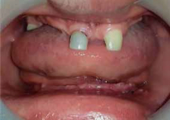
Stomatological examination revealed bone atrophy of alveolar processes in both jaws, true anodontia of teeth number: 11, 12, 13, 14, 15, 16, 18, 22, 23, 24, 25, 26, 27, 28, 31, 32, 33, 34, 35, 36, 37, 38, 41, 42, 43, 44, 45, 46, 47, 48, presence of cone-shaped, primary tooth 51 with mobility, moreover, there was presence of upper frenulum over-insertion. Alveolar ridges of upper and lower jaw were arranged as a class III according to Seibert’s classification. Mild marginal gingivitis associated to generalized dental plaque was diagnosed (Figure 2).
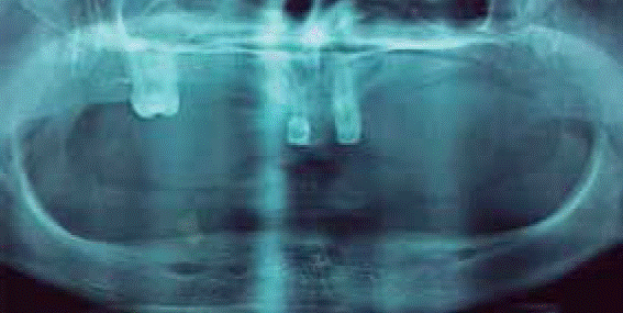
Radiographic examination revealed class II Seibert upper and lower ridge collapse, presence of a root remnant at the level of tooth 43, and radio-opaque areas at level of tooth 51 which were compatible with filling (Figure 3).
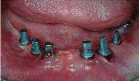
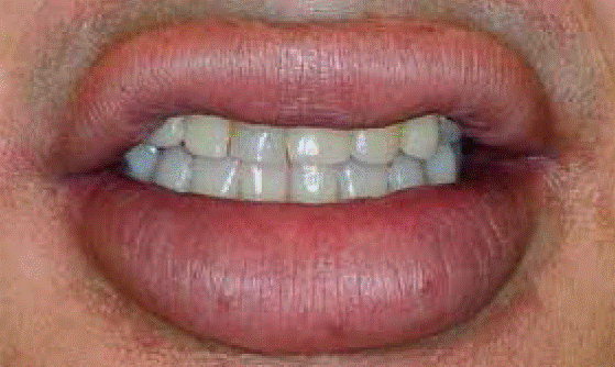
Comprehensive dental treatment included a hygiene phase, where patient was motivated and instructed on suitable oral hygiene habits, plaque control, prophylaxis and assessment of tissue response. Root remnant located at the level of tooth 43 was extracted. Rehabilitation phase included design and adaptation of a full denture in the upper jaw, necessary hygiene indications were administered, as well as instruction on denture care. It was planned to rehabilitate the lower jaw with implants, therefore, patient was referred to the Implantology Department of the School of Dentistry of the Cartagena University for evaluation and final treatment. Bicon® type implants were placed: five 4.0 × 6 mm-2.5 mm-well implants, and one 5.0 × 6.0 mm well implant. After this, rehabilitation was completed with an implant-supported prosthesis (Figures 4 and 5). It is worth mentioning that the patient was receptive all along treatment, showing interest and commitment during the different designated dental appointments.
The patient’s legal representative was requested to sign an informed consent form covering treatment execution, photo shooting and their subsequent publication.
Discussion
Hypohydrotic X chromosome-linked ED is the most common ectodermal dysplasia form. Also known as Christ-Siemens-Touraine syndrome, it is the most frequent form out of the approximately 150 types of ectodermal dysplasia. It exhibits a frequency of 1 per 100,000 live births.11
Christ-Siemens-Touraine syndrome manifests with a characteristic triad consisting of: reduction of hair amount (hypotrichosis), absence of sebaceous glands (asteatosis) and absence of sweat glands (anhidrosis). Cases can be mild or severe. Oral anomalies common to this disease have already been well described, among them we can mention: hypodontia (anodontia and oligodontia), in both jaws in primary and permanent dentition, alterations in tooth size and shape (cone or peg-shaped teeth)., delays in eruption of permanent teeth, bone atrophy in alveolar ridges, malocclusions, decreased vertical dimension, high palatal arch or cleft palate, salivary gland hypoplasia and absence of oral accessory glands, giving rise to xerostomia and dry and cracked lips.12,13
It is of the utmost importance for a dentist to be familiar with all the aforementioned physical and stomatological characteristics, so as to be able to conduct needed referrals, support medical and dental diagnosis and offer, together with the medical team, comprehensive treatment to ED patients.14 It has already been determined that dental appearance is of the utmost importance for the social and psychological development of these patients, since their self-esteem can be affected.
Presently there are different treatment options to facilitate oral functional rehabilitation and esthetics, thus allowing for the patient to feel comfortable and enjoy better quality of life. Among these options we can mention the following: fixed prosthesis, removable prosthesis, full prosthesis, and in recent years dental implants have being recognized as an alternative for denture support and retention, always first considering patient’s age and their potential growth.13,15
Presently, dental approach can be varied and it is conducted bearing in mind the subject’s age, growth and development of stomatognatic system, dental agenesis, degree of tooth malformation as well as motivation. Additionally, it is very important to plan multi-disciplinary treatments involving areas such as psychology, due to the important psychosocial component faced by these patients caused by their physical characteristics.9
Case report described in the present article represents a complex case, since the patient exhibited anodontia of practically all teeth. Treatment therefore consisted of design and adaptation of a partial prosthesis supported by teeth and mucosa in the upper jaw, and as many dental implants in the lower jaw as were necessary to stabilize a denture supported by implants and mucosa. Reasons which prompted selection of this treatment were: patient´s age, 18 years, when facial development and growth were already completed, degree of agenesia present in the patient, alterations in the shape and position of teeth, as well as degree of alveolar resorption in both jaws. In follow-up appointments, patient reported being satisfied with respect to treatment evolution.
Aydinbelge et al, equally reported a successful case of oral rehabilitation with dental implants, in a seven year old patient afflicted with hydrohydrotic ectodermal dysplasia. Even though dental implants are the treatment of choice in cases where facial growth has been completed, these authors developed a satisfactory treatment. On the other hand, Deshraj et al treated a case of HED, with oral characteristic similar to those here described, in a 13 year old patient with partial anodontia in both jaws, underdeveloped alveolar ridges and cone-shape incisors. Nevertheless, for this case, they decided to design and adapt a full upper denture and an over-denture in the lower jaw.16 Use of dentures is the most frequent rehabilitation used for patients with ED, nevertheless, there can be intra-oral characteristics, such a decrease of mandibular arch size and high, blade-shaped, alveolar bone which might complicate prosthesis construction and biomechanics during function .Suitable masticatory function can only be achieved when adequate stability and retention of the denture is provided, this requirement is fully met by dental implants.17
In conclusion, with treatment described in the present article, absolute improvement was obtained in mastication process, as well as increase of vertical dimension, improvement in the appearance of the last facial third; in addition to achieving esthetic smile, satisfactory to the patient and his legal guardian who were grateful for the comprehensive treatment provided by the School of Dentistry of the University of Cartagena, since the patient had not exhibited up to this point functional and aesthetic conformity.
REFERENCES
1. Vasconcelos CM, Romero SS, Paiva CM, Fonseca FT, Nunes SA, Carvalho S et al. Hypohidrotic and hidrotic ectodermal dysplasia: a report of two cases. Dermatol Online J. 2013; 19 (7): 18985. [ Links ]
2. Jananee J, Satishkumar M, Balaji S. Ectodermal dysplasia-a case report. Indian J Multidiscip Dent. 2012; 2: 465-467. [ Links ]
3. Masís PC, Montero SO, Gomez FA. Diagnóstico y manejo odontológico del paciente infantil con displasia ectodérmica anhidrótica: Síndrome de Christ Siemens Touraine. Rev Cient Odontol. 2010; 6 (1): 14-19. [ Links ]
4. Marín BM, Espinal BG, Arroyo FT, Posso ZM, David PM, Castañeda PD, Sierra PJ. Displasia ectodérmica hipohidrótica: Reporte de casos. Av Odontoestomatol. 2013; 29 (1): 11-23. [ Links ]
5. Wright JT, Morris C, Clements SE, D'Souza R, Gaide O, Mikkola M et al. Classifying ectodermal dysplasias: incorporating the molecular basis and pathways. Am J Med Genet A. 2009; 149A (9): 2062-2067. [ Links ]
6. Shigli AN SP. Prosthodontic management of patients with Christ-Siemens-Touraine syndrome. BMJ Case Rep. 2012; 10: 1-7. [ Links ]
7. Jalili VP, Neema HC. Ectodermal dysplasia. A cephalometric appraisal. J Pierre Fauchard Acad. 2013; 27 (2): 41-48. [ Links ]
8. Alves N, De Oliveira RJ, Figueiredo ND. Displasia ectodérmica hidrótica. Un síndrome de interés para la odontología. Int J Odontostomat. 2012; 6 (1): 45-50. [ Links ]
9. Nascimento SA, Ribeiro LM, Oliveira MS, Barbosa DS, Barros ML, Júnior MH. Orofacial features of hypohidrotic ectodermal dysplasia. Head Neck Pathol. 2012; 6 (4): 460-466. [ Links ]
10. Vallejo PA, López-Arranz ME, González GM. Tratamiento odontológico en la displasia ectodérmica: actualización. Av Odontoestomatol. 2006; 22 (3): 171-176. [ Links ]
11. García-Martín P, Hernández-Martín A, Torrelo A. Displasias ectodérmicas: revisión clínica y molecular. Actas Dermo-Sifiliográficas. 2016; 104 (6): 451-470. [ Links ]
12. Mokhtari S, Mokhtari S, Lotfi A. Christ-siemens-touraine syndrome: a case report and review of the literature. Case Rep Dent. 2012: 586418. [ Links ]
13. Kishore M, Panat SR, Aggarwal A, Agarwal N, Upadhyay N, Ajai K et al. Hypohidrotic ectodermal dyplasia: a case series. J Clin Diagn Res. 2014; 8 (1): 273-275. [ Links ]
14. Halai T, Stevens C. Ectodermal dysplasia: a clinical overview for the dental practitioner. Dent Update. 2015; 42 (8): 779-780, 783-784, 787-788. [ Links ]
15. Maroulakos G, Artopoulou II, Angelopoulou MV, Emmanouil D. Removable partial dentures vs overdentures in children with ectodermal dysplasia: two case reports. Eur Arch Paediatr Dent. 2015; 17 (3): 205-210. [ Links ]
16. Report C. Hypohidrotic ectodermal dysplasia: prosthetic and endodontic management. Int J Clin Pediatr Dent. 2010; 3 (1): 63-67. [ Links ]
17. Vilanova LS, Sánchez-Ayala A, Ribeiro GR, Campos CH, Farias-Neto A. Conventional complete denture in patients with ectodermal dysplasia. Case Rep Dent. 2015; 2015: 714963. [ Links ]
*This article can be read in its full version in the following page: http://www.medigraphic.com/facultadodontologiaunam
Received: June 01, 2016; Accepted: August 01, 2017
 Este es un artículo publicado en acceso abierto bajo una licencia Creative Commons
