


Services on Demand
Journal
Article
 text in
  English (pdf)
 Article in xml format
 Article references
 Send this article by e-mail
Indicators
-
 Cited by SciELO
 -
 Access statistics
Related links
-
 Similars in
SciELO
Similars in
SciELO
Share

 Permalink
Revista odontológica mexicana
Print version ISSN 1870-199X
Rev. Odont. Mex vol.21 n.4 Ciudad de México Oct./Dec. 2017
Literature review
Fascia lata as an alternative in dental treatments
1DDS, Graduate. School of Dentistry, National University of Mexico (UNAM).
2Professor, Implantology and Periodontics Department, Graduate and Research School National. School of Dentistry, National University of Mexico (UNAM).
3Professor of the Research Seminar of the Implantology and Periodontics Department, Graduate and Research School, and the Professional studies Division. School of Dentistry, National University of Mexico (UNAM).
Introduction:
Fascia lata is the most extreme section of the thigh's aponeurosis. It is a thick and resistant membrane possessing elasticity, flexibility and memory. It is presently used in the medical world to treat abdominal defects, urinary incontinence, and palpebral ptosis. In dentistry it is used in guided tissue regeneration, root coverage, ridge increase and socket (alveolus) preservation.
Method:
An electronic search was conducted in the following databases: Medline, PubMed and Sei ELO, with the term fascia lata. Full texts in English and Spanish were included in timeline spanning from 1983 to 2015. Main characteristic selected for these texts was they explored use of fascia lata and medical and dental areas.
Discussion:
Limiting situations arose during the execution of this project due to the scarcity found in articles and research papers and documented clinical cases targeting use of fascia lata ion dental areas.
Conclusions:
Fascia lata is a resorbable, biocompatible material, well tolerated by the recipient bed; when used in medical and dental specialties it exhibits characteristics of accuracy (security) and long duration.
Keywords: Fascia lata; muscle fascia; dentistry
Introducción:
La fascia lata es la parte más externa de la aponeurosis del muslo. Se trata de una membrana gruesa y resistente que posee elasticidad, flexibilidad y memoria. Actualmente es utilizada en el área médica para el tratamiento de defectos abdominales, incontinencia urinaria, ptosis palpebral y, en odontología, se utiliza para regeneración tisular guiada, coberturas radiculares, aumento de reborde y preservación de alveolo.
Método:
Se ha realizado una búsqueda electrónica en las bases de datos de Medline, PubMed y SciELO con el término fascia lata. Fueron incluidos textos compie-tos en idioma español e inglés que abarcan desde 1983 hasta 2015. La característica principal de estos textos es que exploran el uso de fascia lata en el área médica y odontológica.
Discusión:
Durante la realización de este trabajo surgieron situaciones limitantes debido a la escasa cantidad de artículos, investigaciones y casos clínicos documentados enfocados al uso de la fascia lata en el área odontológica.
Conclusiones:
La fascia lata es un material reabsorbible. biocompatible, que es bien tolerado por el lecho receptor, goza de características de seguridad y larga duración en su uso dentro de las especialidades medicas y odontológicas.
Palabras clave: Fascia lata; fascia muscular; odontología
INTRODUCTION
From the 1990׳S onwards, fascia lata has been used in medical areas as an alternative for treatment of urinary incontinence and to repair abdominal wall defects, and generally as a substitute for varied types of allogeneic and autogenous grafts. In dentistry it is used as a variant for collagen membranes for guided tissue regeneration treatment. It likewise represents an alternative to substitute palatal connective tissue graft in the course of gingival recession treatment and ridge increase, since it is a biocompatible, highly elastic material, available in unlimited.1
In 1908, German surgeon Martin Kirschner performed surgery with Edwin Payr and Paul Leopold Friedrich. They executed their first project using fascia lata for tendon repair and interposing tissue application in articulations.2
In 1925 North American surgeon Barney Brooks used stripes of fascia lata to lígate the neck of an aneurysm, wrap the bag and thus avoid its growth and rupture.3
In 1933 Price was the first to describe the use of a fascia lata sling in a female patient suffering from sacral agenesis and urinary incontinence.4 In 1934, surgeon Wargensteen OH was the first to report use of fascia lata to treat wide umbilical defects.5,6 He additionally informed that even in patients with long femurs, fascia lata was not sufficient to cover subcostal defects.6
In 1940, J.A. Bigger achieved with fascia lata almost full neck occlusion of an aortic aneurysm, complementing it with endoaneurysmorrhaphy.3
In 1950, thoracic surgeons Belsey and Churchill reported the case of two patients afflicted with cystic adenoid carcinoma who survived over five years after having been subjected to an extensive intra thoracic tracheal circular resection. The aforementioned tracheal defect was repaired using fascia lata supported with stainless steel wire.7 In 1951 E.J. Wylie, E. Kerr and o. Davis combined several procedures such a vessel wrapping with fascia lata, followed by thromboendarterectomy and endoaneurysmorrhaphy.3
Towards 1983, traumatologist Watson Jones described fascia lata extension technique up to the knee to reconstruct supra-umbilical defects (Figure I).6

As can be observed, some of the fascia lata benefits are known for their reconstructive ability which allows tissue revascularization and its integration to receptor tissue. It exhibits lesser complication risks, minimum risk of disease transmission as well as very low rejection rate. Due to all the aforementioned reasons, it enjoys wide range of use in medical specialties, such as ophthalmology, orthopedics, urogynecology, neurosurgery and in dentistry, as we will see in the present article.8
Fascia lata possesses a main function which consists on decreasing friction between muscles as well as transmitting forces elicited by the musculoskeletal system.9
Fascia lata allograft provided by a corpse is a resorbable and biocompatible material (Figure 2) well tolerated by the receptor bed.10 Moreover, this type of allograft prevents tissue invagination since it acts by prevening evolution of cells with different characteristics.11 It possesses reconstructive ability, allowing graft revascularization through the receptor bed by means of surrounding vessels and finally, it enjoys characteristics of reliability and long duration, thus rendering unnecessary a second surgical event to remove it.8

HISTOLOGICAL CHARACTERISTICS
Fascia lata is a cellular structure-poor tissue, similar to tendons and ligaments. It mainly possesses dense connective tissue which, even in rest position, exhibits some mechanical tension. It is composed of fibroblasts, which exert important function in structure architecture, rigidity control, formation and maintenance. Moreover, it produces collagen and elastin.
Mast cells are another component, this is important in mediation processes in allergic diseases and healing. In a lesser amount, it is also composed of myofibroblasts, which exert a valuable role in tension during wound healing. Its main substance is extracellular matrix.9
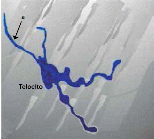
Telocytes are one of the cells participating in tissue repair and regeneration; they contain a small Golgy apparatus, cytoskeleton and are elements of the smooth and rugged endoplasmic reticulum (Figure 3). This type of cells are mainly found among collagen fibers, close to blood vessels and nerve endings. They possess variable thickness, and generally are thicker than collagen fibers possessing different structure to all the other present cells due to the fact they show very long prolongations called telopodes (similar to neurons' axons).9
FASCIA LATA PROCUREMENT
Fascia lata procurement depends on the graft to be used. In medical areas, it is common to use fascia lata autologous grafts. These procedures are conducted in adult patients and older children in order to obtain grafts of suitable length, although these grafts can also be donated by parents of a small child. Procedures can be accomplished under local or general anesthesia, and once the graft is harvested, it must remain in saline solution until placement in the receptor bed.13
In dental areas, allogen grafts provided by corpses are used. Normally, they lack irrigation conservation, exhibit a whitish, iridescent shiny and tense appearance. Once the grafts are procured, they are treated under aseptic circumstances, and are later preserved by means of freezing, freeze-drying, or treatment with solvents. Graft sterilization through gamma radiation is optional.14,15 Human fascia lata allografts are obtained from tissue banks which must be recognized by the National Association of Tissue Banks.
Tissue banks require patient exploration conducted by means of clinical history. Donors are serologically assessed in order to avoid viral diseases such as HIV, syphilis, hepatitis B and C, antigens VIO, cytomegalovirus, human T-lymphotrophic virus. The aim of this assessment is to increase allograft security using different methods and protocols. Tissues exhibiting high contaminant and microbial levels are discarded.14,16
Physical properties of allografts are almost equivalent to those of autografts, although their resistance to fraction can be decreased after gamma sterilization and freezing.17 It has been shown that most appropriate fascia lata material is a dehydrated spescimen with commercially available solvents, differing thus from freeze-dried samples from tissue banks.17 For both cases procurement methods are the same.
FASCIA LATA PROCUREMENT METHODS
The leg is prepared with a slightly flexed knee, and then the leg is leaned on some type of support with the aim of tensing the fascia and rendering extraction easier. An 8 cm incision is performed above the tibia's lateral condyle, incision diameter depends on the amount of fascia lata required for the treatment type. Surgeons seek the support of fasciometers, which are instruments that cut the fascia and facilitate its extraction. With these devices, a sufficient fascia lata stripe can be harvested from a small incision. In cases when no fasciometer is available, a second incision is performed at a location of 10 cm above the first incision. With Metzenbaum's scissors, subcutaneous cell tissue is dissected until reaching the fascia. With blunt dissection, a tunnel is dissected between both incisions underneath the skin and above the fascia lata. Parallel cuts are performed with a scalpel in order to obtain a piece of fascia, which is then placed on a sterile surface to be cleansed from any fat and muscle tissue.13,15
ALLOGRAFT STERILIZATION
FDA (Federal Drug Administration) establishes no specific technique for sterilization. Tissue banks use diverse patented techniques, among these we can mention use of irradiation, soaking and washing with antibiotics, lyophilization and cryopreservation. Once these protocols are accomplished, terminal sterilization is conducted by means of gamma irradiation or ethylene oxide.16
The main purpose of any de-cellularization protocol is to remove cellular material and deactivate infectious microorganisms without harming the remaining extracellular membrane's physical integrity, biological activity and constitution. This can be achieved with sonification, agitation freezing and thawing processes. At a later point the membrane is washed and cell remnants are removed.16
ALLOGRAFT MANIPULATION DURING SURGICAL PROCEDURE IN DENTISTRY
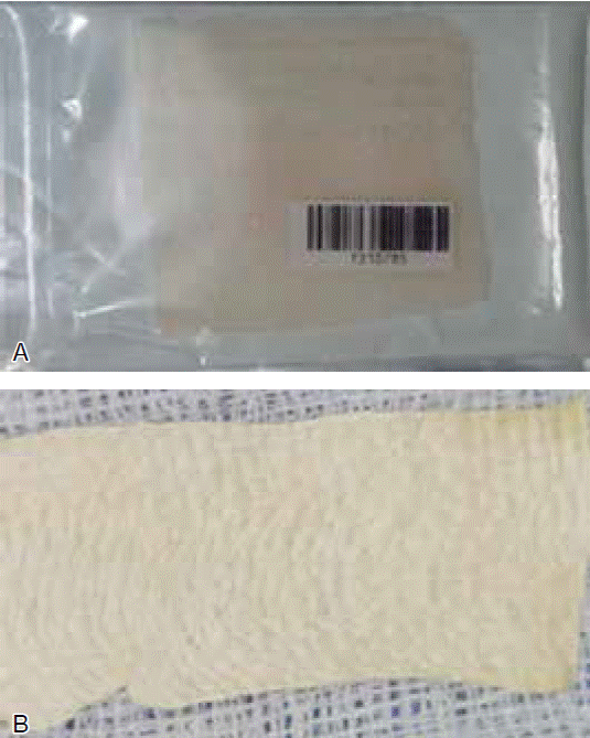
1. Remove fascia lata from package (Figure 4).
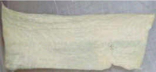
2. Hidration. Before use, material needs to be hydrated in sterile saline solution for 30-40 minutes in order to avoid continuous expansion once placed, and thus avoid exposition risk caused by its persistent memory (Figure 5).1
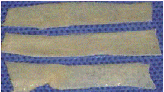
3. Size and shape adaptation of the material according to the treatment to be conducted (Figure 6).1
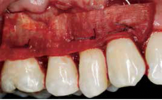
4. Fascia lata placement and fixation by means of resorbable suture points on itself and fixation to the receptor bed, thus guaranteeing immobilization (Figure 7).1
FASCIA LATA APPLICATIONS IN DENTISTRY
Literature reports use of fascia lata in guided tissue regeneration, ridge increase, socket preservation and gingival recessions. This last use is the one we are interested in documenting. Gingival recession is defined as gingival margin displacement in apical direction with respect to the enamel-cement junction, causing pathological exposition of root surfaces to the oral environment of one or more teeth.10 In this study we are histologically documenting the healing process taking place when there is a gingival recession coverage with fascia lata.
Three months after surgical procedure, the fascia lata graft can be observed clearly circumscribed, and separated from the mucosa of the receptor site. No foreign body reaction was observed or any rejection characteristic between fascia lata contact and gingival tissue. No inflammatory infiltration was observed above the fascia lata graft. Lesser blood extravasation and lymphocyte infiltration were observed. Finer collagen fibers could be observed in a more wavelike and less dense arrangement, as well as angiogenesis characteristics in mucous membrane such as grafted fascia lata fragment and a small number of lymphocytes, nevertheless, no inflammatory infiltration was observed.10
Six months after surgical procedure, delimitation between fascia lata and receptor site's connective tissue can still be observed. Similar numbers of fibroblasts can be observed, both in tissue fragments and blood vessels which intertwine connective tissue of the receptor site with the fascia lata graft. There is presence of lymphocytes and absence of inflammatory infiltration. In the fascia lata graft, two types of collagen fibers can be observed, fibers with a more regular disposition with greater number of fibroblasts, as well as numerous capillaries through the whole fascia lata fragment.10
Nine months later, the fascia lata graft is strongly bonded to the mucous membrane of the receptor site. Collagen bundles and fibroblast distribution in the fibers are identical, in the receptor site as well as in the fascia lata graft. Numerous capillaries penetrating into the fascia lata graft are visible at the frontier between both areas.10
Twelve months later, a strong bond can be observed between collagen fibers of the receptor's site connective tissue and fascia lata graft. These fibers have a thick appearance. Great amounts of blood vessels can be seen. There are no characteristics of inflammation or foreign body reaction in the fascia lata graft fragment. Connective tissue's architecture indicated full incorporation of fascia lata graft into receptor site (Table 1).1

Advantages in dentistry
Fascia lata allograft does not promote presence of immunological system cells directed to foreign bodies.10
When fascia lata is exposed, it can be trimmed without having to remove it from the surgical site.1
In the field of periodontics, it represents a treatment alternative to correct Seibert type 1 ridge defects in the anterior sector, since it exhibits thickness growth similar to that of connective tissue.1
Shows better handling with pontics, enabling greater papilla filling when compared to connective tissue.1
It exceeds the palate's anatomical limitations with respect to thickness and size of the donor area.1
It avoids soft tissue invagination and is effective in guided regeneration.11
It exerts the function of biological barrier between bone and mucous tissue during bone reparation times.11
Joint use of fascia lata and bone powder facilitate osteoinduction.11
Allows for new bone formation.11
Estimated risk of contracting infection of human immunodeficiency virus when employing fascia lata harvested from corpses is 1 in 1,667,600.4
DISCUSSION
Some limitations arose when the present study was conducted; this was due to the scarcity of documented articles, research papers and clinical cases devoted to fascia lata use in dental use. This has prevented extensive research, but nevertheless points out important aspects for the dental student and dental professional with respect to characteristics, procurement manipulation and healing of fascia lata. It is worth mentioning that obtained data were reliable, since their recompilation was conducted from articles published in internationally recognized journals. Data from this research indicate that fascia lata use in dentistry has increased in recent years, but they point out the need to further research and publication on this material.
CONCLUSION
Fascia lata is a resorbable and biocompatible material, moreover, it is well tolerated by the receptor bed; it additionally enjoys safety and long term characteristics.
Even though there have been reports on fascia lata use since 1908 within the realm of medical specialties, unfortunately, in the dental environment, the scant literature and scarce research found on characteristics of fascia lata handling and behavior in dental treatments limit the possibility to understand and apply this material. Even though it was discovered long ago, its use is still limited or insufficient due to lack of information. It is therefore of the utmost importance to conduct research on different types of dental treatments, at different follow-up intervals and in different populations. It is also important to mention the worth of elaborating and documenting histological studies in all treatments, so as to allow comparison and clearer knowledge of fascia lata behavior and healing time.
REFERENCES
1. Pazos-Ruiz A, Vargas-Quesada A, Pereira-Ebratt R, Serrano-Álvarez JJ. Comparación de injerto de tejido conectivo y fascia en el tratamiento de defectos de reborde alveolar. Universitas Odontológica. 2010; 29 (62): 27-37. [ Links ]
2. Fernández-Vázquez JM, Camacho-Galindo J. Martin Kirschner (1879-1942). Acta Ortop Mex. 2007; 21 (1): 45-46. [ Links ]
3. de la Garza-Villaseñor L. Los aneurismas en el tiempo. Cir Gen. 2000; 22 (3): 264-271. [ Links ]
4. Juarranz-Sanz M, Terrón-Barbosa R, Roca-Guardiola M. Soriano-Llora T, Villamor-Borrego M, Calvo-Alcántara MJ. Tratamiento de la incontinencia urinaria. Aten Primaria. 2002; 30 (5): 323-332. [ Links ]
5. Feijóo L, Martín ML, Villarreal C, Lomas M. Reconstrucción de pared abdominal inferior usando el colgajo miocutáneo tensor de la fascia lata. Cir Esp. 2002; 71 (1): 37-39. [ Links ]
6. Cavadas PC. Abdominal wall reconstruction in an electrical burn with a myocutaneous tensor fasciae latae island flap. Case report. Ann Burns Fire Disasters [Internet]. 1999; 12 (4). Disponible en: http://www.medbc.com/annals/review/vol_12/num_4/text/vol12n4p221.htm [ Links ]
7. Villar-Álvarez F, Jareño-Esteban J, Álvarez-Sala Walther R. Patología respiratoria. Manual de procedimientos de diagnóstico y control. Gráficas Enar, S.A.; 2007. Disponible en: http://www.neumomadrid.org/descargas/manualprocedimientosbaja.pdf [ Links ]
8. Peláez-Mata D, Alvarez-Zapico JA, Gutiérrez-Segura C. Fernández-Jiménez I, García-Saavedra S, González-Sarasúa J et al. Injerto de fascia lata de donante cadáver en la reconstrucción de defectos de pared abdominal en niños. Cir Pediatr. 2001; 14 (1): 28-30. [ Links ]
9. Dawidowicz J, Szotek S, Matysiak N, Mielanczyk L. Maksymowicz K. Electron microscopy of human fascia lata: focus on telocytes. J Cell Mol Med. 2015; 19 (10): 2500-2506. [ Links ]
10. Zurek J, Dominiak M, Tomaszek K, Botzenhart U, Gedränge T, Bednarz W. Multiple gingival recession coverage with an allogeneic biostatic fascia lata graft using the tunnel technique--A histological assessment. Ann Anat. 2016; 204: 63-70. [ Links ]
11. Rodríguez PA, Lenarduzzi AL, Fernández-Solari J, Elverdin JC. Utilización de membrana de fascia lata y hueso en polvo liofilizado en cirugía apical: caso clínico. Rev Fac de Odón UBA. 2012; 27 (62): 11-15. [ Links ]
12. Torralba-lglesias EG. Tendinopatía de cintilla iliotibial (Resumen) [Internet]. Disponible en: http://quiquetorralba. blogspot.mx/2011/03/tendinopatia-de-cintilla-iliotibial.html [ Links ]
13. ARDITO Oculoplástica. Cirugía de ptosis: suspensión al frontal con fáscia lata [Internet]. 2012. Disponible en: http://draarditocirugiaplasticaocular.blogspot.mx/2012/10/suspension-al-frontal-con-fascia-lata.html [ Links ]
14. Fitzgerald MP, Mollenhauer J, Brubaker L. The antigenicity of fascia lata allografts. BJU Int. 2000; 86 (7): 826-828. [ Links ]
15. Haupert A, Lorbach O. Anatomic reconstruction of the medial patellofemoral ligament using the fascia lata as an autograft. Arthrose Tech. 2015; 4 (1): e57-e63. [ Links ]
16. Ardila Medina CM. Propiedades biomecánicas y proceso de esterilización de las matrices alodérmicas usadas en periodoncia. Avances en Periodoncia. 2011 ; 23 (3): 187-193. [ Links ]
17. Kayurapan A, Makadelok S, Waikakul S. Effect of gamma sterilisation and deep-freezing on length and strength of fascia latae. J Orthop Surg (Hong Kong). 2010; 18 (1): 68-70. [ Links ]
18. Zucchelli G. Mucogingival esthetic surgery. Chicago: Quintessence; 2012. [ Links ]
*This article can be read in its full version in the following page: http://www.medigraphic.com/facultadodontologiaunam
Received: October 01, 2016; Accepted: February 01, 2017
 Este es un artículo publicado en acceso abierto bajo una licencia Creative Commons
