


Serviços Personalizados
Journal
Artigo
 texto em
  Inglês (pdf)
 Artigo em XML
 Referências do artigo
 Enviar este artigo por email
Indicadores
-
 Citado por SciELO
 -
 Acessos
Links relacionados
-
 Similares em
SciELO
Similares em
SciELO
Compartilhar

 Permalink
Revista odontológica mexicana
versão impressa ISSN 1870-199X
Rev. Odont. Mex vol.21 no.1 Ciudad de México Jan./Mar. 2017
https://doi.org/10.1016/j.rodmex.2017.02.012
Case report
Pre-bacute lymphoblastic leukemia: case report and literature review
* Full time Professor, Metropolitan Autonomous University (UAM), Xochimilco Campus.
Leukemia is one of the most common oncological disorders found in the first decade of life. It is characterized by the excessive production of non-functional immature lymphocytic cells, called blasts, which invade the bloodstream causing fatal consequences. Unless timely treated, it is a deadly, severe condition. Its more common manifestations are the following: generalized paleness, spontaneous hemorrhaging, vascular lesions such as bruises and petechiae, general malaise, weight loss, and specific stomatological manifestations such as oral mucosa paleness, leukocyte infiltrate gingivorrhageor apparition of petechiae in some locations of the mouth. The present article documents the case of a five year old girl, diagnosed with B-cell precursor acute lymphoblastic leukemia. The child was subjected to oral rehabilitation under general anesthesia due to the fact that she exhibited multiple infectious foci in her teeth which contraindicated initiation of chemotherapy treatment.
Key words: Hospital dentistry; leukemia; chemotherapy; oncologic cancer patients in dentistry
La leucemia se encuentra entre los trastornos oncológicos más comunes en la primera década de vida. Ésta se caracteriza por la producción excesiva de células inmaduras linfocíticas no funcionales denominadas blastos que invaden el torrente sanguíneo produciendo consecuencias fatales. Se trata de un padecimiento grave y mortal si no es tratado oportunamente. Entre sus manifestaciones más comunes se encuentran: la palidez generalizada, las linfadenopatías, hemorragias espontáneas, lesiones vasculares como moretones o petequias, malestar general, pérdida de peso y manifestaciones estomatológicas específicas como palidez de mucosa oral, los infiltrados leucocitarios, gingivorragias o aparición de petequias en algunos sitios de la boca. En el presente artículo se informa el caso de una niña de cinco años de edad diagnosticada con leucemia aguda linfoblástica precursora de células B, quien fue sometida a rehabilitación bucal bajo anestesia general, pues presentaba múltiples focos infecciosos en sus dientes que contraindicaban el inicio de su tratamiento de quimioterapia.
Palabras clave: Odontología hospitalaria; leucemia; quimioterapia; pacientes oncológicos en odontología
INTRODUCTION
Leukemia is the most common oncologic disorder found in childhood. It is characterized by a bone marrow disorder which generates excessive production of immature cells called blasts. In order to be classified, leukemia exhibits great diversity of characteristicsbased on type of producing cells or maturation degree of these cells.
According to producing cell lineage leukemia can be lymphoblastic or myeloblastic.1
Lymphoblastic leukemia is that whose cellularity derives from the lymphoid lineage, that is to say, Lymphocytes B and T respectively, whereas myeloblastic leukemia derives from myeloid lineage cells such as red blood cells, neutrophils, basophils ,eosinophils and platelets (Figure 1). Thus, according to cell functionality, they can be differentiated into acute and chronic.

Acute leukemia is characterized by possessing a non-functional cell population, since cells are totally immature, differing from chronic leukemia where cells exhibit greater maturation degrees. From this premise we can infer that acute leukemia is generally more aggressive than chronic leukemia. The origin of this condition is strongly associated to genetic syndromes such as Down’s syndrome,2 Lifraumeni’s syndrome, Klinefelter syndrome, Wiskitt Aldrich disease , Fanconi anemia, early age exposition to ionizing radiations and chemical agents as well as specific cytogenetic predisposition such as presence of Philadelphia chromosome.1,2 Lymphoblastic acute leukemia precursor of B cells is the most commonly found in Mexican pediatric population.
CLINICAL CASE
A five year old female, coming from León, Guanajuato, with previous diagnosis of B cell precursor acute lymphoblastic leukemia was referred to receive dental treatment. The oncologist deemed dental treatment urgent so as to achieve dental rehabilitation previous to initiation of chemotherapy treatment (Figure 2). Medical history revealed that the patient had begun with generalized paleness, weakness, bone-ache, generalized petechiae in the back, thorax and limbs. After initial pediatric assessment she was immediately referred to the oncologist on suspicion of leukemia. Due to the nil cooperation of the patient, to the complexity of her condition, as well as the urgency present, it was decided to rehabilitate the patient under general anesthesia (Figure 3).

Figure 2 Initial photograph of the patient treated in this case, diagnosed with B-lymphoblastic acute leukemia.
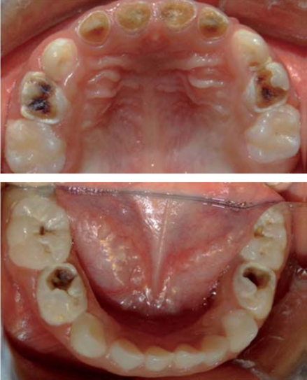
Figure 3 Patient’s initial circumstances in the mouth, favoring the decision of an intervention under general anesthesia.
Before dental treatment under general anesthesia, relevant studies were conducted to prepare the patient pre-operatively. Laboratory tests of blood count and clotting times were requested.
Blood count tests revealed anemia with 9 g/dL hemoglobin, neutropenia with neutrophil count of 500/mm3 and thrombocytopenia with platelet count of 50,000/mm3.
Previous to procedure initiation, the patient received a platelet apheresis to obtain maximum regulation of platelet levels, achieving elevationto up to 90,000/ mm3. Once in the operating theatre, dosed antibiotic prophylaxis was administered (20 mg/kg/dose) with intravenous clindamycin. The patient was intubated bynaso-tracheal route.
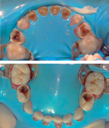
Sterile fields were conventionally placed and arches were fully isolated with a rubber dam (Figure 4). The following dental treatments were performed: pulpotomies of upper first molars, stainless steel crowns in upper and lower first molars, resin restoration in lower secondmolars, and pit and fissure sealers in upper second molars.
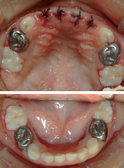
In views of the fact that the patient was to be subjected to a chemotherapy treatment which would significantly immunosuppress her, it was decided to preventively extract upper lateral and central incisors, so as to avoid latent risk of odontogenic infection which could aggravate her health circumstances. At that time, it was decided against placement of esthetic devices to replace extracted teeth, since appliances could act as bacteria reservoir, which at a given time could complicate the patient’s clinical evolution (Figure 5).
Because of preventive care exerted before treatment, the patient evolved satisfactorily, and was remitted to the recovery room in order to monitor vital signs.
Once the procedure was completed, the patient was ready to initiate chemotherapy as soon as possible, after healing of wounds caused by extractions.
DISCUSSION
All leukemia types belong to the group of proliferative malignant diseases. They represent the most common cancer disorders found in childhood, in addition to retinoblastoma, osteosarcoma and neuroblastoma.3 As mentioned before, leukemia can be classified according to producing cell lineage and to functionality and cell differentiation degree.1-3
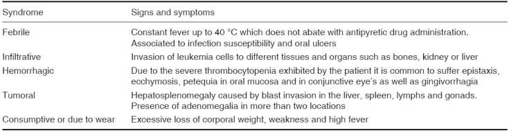
According to statistical studies conducted by the Health Ministry (Secretaría de Salud) in 2001, approximately 13,500 patients were admitted in hospitals with leukemia diagnosis. These cases were fatal in 232 patients aged one to four years, and 558 fatalities were recorded in the group of children aged five to fourteen years.4,5 Acute lymphoblastic leukemia is characterized by having its onset in the first decade of life, nevertheless, it is possible that the frequency might increase in senior citizens. Immature cells, which under normal circumstances would differentiate from any cell of lymphoid or myeloid lineage are called blasts.6 These cells, being non-functional, should not be found in the bloodstream, nevertheless, when the patient first displays leukemia, an excessive blast population appears which invades the bone marrow of long bones, preventing thus normal reproduction of other well-differentiated cells and their installation in the space within the bone marrow, generating thus in the patient a decrease in cell counts (pancytopenia) as well as pain in arms and legs. Since platelets and hemoglobin experience a sharp decrease, the patient experiences anemia and thrombocytopenia, which clinically translates into generalized paleness, asthenia, adynamia, and vascular lesions in the skin and oral mucosa.5-7 All patients first experiencing leukemia suffer certain associated syndromes, which can be: febrile, infiltrative, hemorrhagic, anemic, tumor and consumptive syndromes (Table I).
Since this is a disease derived from white blood cells, it is common to find leukocyte infiltrate in the oral mucosa; this represents the invasion of leukocyte neoplastic cells which penetrate into the oral mucosa to then destroy it. Clinically, they are observed as whitish lesions that do not detach when scraping. Leukocyte infiltrate can be a subclinical manifestation that a patient is suffering leukemia (Figure 6).
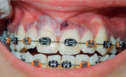
Figure 6 Leukocyte infiltrate in the gingival mucosa of a 15 year old patient who started with acute B-lymphocytic leukemia. All orthodontic appliances were removed from this patient before initiation of chemotherapy treatment.
Medical diagnosis of leukemiais based on characteristic symptomatology as well as laboratory studies prescribed for the patient (Table II), Bone marrow aspiration is generally indicated to corroborate diagnosis, in order to assess whether blast invasion is absent or present; the aforementioned diagnosis might be emitted in cases where there is medullar blastosis exceeding 30% of total cell population. Beside cell counts revealed by laboratory tests, characterized by exhibiting high leukocyte numbers (exceeding 50,000 mm3)) as well as reported presence of blasts, there is important decrease in the count of platelets, neutrophils, hemoglobin and erythrocytes.

Cytogenetics is very important to determine leukemia prognosis. When cell with larger numbers of chromosomes (calledhyperdiploid) are observed in a cytogenetic study, patients enjoy greater possibilities of healing, differing from those patients with lesser amounts of chromosomes (hypodiploid) who generally suffer worse prognosis. Likewise, the presence of the so-called Philadelphia chromosome is a factor which determines a poor prognosis for this disease.4,5,7
Nevertheless, besides cytogenetics there are some other factors which are related with leukemia prognosis (Table III).

Once the patient has been diagnosed,chemotherapy treatment must be undertaken as soon as possible, nevertheless this treatment cannot be initiated in cases when the patient presents infectious foci in the mouth, therefore, the dentist plays an essential role in the treatment of these patients.8
Dentists must be familiar with chemotherapy phases to which the patient with this disease will be subjected to, since, at each given phase there are specific drugs an sequels which might contraindicate of warrant a modification in the dental treatment course chosen for the patient. When the child has not been subjected to chemotherapy treatment, the dentist must evaluate blood count and coagulation (clotting) times to assess whether the patient requires platelet apheresis, or prophylactic antibiotic administration before invasive dental treatments due to the important decrease of defense cells. At this stage it is also necessary to completely remove any orthopedic or orthodontic device worn by the patient, since they could become a plaque reservoir which might affect the patient’s evolution once immunosuppression is triggered by chemotherapy treatment.
The first immunotherapy phase is called remission induction.2,3,8During this phase, the patient receives high doses of corticosteroids, such as methylprednisolone, with the sole aim of immunosuppressing him and initially arrest the disease’s evolution; if the patient evolves satisfactorily, the next stage will be undertaken. This stage is called consolidation or intensification stage,2,8 and in it, in addition to administering corticosteroids, chemotherapy drugs are also administered, among these drugs we can count vincristine, citacarabine, daunorubicin, L-asparginase, etoposides, cyclophosphamide, and methrotrexate. After having ingested these drugs, the patient experiences specific sequels such as anagen effluvium, weight loss, nausea, vomit, and generalized paleness (Figure 7). After a week of ingesting these drugs, the patient will experience an ailment called seventh day neutropenia, or nadir phase, which is characterized by under 200 neutrophil counts, sometimes even reaching 0 neutrophils per cubic millimeter.
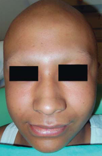
Figure 7 Manifestations caused by chemotherapy in a leukemia patient. To be noted is the tegument paleness and anagen effluvium characterized by total loss of hair and eyebrows.
During this phase the patient might exhibit the most painful chemotherapy sequel in the mouth, called mucositis .Mucositistends to appear as a consequence of methotrexate administration and is characterized by ulceration and desquamation of all the digestive tract mucosa, from the mouth to the anus. The patient experiences decrease to oral route tolerance in a hundred per cent, the dentist should prescribe use of palliative treatment to help the patient tolerate oral intake. The treatment can be mucosa coaters or topical antihistamines which will decrease local inflammation of the ulcerated oral mucosa (Figure 8).9-12
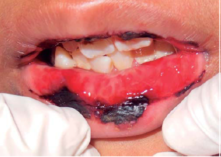
Figure 8 Oral mucositis as a consequence of methotrexate administration in an 8 year old leukemia patient.
It is at this precise moment due to the fragile circumstances of the child, when the dentist must suppress all treatment. Any ailment must be treated palliatively (pain killers, antibiotics or antifungal in necessary cases) waiting for a normalization of cell count in the following days.10,13
Once this phase is overcome, the patient is subjected to a new bone marrow aspiration and another cell count, where important reduction of blast cells is expected, which would indicate suitable response to the treatment.
After this, the patient passes to the following and next to last phase called maintenance phase. In it corticosteroid and chemotherapeutic agents are periodically administered in order to control the disease. A patient can last several years in this phase, and obviously under sporadic laboratory control studies (blood count, bone marrow aspiration and lumbar punctures). This patient can be treated in the dental office, always taking into account possible sequels to administered dugs, antibiotic prophylactic coverage might be required in some cases.
When the patient has overcome the disease, and having spent a minimum of two years with low doses of drugs and total absence of blasts (as revealed in periodical tests), the last phase of the oncological treatment is undertaken, this is the surveillance (monitoring) phase. All subjects in their monitoring phase must undergo yearly periodical revision for the rest of their life. Nevertheless, it must be pointed out that a patient in monitoring phase, can be treated as a healthy patient from the dental point of view.
Leukemia, once overcome, exhibits an important relapse risk; this is due to specific situations which must be foreseen before initiating chemotherapy treatment. Leukemic cells tend to penetrate into the so-called «sanctuary organs», which are the central nervous system and the testicles in males. These are zones where chemotherapeutic drugs are ineffective, and thus become the anatomical areas suffering higher risks of relapse.2,3,14 Therefore intrathecal therapy is mandatory in the treatment of a leukemia patient, as well as performance of several lumbar punctures during treatment, conducted with the aim of discarding blast invasion in the central nervous system.14,15
CONCLUSIONS
Dentists play a key role in the diagnosis and treatment of a leukemia patient. Many leukemiaindicating early lesions can appear in themouth, for this reason, dental personnel is obliged to be familiar with them. Dental intervention is of the utmost importance due to the need to eradicate infectious foci before initiating cancer therapy, as well as treating oral and dental problems whichmight arise as a consequence of chemotherapy. Knowledge of the chemotherapy phases to which a patient will be subjected, to a great extent determines the dental and stomatological conduct that must be followed.
REFERENCES
1. Aster JC. Enfermedades de los leucocitos, los ganglios linfáticos, el bazo y el timo. En: Kumar V, Abbas AK, Fausto N. Patología estructural y funcional. Ed. Madrid, España: 2005, pp. 670-680. [ Links ]
2. Zipursky A, Poon A, Doyle J. Leukemia in Down syndrome: a review. Pediatr Hematol Oncol. 1992; 9 (2): 139-149. [ Links ]
3. American Cancer Society. Cancer facts & figures 2015. Atlanta, GA: American Cancer Society; 2015. [ Links ]
4. Protocolo de la atención para leucemia linfoblástica. Guía Clínica y Esquema de Tratamiento. Instituto Nacional de Salud Pública. [ Links ]
5. Labardini MJ, Cervera CE, López NO, Corrales AC, Balbuena MM, Barbosa IA y cols. Oncoguía. Leucemia aguda linfoblástica. Cancerología. 2011; 6: 111-115. [ Links ]
6. Hoffman, R. Furie B, McGlave , et al. Hematology: basic principles and practice. 2008; (4ta ed). 63: 1-5. [ Links ]
7. National Comprehensive Cancer Network. Practice Guidelines in Oncology-v.2.2013. Acute Lymphoblastic Leukemia. [Accessed 18 December 2013] Available in: Available in: http://www.nccn.org/professionals/physician_gls/pdf/all . [ Links ]
8. American Academy of Pediatric Dentistry. Guideline on dental management of pediatric patients receiving chemotherapy, hematopoietic cell transplatation, and/or radiation. Pediatric Dentistry. 2012; 34 (6): 280. [ Links ]
9. De la Teja Ángeles E, Niembro-Zermeño A, Durán Gutiérrez A. Mucositis bucal. Acta Pediatr Mex. 2011; 32 (4): 255-256. [ Links ]
10. López CF, Oñate SER, Roldán CR, Cabrerizo MMC. Measurement of secondary mucositis to oncohematologic treatment by means of different scale. Med Oral Patol Oral Cir Bucal. 2005; 10: 412-421. [ Links ]
11. Hong CH, Brennan MT, Lockhart PB. Incidence of acute oral sequelae in pediatric patients undergoing chemotherapy. Pediatr Dent. 2009; 31 (5): 420-425. [ Links ]
12. da Fonseca M. Childhood cancer. In: Nowak AJ, Casamassimo PS, ed. The handbook of pediatric dentistry, 4th Edition: Chicago, Ill. American Academy of Pediatric Dentistry; 2011, pp. 225-231. [ Links ]
13. Keefe DM, Schubert MM, Elting LS, Sonis ST, Epstein JB, Raber-Durlacher JE et al. Update clinical practice guidelines for the prevention and treatment of mucositis. Cancer. 2007; 109 (5): 820-831. [ Links ]
14. Little JW, Falace DA, Miller CS, Rhodus NL. Cancer and oral care of the cancer patient. In: Little and Falace’s dental management of the medically compromised patient. 8th ed. St. Louis, Mo: Elsevier-Mosby; 2012, pp. 459-492. [ Links ]
15. Smith MA, Ries LA. Childhood cancer: incidence, survival, and mortality. In: Pizzo PA, Poplack DG, eds. Principles and practice of pediatric oncology. 4th edition, Lippincott Williams & Wilkins, Philadelphia, PA, USA, 2002, pp. 1-12. [ Links ]
***This article can be read in its full version in the following page: http://www.medigraphic.com/facultadodontologiaunam
Received: February 01, 2016; Accepted: August 01, 2016
 Este es un artículo publicado en acceso abierto bajo una licencia Creative Commons
