


Services on Demand
Journal
Article
 text in
  English (pdf)
 Article in xml format
 Article references
 Send this article by e-mail
Indicators
-
 Cited by SciELO
 -
 Access statistics
Related links
-
 Similars in
SciELO
Similars in
SciELO
Share

 Permalink
Revista odontológica mexicana
Print version ISSN 1870-199X
Rev. Odont. Mex vol.18 n.4 Ciudad de México Oct./Dec. 2014
Original research
Pre-orthopedic appliance with pins used in alignment of maxillary segments in patients with unilateral cleft lip and palate
Luis González García,* Erika González Rodríguez,II Manuel Yudovich Burak,§ María de la Paz Aguilar Saavedra,II Salvador García López §.¶ y Rosina E Villanueva Arriaga¶
* University Hospital, Monterrey, Nuevo Leon, Mexico.
§ Orthodontics Department, General Hospital <<Dr Manuel Gea González>> UNAM, Mexico City.
II Private practice.
¶ Metropolitan Autonomous University, Xochimilco, Mexico City.
ABSTRACT
The present article describes an orthopedic appliance with pins used in the pre-surgical treatment of fully fissured unilateral cleft lip and palate before lip reparation. This appliance was designed with the aim of transversally correct palatal segments in the posterior area, align the smaller segment in the anterior section towards the facial midline, and decrease the cleft located near the lip so as to improve the shape of the nasal base floor and avoid later surgical compensations of the soft tissues.
Key words: Unilateral cleft palate, lip reparation, pre-surgical orthopedics.
INTRODUCTION
Cleft lip and palate are the most common malformations of the craniofacial region. These malformations affect one out of 700 newborns. Reports inform that frequency varies depending on the type of cleft, ethnicity and gender. Etiology of cleft lip and palate is still subject to debate. Nevertheless, in certain types of deformities clefts occur when mesenchymal connective tissues from the different embryonic structures do not fuse. This lack of fusion apparently takes place during the third month of fetal development. Deformities can be unilateral or bilateral and can extend up to the alveolar process.
Pre-surgical orthopedics consists on placing appliances a few days after birth in order to align maxillary segments. This process is undertaken before a plastic surgeon reconstructs lip and palate. Protocols for cleft treatment can be active or passive, as well as intra- or extra-oral.1 Some of the active maxillary appliances are fixed with surgical pins, others are not. The appliance pre-determinedly displaces alveolar segments of the cleft, by means of controlled forces. Results of this treatment are long-term, physical and financial, quantifiable benefits for the patient.2,3 Several publications have reported benefits of early maxillary orthopedic treatment in cleft lip and palate patients.4-9 The aim of the present article was to describe the designing process for an appliance with pins which could be successfully used during pre-surgical orthopedic treatment in patients with fully fissured unilateral lip and palate.
Case assessment
Traditionally, diagnosis of facial clefts is conducted at birth, during the first physical examination, although an in uterus diagnosis is also possible. Patients presenting a left unilateral lip and palate full cleft, larger than 9 mm, are sent to the Pre-surgical Orthopedic Center (Figures 1 and 2). This formula is necessary since these malformations can cause difficulties during breastfeeding as well as loss of nasal air flow, along with an esthetic factor. Nevertheless, most newborns can be bottle-fed, with a large nozzle which can have a wider opening. Evidently, when parents discover that their child suffers a facial cleft, they become extremely anxious, therefore, timely and suitable handling of this situation must be initiated at an early stage.
Facial cleft treatment
Treatment of a newborn with facial cleft must bear the main objective of improving function, occlusion and appearance, even at the expense of an ideal occlusion, since the main target is to improve appearance and language. During the first visit with the orthodontist, the patient is assessed by two departments: Plastic Surgery and Orthodontics, which will devise a suitable treatment plan. This plan usually involves placing appliances to align maxillary segments during the first days of the patient's life. This is achieved with the help of pre-surgical orthopedics to facilitate surgical repair of the lip, with the purpose of achieving long-term facial growth. This procedure might not have a very promising future, nevertheless, social and personal interests must be taken into account.
All patients with cleft lip and palate must be individually assessed so that devised treatment will depend upon the type and severity of the cleft. The first step is to expand the maxillary segments in the posterior region, later, they are leveled and the smaller segment is rotated towards the face's midline. As a result, the cleft in the anterior segment is decreased by at least 1.5 mm. This procedure is undertaken before repairing the lip.
Proposal of orthopedic appliance for unilateral clefts
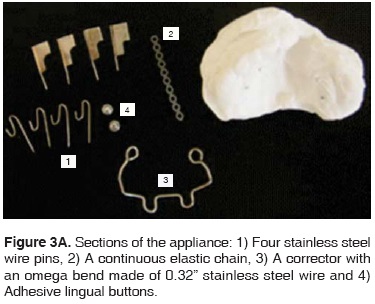
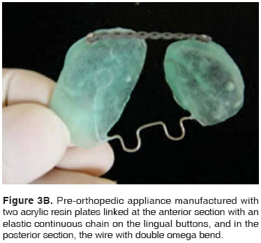
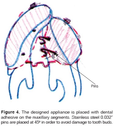
After discussing treatment plan, an impression of the maxilla is taken with a tray which must be specially manufactured for that patient. The impression is taken ensuring that depth details of the hemi-alveolar rims are carefully recorded so as to properly build the appliance. Plaster models are cast from the impression. The rims of the plaster study model must be very accurate so as to allow the appliance's suitable adjustment and retention. The appliance is composed of two palatal plates, built with acrylic resin and linked by a .032 stainless steel wire frame (Dentaurum, Germany) which has a double omega bend to control expansion of the posterior segment and rotation of the smaller segment. Palatal plates count with embedded metallic buttons which are linked with an elastic chain so as to allow stability when the appliance is put into place. At the same time, they provide retention when the movement of the cleft is achieved (Figures 3A and 3B). The appliance is placed on the fissured palate with .032 stainless steel wire pins, with a 45 degrees inclination so as to ensure they do not affect present tooth buds (Figure 4). Moreover, this inclination allows a lighter pressure in the retention of the appliance.
Surgical and orthopedic treatment
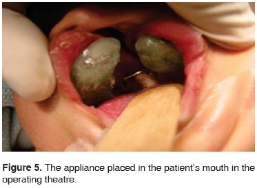
Orthodontists and plastic surgeons coordinate the procedure, which is conducted in an operating theater with the patient under general anesthesia. The appliance is put into place with a dental adhesive. Previously sterilized pins are installed with a band pusher, and acrylic resin is applied to the appliance so as to avoid its slippage (Figure 5).
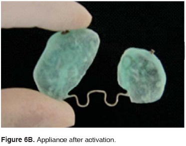
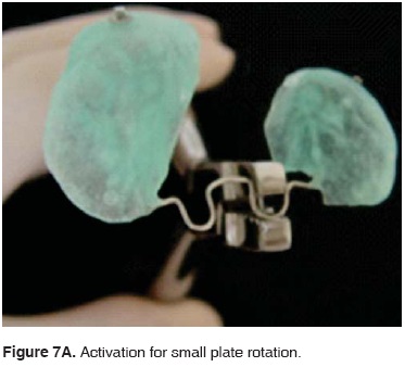
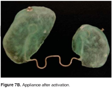
Once the appliance is properly placed, its activation is initiated in the orthodontist's office. The number of required appointments varies according to the width of the cleft. The first step is to expand the palatal segments through activation of the wire located between the two omegas. For this purpose threepronged straight pliers are used (Figures 6A and 6B). The smaller plate must be rotated with the lingual arch pliers by exerting pressure on the omega located closest to the plate. Once the appliance is widened, the plate can be fully and securely rotated (Figures 7A and 7B). In order to decrease the cleft with the appliance , activation is applied to the middle section of the connector, for this purpose three-pronged straight pliers are used (Figures 8A and 8B). Appliance activation must be conducted every 3 days in the dental office. Patient's diet must remain unchanged at all stages of the treatment. This treatment will last for almost 4 weeks, so that segments can be positioned, and the palatal segments can be re-organized, and therefore the anterior region cleft can be reduced to 1.5 mm (Table I). When the aforementioned results are achieved, the treatment is considered successful. In the case here presented, treatment was conducted without encountering any complications (Figure 9).


Clinical changes were observed when aligning the cleft. Among them we can mention shape improvement, since the smaller segment was forwardly displaced into the proper position, that is, the face's midline. This process decreased by several millimeters the width of the cleft (Figures 10A and 10B). The anterior section cleft was decreased from 9 to 1.5 mm. The expansion in the posterior section was performed with activation of the appliance. This procedure improved bone structure in preparation for later lip repair procedures. A Hotz-type retention appliance was placed in order to achieve nasoalveolar remodeling before lip closure (Figure 11).


Length of treatment and surgical procedure depend on the patient's health as well as the surgeon's experience. Different procedures can be used to repair the lip, the aim is to create a suitably long lip, not too tense and with satisfactory functions. When the maxillary segment's pre-surgical orthopedic alignment is conducted, the lip is practically repaired in about three months (Figure 12).

DISCUSSION
The appliance described in the present article was developed to be used in pre-surgical orthopedics in patients with full unilateral cleft lip and palate. The appliance was intended to align displaced maxillary segments, move them along soft tissue margins towards the face midline and thus decrease the size of the cleft in the anterior segment before the surgical repair of the lip. Alignment of the smaller segment is of the utmost importance, since, due to its size, it could become unstable and could rotate and collapse. This process entails functional consequences since, when some patients are operated on without previously having aligned segments of the palate, the result could be asymmetry in the nasal floor, causing thus a depression in the alar base. Furthermore, unilateral cleft lip and alveolar deformities are associated with significant anomalies of the nasal cartilage morphology and the asymmetry of alar base and columella. After performing pre-surgical orthopedics, deformities of nasal cartilage and soft tissue are corrected through naso-alveolar remodeling.10,11 Although the concept of timely maxillary orthopedics in infants with cleft lip and palate was already known in the 50's,4 great diversity of active and passive appliances have since then been developed.7,12-15 Nevertheless, presently there is some degree of controversy with respect to treatment of patients with unilateral cleft lip and palate16-18 as well as in time invested in treatments.19 Likewise, other clinical operators have stated there is no difference in facial growth in cleft lip and palate patients treated with pre-surgical orthopedics when compared to those who received no treatment prior to the primary gingivoperiosteoplasty20-22 as was described by Drs Millard and Latham.23 Avantgarde clinical methods must be assessed with the help of prospective, multicenter studies in order to ascertain benefits achieved with pre-surgical pediatric orthopedics. These studies must per force be random and controlled in order to be able to judge therapy effectiveness24 based on the ability to achieve long term positive results to improve balance between hard and soft tissues.25
CONCLUSIONS
The designed pre-orthopedic appliance achieved transverse and anterior-posterior correction of maxillary segments before lip repair. Results must be assessed in the long term.
REFERENCES
1. Huebener DV, Liu JR. Maxillary orthopedics. Clin Plast Surg. 1993; 20: 723-732. [ Links ]
2. Maull DJ, Grayson BH, Cutting CB, Brencht LL, Bookstein FL, Khorrambadi D et al. Long-term effects of naseoalveolar moulding on three-dimensional nasal shape in unilateral clefts. Cleft Palate Craniofac J. 1999; 36: 391-397. [ Links ]
3. Pfeifer T, Grayson BH, Cutting CB. Gingivoperiosteoplasty versus alveolar E bone graft: an outcome analysis of costs in the treatment of unilateral cleft alveolus. Presented at the 55th. Annual Meeting of the American Cleft Palate-Craniofacial Association; April 1998; Baltimore, MD: 1998. [ Links ]
4. McNeil CK. Orthodontic procedures in the treatment of congenital cleft palate. Dent Record. 1950; 79: 126-132. [ Links ]
5. Hotz M, Gnoinski W. Comprehensive care of cleft lip and palate children at Zurich University: a preliminary report. Am J Orthod. 1976; 70: 481-504. [ Links ]
6. Latham RA. Orthodontic advancement of the cleft maxillary segment: a preliminary report. Cleft Palate J. 1980; 17: 227-233. [ Links ]
7. Huebener DV, Marsh JL. Alveolar molding appliances in the treatment of cleft lip and palate patients. In: Bardach J Morris HL, eds. Multidisciplinary management of Cleft Lip and Palate. Philadelphia: WB Saunders; 1990. 601-607. [ Links ]
8. Grayson BH, Cutting CB. Presurgical nasoalveolar orthopedic molding in primary correction of the nose, lip and alveolus of infants born with unilateral and bilateral Clefts. Cleft Palate Craniofac J. 2001; 38: 193-198. [ Links ]
9. Suri S, Thompson BD. A modified muscle-activated maxillary orthopedic appliance for presurgical nasoalveolar molding in infants with unilateral cleft lip and palate. Cleft Palate Craniofac J. 2004; 3: 225-229. [ Links ]
10. Grayson BH, Cutting CB, Wood R. Preoperative columella lengthening in bilateral cleft lip and palate. Plast Reconstr Surg. 1998; 101: 630-639. [ Links ]
11. Grayson BH, Santiago P, Brecht L, Cutting CB. Presurgical naso-alveolar molding in patients with cleft lip and palate. Cleft Palate Craniofa J. 1999; 36: 486-498. [ Links ]
12. Burston WR. The early treatment of cleft palate conditions. Dent Pract. 1958; 9: 41-56. [ Links ]
13. Georgiade NG, Mladlick RA, Thorne FL. Positioning of the pre-maxilla in bilateral cleft lips by oral pinning and traction. Plast Reconstr Surg. 1968; 41: 240-243. [ Links ]
14. Georgiade NG, Latham RA. Maxillary arch alignment in the bilateral cleft lip and palate infant using the pinned coaxial screw appliance. Plast Reconstr Surg. 1975; 56: 52-60. [ Links ]
15. Figueroa AA, Reisberg DJ, Polley JW, Cohen M. Intraoral-appliance modification to retract the premaxilla in patients with bilateral cleft lip. Cleft Palate Crabiofac J. 1996; 33: 497-500. [ Links ]
16. Winters JC, Hurtwitz DJ. Presurgical orthopedics in the surgical management of unilateral cleft lip and palate. Plast Reconstr Surg. 1995; 95: 755-764. [ Links ]
17. Kuijpers-Jagtman AM, Long RE Jr. The influence of surgery and orthopedic treatment on maxillofacial growth and maxillary arch development in patients treated for orofacial clefts. Cleft Palate Craniofac J. 2000; 37: 527. [ Links ]
18. Prahl-Anderson B. Dental treatment of predental and infant patients with clefts and craniofacial anomalies. Cleft Palate Craniofacial J. 2000: 37; 528-532. [ Links ]
19. Gnoinski WM. Infant orthopedics and later orthodontic monitoring for unilateral cleft lip and palate patients in Zurich. In: Bardach J, Morris HL, eds. Multidisciplinary management of cleft lip and palate. Philadelphia: WB Saunders; 1990. 578-590. [ Links ]
20. Wood R, Grayson BH, Cutting CB. Gingivoperiosteoplasty and growth of the midface. Surg Forum. 1993; 16: 229. [ Links ]
21. Wood R, Grayson BH, Cutting CB. Gingivoperiosteoplasty and midfacial growth. Cleft Palate Craniofac J. 1997; 34: 17-20. [ Links ]
22. Lee C, Grayson BH, Lin WY, Cutting CB. Long term study of midface growth in unilateral cleft lip and palate patients following gingivoperiosteoplasty. Presented at the American Cleft Palate-Craniofacial Association; April 1999; Scottsdale, AZ: 1999. [ Links ]
23. Millard DR, Latham RA. Improved primary surgical and dental treatment of clefts. Plast Reconstr Surg. 1990: 86; 856-871. [ Links ]
24. Roberts CT, Semb G, Shaw WC. Strategies for the advancements of surgical methods in cleft lip and palate. Cleft Palate Craniofac J. 1991: 28: 141-149. [ Links ]
25. Smith WP, Markus AF, Delaire J. Primary closure of the cleft alveolus: a functional approach Br J Oral Maxilo Fac Surg. 1995; 3: 156-165. [ Links ]
Note This article can be read in its full version in the following page: http://www.medigraphic.com/facultadodontologiaunam Mailing address:
Salvador García López
E-mail: salgarlop@hotmail.com

