


Servicios Personalizados
Revista
Articulo
 texto en
  Inglés (pdf)
 Artículo en XML
 Referencias del artículo
 Enviar artículo por email
Indicadores
-
 Citado por SciELO
 -
 Accesos
Links relacionados
-
 Similares en
SciELO
Similares en
SciELO
Compartir

 Permalink
Revista odontológica mexicana
versión impresa ISSN 1870-199X
Rev. Odont. Mex vol.17 no.4 Ciudad de México oct./dic. 2013
Original research
Effect of lipoteichoic acid on gene expression in mice (H9c2) cardiomyocite
Ilayali Adam Bustamante,* Gloria Gutiérrez Venegas,§ Amalia Ballesteros VizcarraII
* Student, Endodontics Specialty, Graduate School, National School of Dentistry, National University of Mexico.
§ Tutor, Head of the Biochemistry Laboratory, Graduate and Research School, National School of Dentistry, National University of Mexico.
II Consultant, Professor of Endodontics, Graduate and Research School, National School of Dentistry, National University of Mexico.
ABSTRACT
Most dental pulp diseases and diseases of tissues surrounding the root are somehow related to micro-organisms. Peptidoglycans and lipoteichoic acid are two of the main Gram-positive bacteria components with activities related to sepsis development. When tissues sustain microbial invasion the host responds with both unspecific inflammatory defenses and specific immunological reactions. Surgical and non surgical endodontic treatments are essentially debridement procedures intended to destroy and eliminate the microbial eco-system associated to the pathological process. It is essential for clinicians to understand the intimate relationship existing between micro-organisms and endodontic disease, so as to be able to tailor a rational and effective treatment especially in subjects susceptible to infective endocarditis processes. In the present study research was conducted on TNFα, IL-1 COX-2 expression through the effect of lipoteichoic acid (LTA) of Streptococcus sanguinis by characterizing intra-cellular signals involved in H9c'' cardiomyocytes. The cell line was treated with LTA at different concentrations during 30 minutes. When compared to control group, responses to LTA treatment were dependent on dosage. That expression was assessed by means of a One Step RT-PCR (Invitrogen) analysis. It was noted that the aforementioned expression resembled the organisms's physiological response during an infective endocarditis episode and to exacerbation observed during an endodontic procedure.
Key words: Lipoteichoic acid, H9c2 cardiomyocytes, Streptococcus sanguinis , RT-PCR.
INTRODUCTION
The traditional concept to illustrate bacterial process in human beings suggests the fact that disease is caused as a result of harmful micro-organisms invasion which fight against the host defenses and trigger mechanisms which release antibodies and immune cells. The impact of this approach generates a tendency to research the most dangerous micro-organisms which could cause most severe damage in the host.1
Root canal infections possess a nature different from caries and periodontitis, since they have originally settled in sterile compartments of the oral cavity. In many cases, this leads to the idea that the etiology of infections of the root canals imply one single pathogen. Recently, the frequent presence of Enterococus faecalis in root canals associated to persistent infections has renewed interest to research this bacteria. This bacteria has become the ideal micro-organism to test different irrigating agents , drugs and antiseptic solutions used in vitro in the endodontic field. Results of this research tend to point out the bacteria's capacity for innate resistance. Nevertheless, the great interest poured on E. faecalis has resulted in far less information on the existence of other organisms in these infections, which might present similar tolerance characteristics as E. faecalis and which could reveal the existence of persistent poly-microbial communities.2
Poly-microbial infections extend from the root canal to adjacent tissues surrounding the root. Endodontic abscesses are mixed infections, all of them host different bacterial strains. During root canal treatment, acute exacerbation of the disease could appear after initiating or continuing root canal treatment. Incidence thereof varies from 1.4 to almost 45%.1
Lipoteichoic acid (LPA) is a unique component of Gram-positive bacteriae cellular walls. It is a glycerol-phosphate polymer containing sugar and two acyl groups which confer the property of anchoring in the cellular membrane. A non-acetylated form of LTA is the teichoic acid which is covalently linked to polyglican of the cellular wall of Gram positive bacteriae.3,4
It has been mentioned that a possible function of the lipoteichoic acid could be autolysin function regulation. Another LPA characteristic is its polyanyonic nature, which allows to play a key role in the maintenance of the cation-divalent balance on the cellular surface, possibly through the cell wall by means of an ionic change interaction. A possible role it shares with lipoglycans is the participation as mediator in interactions cell-cell, cell-substrate and the consequent bacterial virulence.4,5
LTA has also been identified as the responsible agent for the hydrophobicity of group A Streptococcus ' surface, in which participates adherence of the bacteria to the fibronectin in the surface of endothelial cells, as well as adherence of Staphylococcus saprophyticus to the uro-epithelial cells; in Staphylococcus epidermidis adherence takes place to fibrin-platelet clots and in Staphylococcus aureus with epithelial and mucosa cells.
It has recently been detected that LTA shares with lipopolysaccharide (LPS) many physio-pathological properties, and that it acts as a potent antagonist with anti-inflammatory properties, besides playing a key role in septic shock.5,6
LTA is synthesized in great amounts when cariogenic bacteria find saccharose as available carbonated source. LTA is anchored by hydrophobic forces to the membrane of Gram-positive bacteria such as streptococci. When bacteria divide or decrease pH, LTA is exported to the extracellular matrix where it induces pro-inflammatory citokines expression.5 In vitro studies demonstrated the fact that monocytes express interleukin1β (IL-1β), interleukin -6 (IL-6) and tumor necrosis factor (TNFα) as response to treatment with LTA.7-9
Infective endocarditis is a term preferred to bacterial endocarditis, since the disease can be caused by several micro-organisms. The spectrum of afflicted patients has changed in recent years. In former years, infective endocarditis affected mainly patients with congenital heart disease or rheumatic valve disease. Presently there is a rise in the mean age of patients and heart disease is no longer common history, by contrast degenerative valve changes play an important role in aged patients.
Presently, it is important to also consider the iatrogenic causes of infective endocarditis, the most important of which still are gingival and dental infections, respiratory and urinary tract infections, as well as skin infections.
Infections of oral tissues causing diseases such as periodontitis and periapical abscesses allow, through blood circulation, entrance and dissemination of a mix of oral pathogens and commensal bacteria. These poly-microbial bacteremia can spontaneously appear in the midst of innocent activities such as routine oral hygiene practices; they can also be the result of invasive dental procedures. Once in the blood, even micro-organisms which are harmless in the oral cavity can behave as pathogens and infect susceptible cardiac valves.10
METHODS
The study population was the cell line H9c2 (ATCC:CLR-1446TM) from mice cardiomyocyte. Approximately 1,000 cells were placed in each well. Inclusion criteria were: mice cardyomiocyte, tapered shaped cells with large oval central nuclei. Exclusion criteria were: Mice cardiomyocite contamination of medium cells. Elimination criteria: Mice cardiomyocite. Confluent.
VARIABLES
• Dependent: TNFα, IL-1 and COX-2. Defined by the presence of bands corresponding to the following molecular weights of the following proteins: IL-1 275.5 base pairs (bp), COX-2 .309 bp and TNFα: 500bp
• Presence of bands whose optical density was measured taking as reference the control group; it was observed with ultraviolet light camera. Independent lipoteichoic acid of S. sanguinis (1 μg/mL).
• Cell treatment: Cardiomyocyte stimulation was conducted at different concentrations with LTA of S. sanguinis .
RT-PCR ESSAY
Total RNA (1 mg) was subjected to reverse form (RT) using equipment One Step RT-PCR (Invitrogen). Polymerase chain reaction (PCR) was conducted using the primer for COX-2 (5' to 3') TTC AAA TGA GAT TGT TTTAAA ATT GCT, COX-2 (5' to 3') AGA TCA TCT CTG CCT GAG TAT CTT, TNFα (5' to 3')
ATT GGT CCC AAC AAG GAC GAT, TNFα (5' to 3') GGA CTC CGT CAT GTC TAA GTC, IL-1 (5' to 3') GGC TGC AGT TCA GTG ATC GTA CAG G y IL-1 (5' to 3') AGA TCTAGA GTA CCT GAG CTC GCC AGT GAA. PCR conditions included denaturalization at 94 °C for 1 minute, alignment at 55 °C for 1 minute and extension at 72 °C for 1.5 minutes.
Identity of the fragment was characterized by its apparent size in agarose gels stained with ethidium bromide.
STATISTICS
Experiments were conducted in three separate instances and images were digitalized. At a later point they were analyzed with LabWorks® software. Results were represented as mean ± Standard error.
RESULTS
DOSE RESPONSE OF TNFα IN MICE CARDIOMYOCYTE (H9C2) TREATED WITH LIPOTEICHOIC ACID
Cardiomyocytes were treated with lipoteicoic acid at different dosages. RT-PCR densitometric analysis revealed the fact that TNFα expression was dependent upon concentration of lipoteichoic acid (Figure 1). Basal level was the first, that is to say where no LTA stimulation was present, and therefore, since there was no inflammatory response to the toxin there was no TNFα expression. TNFα expression was observed even at the lowest concentration used in the present study (1 μg/mL) and gradually increased when applying 2.5 and 5 μg/mL. It increased significantly when a 10 μg/mL dosage was applied, at which point it elicited the highest expression levels. At a 15 μg/mL dosage, a decrease in TNFα was observed, which resulted in cell receptors saturation. TNFα expression was determined by optical density and was analyzed with the Lab/Works® program.

DOSE RESPONSE OF IL-1 EXPRESSION IN MICE CARDIOMYOCYTE (H9C2) TREATED WITH LIPOTEICHOIC ACID
As we previously mentioned, cardiomyocytes were treated with lipoteichoic acid at different doses. RT-PCR densitometry analyses revealed the fact that IL-1 expression depended on lipoteichoic acid concentration (Figure 2). IL-1 expression appeared from the moment a minimum dosage of LTA was applied (1 μg/mL). At greater concentrations (2.5 and 5 μg/mL) expression increased proportionally. Maximum level of expression was once more achieved at a dosage of 10 μg/mL, a decrease was observed when the dose increased to 15 μg/mL. This suggested that 10 μg/mL was the most toxic dose, and cell receptor saturation was observed at 15 μg/mL.
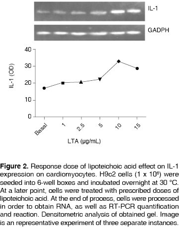
IL-1 expression was determined with the help of optical density and was analyzed with LabWorks® program.
DOSE RESPONSE OF COX-2 EXPRESSION IN MICE CARDIOMYOCITE (H9C2) TREATED WITH LIPOTEICHOIC ACID
H9c2 cells were treated with lipoteichoic acid at different doses. RT-PCR densitometric analysis revealed the fact that, as with other genes COX-2 expression was dependent on lipoteichoic acid concentrations (Figure 3). At basal level no expression was observed. There was COX-2 expression from the minimum LTA dose application onwards (1 μg/mL). A gradual increase was observed when higher concentrations were used (2.5 and 5 μg/mL). Maximum expression was once again achieved at a 10 μg/mL dose. When dosage was increased to 15 μg/mL, a decrease in COX-2 was observed, and as was the case for other studied genes there was no receptor availability, and therefore, expression was decreased; therefore, even though LTA dosage was increased, maximum expression was attained at the most toxic dose (10 μ/mL). COX-2 expression was determined through optical density and analyzed with Lab/Works® program.

DISCUSSION
The main role or micro-organisms in the pathogenesis of pulp and periapical diseases has been clearly established since mid-XX century, when Kakehashi, Moller and Sundqvist established the fact that bacterial presence within root canals was a determining factor for the development of endodontic lesions.2
Dental caries is the main entrance way for micro-organisms into root canals, and is mainly associated to Gram positive micro organisms found in lipoteichoic acid as main pathogenic agent.11,12
Once cariogenic microorganisms de-mineralize enamel or cement, they invade dentinal tubules and are recognized by the first antigen-identifying cells, the dendritic cells, which are associated to the odontoblastic layer. An inflammatory response is then triggered in pulp tissues, inasmuch as, if the causing agent is not removed, there is evolution until reaching necrosis of the pulp tissue, giving thus rise to periapical disease.2,12,13
Previous studies have established the fact that LTA and LPS are potent inductors of inflammatory cytokines as well as other factors involved in inflammatory response and tissue destruction. In fact, LPS is recognized as one of the most potent stimuli to induce cytokine formation, which are inflammation chemical mediators as well as metalloproteinases in macrophages, monocytes, periodontal ligament cells and human gingival fibroblasts.14
Nevertheless, information concerning LTA is rather scarce. LTA could be considered the equivalent of LPS in Gram-positive bacteria, which are the first to colonize the oral cavity and establish on dentobacterial plaque. Therefore, in the case of pulp cells and odontoblasts, more emphasis has been given to LPS.14 Previous studies have established vascular endothelial growth factor by LPS induction through TLR4 and CD14 receptors. Nevertheless, Telles15 found that LTA is also capable of inducing VEGF in mice pulp cells, through its TLR2 receptor.
Most pathogenic micro-organisms, as well as their main substrate, necrotic pulp, can be removed during endodontic procedures. Nevertheless, in common clinical practice this cannot be completely achieved due to the limitations imposed by the complex anatomy of the root. Microorganisms can lodge within dentinal tubules, ramifications, apical deltas or isthmus and thus re-establish infection.16-18
Microorganisms associated to persistent periapical lesions are characterized by being limited to a small number of microbial species (generally only one species). Prevalence of facultative anaerobic Gram positive microorganisms is observed.19-21 That is the case of Enterococus faecalis , a Gram positive coccus which possesses a cellular wall with group D antigens (lipoteichoic acid intracellular glycerol associated to the cytoplasmic membrane). This microorganism is preponderant in persistent endodontic infections, and, by virtue of its phenotypical and microbiological specific characteristics is capable to survive in nutrient and oxygen poor, arid environments. It is also capable of forming biofilm among same-species microorganisms or with other microorganisms, and is equally able to survive when faced to irrigation, intra-canal medication and obturation materials protocols.22,23
In root canal non surgical treatment, bacteremia incidence is low, but there is a possibility for micro-organisms to extend beyond the root apex. Since infective endocarditis is a very serious disease, susceptible patients, before being subjected to dental procedures capable of causing baceteremia, must receive antibiotic coverage, according to current AHA guidelines. The purpose for this procedure is to attempt to clean the circulation of bacteria which settle within damaged cardiac valves.2 Percentages of reported bacteremia caused by dental procedures varies according to the procedure. In endodontic procedures, it varies from 9-32% when observing complete isolation, joined to > 20% when performing endodontic procedures. The aforementioned calculation does not include infection variables.24
Focal infection theory concerning teeth with non-vital pulp as well as endodontically treated teeth has been disapproved. Nevertheless, there is increasing evidence of clear relationship between oral and general disease. Recent case-based epidemiological studies have shown a positive relationship between periodontitis and both cardio-vascular and cerebral-vascular diseases. Periodontitis has also been related to premature, low weight births. In 1993, De Stefano & al reported the fact that subjects afflicted with periodontitis experienced a 25% increase in coronary heart disease risk, when compared to subjects experiencing minimal periodontal disease.2
Meyer and Taylor report the fact that inflammatory mediators such as alpha tumoral necrosis factor (TNFα), interleukin 1 β (IL-1β) and prostaglandins play an important role in heart disease. Bacterial toxins initiate IL1β expression. This process prevents fibrinolysis, but favors clotting and thrombosis. Aorta diseases are associated to TNFα and IL-β secretion. These cytokines favor cholesterol accumulation and smooth muscle proliferation, which presumably results in thinning of vascular walls. Thus, the LTA way, which activates inflammatory mediators is involved as an important mechanism in heart disease and atherosclerosis pathogenesis.25
We are acquainted with the fact that dental pulp inflammation is similar to inflammation of other connective tissues and is mediated by cellular and molecular factors. One of the main molecules involved in inflammatory response are prostaglandins such as PGE2, PGF2a, which have been widely associated to pulp pain and tissue destruction caused by regulation of cytokines and enzymes such as metalloproteinases.14
General systemic responses have been observed in endodontic treatments. These can be due to penetration of bacteria into the tissues, as well as transient bacteremia. It has also been shown that the endotoxin of microorganisms is able to penetrate into periapical tissues, and is present in sufficient amounts so as to induce diseases like infective endocarditis. Evidence supports the assumption that periapical and pulp disease lead to a general exposition to oral bacteria, and thus a potential source of systemic inflammatory mediators able to initiate or exacerbate conditions associated to cardiac disease.24
The aim of the present study was to characterize TNFα, IL-1 and COX-2 on H9c2 treated with LTA. With this purpose in mind, we achieved a H9c2 cells cellular culture where we conducted experiments of the type dose-response so as to characterize gene induction through RT-PCR.
Our results showed that TNFα, IL-1 and COX-2 expression appeared from the moment a minimal dose was applied (1 μg/mL). Nevertheless, we found a more intense induction when a 10 μg/mL dose was applied. This dose induced higher expression levels. The aforementioned expression decreased when a 15 μg/mL dose was applied, this was probably due to cellular receptors' saturation or toxicity induced cellular death.
Peptidoglycans and lipoteichoic acid act like ligands of TLR-2 receptors, and although both agents induce pro-inflammatory molecules expression it has been demonstrated that these ligands elicit different biological responses. No molecular bases have been established to explain in which way peptodoglycans and lipoteichoic acid act synergistically. This would allow the determination of which are the signaling events which result in organ failure, and thus propose new therapies to counter-act its effects. Further studies are needed to support the role of lipoteichoic acid as an important virulence factor.
CONCLUSIONS
The present study revealed the following: a) lipoteichoic acid induced genetic expression of H9c2 cells in a dose-dependent proportion. b) lipoteichoic acid is considered the main factor for Gram positive bacteria, c) lipoteichoic acid functions as adhesion molecule which enables bacteria association to cells, colonization and tissue invasion.
In turn, the present research project showed the possible consequences of deficient endodontic treatment which could lead to activation of biological processes caused by the interaction of microorganisms with host cells. This could elicit, not just the untoward evolution of the treatment, it could also foster the development of a systemic disorder such as infective endocarditis.
The American Heart Association (AHA) has updated their recommendations for prophylactic antibiotic coverage of medically compromised patients. These guidelines are not based upon controlled clinical studies, but rather on relevant articles analysis. They are offered as adjuvants for clinicians and certainly not as a treatment norm or clinical judgment substitute. In case of any doubt on the need to use antibiotic prophylaxis, the clinician should consult with the patient's physician.
Moreover, it is important to develop further clinical studies which could allow abating the bacterial lysis in the bloodstream as well as prevent LTA and peptodoglycans release, and thus decrease risk of infections such as infective endocarditis.
There are still many questions to be answered on the precise role of oral bacteria at the initiation and progression of systemic disorders Clarification is needed on whether there is indeed a cause-effect relationship between periodontal and systemic disease. Nevertheless, there is no scientific evidence to support the idea that bacteria remaining in dentinal tubules after a properly conducted endodontic treatment act as infection foci able to trigger chronic systemic disease.
REFERENCES
1. Chávez de Paz L. Redefining the persistent infection in root canals: possible role of biofilm communities. JOE . 2007; 33 (6). [ Links ]
2. Burns RCS. Vías de la pulpa . 7a. ed. Harcourt. 1999. [ Links ]
3. Peña L. Inmunología . Manual Moderno. España. 1999. [ Links ]
4. Venegas G, Cardoso P. Ácido lipoteicoico: receptores y mecanismo de transducción. REB . 2006; 25: 002. [ Links ]
5. Seung HH, Je Hak K, Martin M, Michalek S, Nahm M. Pneumococcal lipoteichoic acid (LTA) is not as potent as staphylococcal LTA in stimulating toll like receptor 2. Infection and Inmunity . 2003; 71: 10. [ Links ]
6. Guyton and Hall. Manual del tratado de fisiología médica . McGraw Hill Interamericana. México. 1999. [ Links ]
7. Swantek JL, Tsen MF, Cobb JA. IL-1 receptor associated kinase modulates host responsiveness to endotoxin. J Inmunol . 2000; 164: 4301. [ Links ]
8. Woodcock EA, Matkovich SJ. Cardiomyocytes structure, function and associated pathologies. Int J Biochem Cell Biol . 2005; 37. [ Links ]
9. Beshay NM, Zordoky OS. H9c2 cell line is a valuable in vitro model to study the drug metabolizing enzymes in the heart. Journal of Pharmacological and Toxicological Methods . 2007; 56. [ Links ]
10. Anderson R, Becker A. El corazón. Estructura normal y patológica . Mosby Doyma. México. 1994. [ Links ]
11. Weine F. Tratamiento endodóncico . 5a. ed. Madrid. España. Harcourt. 2000. [ Links ]
12. Hargreaves KM, Goodis HE. Seltzer and Bender's dental pulp . Quintessence Books. 2002. [ Links ]
13. Murray P, Smyth T et al. Analysis of pulpar reactions to restorative procedures, materials, pulp capping and future therapies. Crit Rev Oral Bio Med . 2002; 13 (6): 509-520. [ Links ]
14. Ontiveros AG, Gutiérrez G, Lazo MR. Efecto del lipopolisacárido (LPS) y el ácido lipoteicoico (LTA) sobre la ciclooxigenasa-2 (COX-2) en células pulpares humanas. Revista Odontológica Mexicana . 2008; 12 (4): 177-183. [ Links ]
15. Telles, P. Hanks, C. lipoteichoic acid up regulates VEGF expression in macrophages and pulp cells. J Dent Res . 2003; 82: 466-470. [ Links ]
16. Siren E, Haapasalo M, Ranta K, Salmi P, Kerosuo E. Microbiological findings and clinical treatment procedures in endodontic cases selected for microbiological investigation. Int Endod J . 1997; 30 (2): 91-95. [ Links ]
17. Siqueira J. Aetiology of root canal treatment failure: why well-treated teeth can fail. Int Endod J . 2001; 34: 1-10. [ Links ]
18. Sjogren U, Figdor D, Persson S, Sundqvist G. Influence of infection at the time of root filling on the outcome of endodontic treatment of teeth with apical periodontitis. Int Endod J . 1997; 30 (5): 297-306. [ Links ]
19. Chavez De Paz L, Dahlen G, Molander A, Moller A, Bergenholtz G. Bacteria recovered from teeth with apical periodontitis after antimicrobial endodontic treatment. Int Endod J . 2003; 36 (7): 500-508. [ Links ]
20. Peciuliene V, Balciuniene I, Eriksen HM, Haapasalo M. Isolation of Enterococcus faecalis in previously root-filled canals in a Lithuanian population. J Endod . 2000; 26 (10): 593-595. [ Links ]
21. Peciuliene V, Reynaud A, Balciuniene I, Haapasalo M. Isolation of yeasts and enteric bacteria in root-filled teeth with chronic apical periodontitis. Int Endod J . 2001; 34 (6): 429-434. [ Links ]
22. Siqueira JF, Rocas I, Souto R, de Uzeda M, Colombo A. Actinomyces species, streptococci, and Enterococcus faecalis in primary root canal infections. J Endod . 2002; 28 (3): 168-172. [ Links ]
23. Rocas I, Siqueira J, Santos K. Association of Enterococcus faecalis with different forms of periradicular diseases. J Endod . 2004; 30: 315-20. [ Links ]
24. Wilson, Walter et al. Prevention of infective endocarditis. Journal of American Heart Association . 2007; 116: 1736-1754. [ Links ]
25. Meyer, D. Taylor, P. Oral Pathogens: from dental plaque to cardiac disease. Current Opinion in Microbiology . 1998; (1): 88-95. [ Links ]
Note This article can be read in its full version in the following page http://www.medigraphic.com/facultadodontologiaunam Mailing address:
Gloria Gutiérrez Venegas
E-mail: gloria@fo.odonto.unam.mx

