


Services on Demand
Journal
Article
 text in
  English (pdf)
 Article in xml format
 Article references
 Send this article by e-mail
Indicators
-
 Cited by SciELO
 -
 Access statistics
Related links
-
 Similars in
SciELO
Similars in
SciELO
Share

 Permalink
Revista odontológica mexicana
Print version ISSN 1870-199X
Rev. Odont. Mex vol.17 n.3 Ciudad de México Jul./Sep. 2013
Original research
In vitro comparison of anti-microbial activity of AH Plus, RSA and Ledermix against Enterococcus faecalis
Gisela García Ávila,* Raúl Luis García Aranda,§ Luis Manuel Perea MejíaII
* Endodontics Specialty Graduate, Research and Graduate School, National School of Dentistry, National University of Mexico (UNAM).
§ Professor, Endodontic Specialty, Research and Graduate School, National School of Dentistry, National University of Mexico (UNAM).
II Professor, Public Health Department, School of Medicine, National University of Mexico (UNAM).
ABSTRACT
Aim: In vitro assessment of antimicrobial activity sustained by two root canal sealers: RSA®, AH Plus® as well as LedermixN® paste upon Enterococcus faecalis using three different techniques.
Method: Direct contact test (DCT). Sealers were placed on an acrylic surface. A E. faecalis suspension was inoculated and left in a microtube with 1 mL of BHI broth. Logarithmic dilutions were conducted spreading them in blood agar plates so as to quantify CFU's (colony forming units). Dilution test (DT). Sealers and paste were placed in a plastic cylinder. Bacteriae were inoculated in the culture broth and the same quantification procedure was undertaken. Agar dilution test (ADT). On a blood agar plate three wells were manufactured: they were filled with both cements. On the LedermixN paste surface a E. faecalis suspension was inoculated so as to assess growth inhibition areas.
Results: In the Direct contact test, LedermixN paste showed higher antimicrobial activity percentage. Neither of both cements nor the paste presented antimicrobial activity in dilution and Agar dilution test. In the Agar dilution test, AH Plus sealer and LedermixN paste exhibited a hemolysis halo in the blood agar plates.
Conclusions: Direct contact test technique was considered the most appropriate to assess antimicrobial effects of cements.
Key words: Enterococcus faecalis, ADT (agar diffusion test), DCT (direct contact test), sealer cement, antimicrobial activity, LedermixN.
INTRODUCTION
When performing root canal treatment, the main cause of failure is micro-organism survival.
Conditions are uncertain in a necrotic pulp, especially in cases where all pulp space has become infected.
Periapical lesions contain certain variety of bacterial populations. These include Gram-negative anaerobic bacilli, Gram-positive anaerobic cocci and facultative anaerobic streptococci.1,2 Bacteriae are not only present in acute periapical lesions, they have also been found in asymptomatic periapical lesions.3
Dahlen and Bergenholtz4 confirm the presence of a strong association among LPS levels and Gram-negative bacteria prevalence in necrotic pulps. Experiments conducted in animals have demonstrated the fact that endotoxins placed in sterile, empty root canals produce periapical lesions. Schonfeld & al5 correlated the presence of toxins to inflammation in the periapical region. Nevertheless, Yamasaki & al6 have pointed out that inflammatory apical lesions develop long before the pulp becomes totally necrotic. These findings offer great support to the concept that bacterial metabolites and degradation products play a relevant role in the pathogenesis of apical periodontitis.
Enterococcus faecalis is the most frequently isolated microorganism in endodontic failures (80-90%). This fact would lead to suggest that it is a pathogen whose persistence in the root canal represents a significant therapeutic problem.7
E. faecalis is associated to several periapical diseases, including primary infection as well as persistent infections. In the field of primary infections, it is more frequently associated to asymptomatic, chronic periapical lesions than to acute apical periodontitis or acute periapical abscess. It is found in 40% of all endodontic primary infections, with higher frequency in persistent periapical lesions. In fact, E. faecalis has been found nine times over in failed root canal treatment cases than in primary infection cases.8
In order to eliminate infection from the root canals system, it is necessary to cleanse and conform them, as well as achieving a tri-dimensional sealing, eliminating thus empty spaces which potentially could be infected or re-infected.
In the process of sealing root canals, the sealer cement is of paramount importance: it fills spaces unreached by gutta-percha or irrigant agents. This is due to the fact that cements possess components with anti-microbial characteristics which persist long after completion of root canal preparation process.
MATERIALS AND METHODS
Two cements were used in the present study: AH Plus cement, based on epoxy resin (Dentsply-Maillefer Swz) and RSA-Roeko Seal Automix silicon based cement (Whaledent International, USA). LN- Ledermix medicated paste with corticosteroids and broad spectrum antibiotics (Leder, Germany) was equally used in the present study.
Sealer cements and paste were mixed according to manufacturers indications.
ATCC 29212 Enterococcus faecalis strain was used. This was provided by the Public Health Department, School of Dentistry, National University of Mexico (UNAM).
DIRECT CONTACT TEST (DCT)9
Squares of 1 x 1 cm size and 0.5 cm thickness were manufactured with self-cured acrylic. For each sample five squares were used, and were applied on to the acrylic surface. Once set a 10 µL aliquot of an E. faecalis (1 x 10⁸ CFU/mL) solution was inoculated on the sample's surface. The squares were placed on individual micro-tubes (Eppendorf) with 1 mL of BHI (Bioxon) broth with 6.5% NaCl.
Ten µL of each sample were taken, and 5 logarithmic dilutions were performed in order to assess the number of remaining bacteria in each tube.
A 10 µL aliquot of each solution was spread (direct spreading) on blood agar plates, so as to determine colony forming units (CFU/mL).
An additional technique was used to assess bacterial growth, which in the present study was named «plating». It entailed inoculating 10 µL of each and every solution onto the blood agar plate surface, and then extending it immediately with a glass rod, in order to quantify developed CFU.
Plates were incubated at 37ºC for 24 hours. The next day, the same procedure was undertaken so as to again assess the amount of CFU in each tube.
DILUTION TEST (DT)
Both sealer cements as well as the paste were placed within a plastic cylinder measuring 0.8 cm diameter and 0.5 cm thickness.
The cylinder was placed in a microtube with 1 mL BHI broth with 6.5% NaCl.
Twenty µL of E. faecalis (1 x 10⁸ CFU/mL) were inoculated and then shaken. After four hours, 8 logarithmic dilutions were performed. In order to quantify the amount of CFU in each tube a 10 µL aliquot of each sample was taken to be then spread in a blood agar plate which was incubated for 24 hours at 37°C.
AGAR DIFFUSION TEST (ADT)10
Three 0.7 cm diameter and 0.5 cm depth wells were manufactured in a blood agar plate. Each well was filled with sealing cements and paste, and materials were allowed to set.
A 10 µL aliquot of E. faecalis (1 x 10⁸ CFU/mL) was inoculated onto the plate's surface and was evenly spread.
The inhibition zone was assessed so as to determine antimicrobial effect of each material.
RESULTS
DIRECT CONTACT TEST (DCT)
Table I shows comparison of percentages of surviving bacteriae in direct contact, at 0 hours. Two different techniques were used to evaluate.
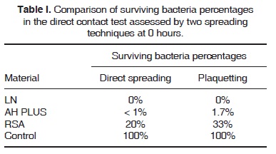
The direct spread column shows the following: LN with no bacterial growth, AH plus with a <1% percentage, and in the case of RSA 20% of total amount of bacteriae survived when compared to the control group.
The «spreading by plates» method revealed similar results: LN prevails with no bacterial development, AH Plus maintains high anti-microbial activity, exhibiting 1.7% of surviving bacteria, with RSA sealer, a 33% bacteria survival rate was observed, thus placing it as the material exhibiting lesser degree of antimicrobial activity when confronted to E. faecalis.
Table II and figure 1 show surviving bacteria in direct contact plate when confronted to bactericidal challenge at 0 and 24 hours. When compared to control sample, LN paste and AH Plus cement presented greater antimicrobial activity with respect to RSA.
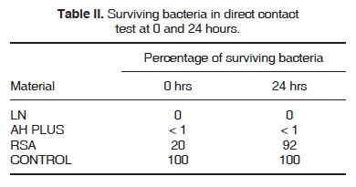
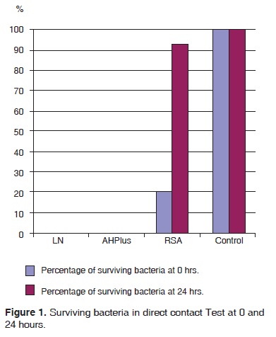
Nevertheless, with the 24 hour incubation period, we were able to confirm the fact that LN eliminated all bacteria; AH Plus exhibited growth lesser than 1%. This would indicate the fact that bacteria which survived direct contact at 0 hrs were able to grow and multiply. With RSA, amount of bacteria increased to 92%; this would indicate existence of a bacteriostatic effect.
STATISTICAL ANALYSIS
CFU data obtained at time 0 were used to conduct a T-test for independent tests: p = 0.130153, therefore p >0.05.
DILUTION TEST
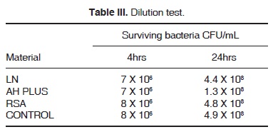
In this test E. faecalis bacterial growth in sealer cements and paste was observed. As a result we could see CFU growth similar to control, this shows absence of antimicrobial activity (Table III).
AGAR DIFFUSION TEST
In this test, neither sealer cement nor paste presented anti-microbial effect, since no inhibition halo was observed on the agar surface. Nevertheless, with LN and AH Plus, a 20 mm hemolysis halo was observed.
DISCUSSION
The aim of the present study was the in vitro evaluation of two sealer cements and one paste against E. faecalis through the use of direct contact test (DCT), agar diffusion test (ADT) and dilution.
Direct contact test assessed cement's anti-microbial effect when bacteria initiate contact with cement surfaces. Sealers examined in the present study exhibited different effects against E. faecalis: LN showed anti-microbial activity, followed by AH Plus; RSA cement took the last place which, in contrast with results obtained with ADT exhibited lesser effect under experimental conditions. It is therefore suggested that due to its rapid polymerization, secondary release substances are minimal, and its excellent sealing ability is due to the expansion it sustains, thus minimizing its dilution.11,12
Funda & al10 also conducted DCT tests with AH Plus and RSA against E. faecalis. In that study, AH Plus did exhibit antimicrobial activity, whereas RSA did not exhibit it. This concurred with our own data. Nevertheless, it must be borne in mind that they measured turbidity and not viability, since turbidity can be a reflection of detached material, and not necessarily be bacteria, which, if present, could be viable or not. In our study we assessed viability in test tubes, and thus were able to demonstrate more accurately the sealer's anti-microbial effect.
In the present test it was also observed that RSA cement exhibited bacterial growth after 24 hours, the percentage grew from 20 to 92%. This led us to consider that this sealer might possess bacteriostatic effect, thus allowing remaining bacteria to grow and multiply to then provide the aforementioned result.
Agar diffusion test (ADT) is the technique commonly used to assess antimicrobial activity of dental materials. This technique not only depends on the toxicity of the material upon the microorganism; it also depends on the high influence of its diffusion in the medium, the concentration of the examined sealer, as well as the size and shape of the antimicrobial agent. Nevertheless, Funda & al10 assessed with this technique RSA and AH Plus sealer cements. In their study, AH Plus showed inhibition to E. faecalis, but the study did not mention the size of the observed inhibition halo. In our study, neither of both cements nor the assessed paste exhibited inhibition halo. Mickel AK & al13 conducted a similar study: they concurred with observations of the present study, that is to say, AH Plus did not show any inhibition for E. faecalis.
In the present study, although neither of both cements or the paste elicited an inhibition halo, with LN and AH plus a hemolysis zone was observed in blood agar plates.
Other researchers, such as Leonardo & al,14 Kouaouzidou & al,15 Peralta & al,16 found that AH Plus did contain formaldehyde during polymerization process, besides the cytotoxicity observed in murine fibroblasts cultures where their death demonstrated high cytotoxicity.
Based on the aforementioned studies, we can infer that the presence of this substance (formaldehyde) in the medium can elicit erythrocyte destruction in the blood agar plates. This was also the case with Ledermix which was described by Pierce & al17 as a paste producing anti-inflammatory effect, and by Chance & al18 as a suitable material for pain attenuation. Lindskog19 indicated that Ledermix acted as a suitable antimicrobial agent. Abbot20 indicated that lesions associated to treatment with Ledermix appeared with apical reparation.
When conducting dilution tests, we observed that sealer cement components were not easily diluted to thus elicit antimicrobial effect. For this reason, no measurable effect could be observed to compare with the control group. It was observed that each technique entailed different characteristics, therefore, one could be more sensitive than the other. Although in most studies we use these techniques, there are other more sensitive techniques to detect E. faecalis: one of them would be PCR (Polymerase Chain Reaction). In this technique, a high 67-77% presence of this microorganism was found when compared to the 24-70% obtained with culture.21,22
CONCLUSIONS
Under the precise conditions of the present study, we might conclude the following:
• In DCT, out of the two studied cements, LN was the one which presented greater antimicrobial activity, RSA presented the lesser amount of activity, and AH Plus exhibited lesser antimicrobial activity than LN but greater than RSA.
• Neither of both sealer cements nor the paste presented antimicrobial activity in the ADT test. AHP exhibited hemolysis halo.
• Direct contact technique was the most suitable to assess the antimicrobial effect of sealer cements
• Antimicrobial activity must not be the only significant parameter to decide upon use of root cement sealer, the associated cytotoxic effect must also be taken into consideration.
REFERENCES
1. Williams BL, Mc Cann GF, Schoenknecht FD. Bacteriology of dental abscesses of endodontic origin. J Clin Microbiol. 1983: 18: 770-774. [ Links ]
2. Lewis MAO, Mac Farlane TW, Mc Gowan DA. Quantiative bacteriology of acute dento-alveolar abscesses. J Med Microbiol. 1986: 21: 101-104. [ Links ]
3. Trope M, Bergenholtz G. Microbiological basis for endodontic treatment: can a maximal outcome be achieved in one visit? Endodontic Topics. 2002. [ Links ]
4. Dahlén G, Bergenholtz G. Endotoxin activity in teeth with necrotic pulps. J Dent Res. 1980; 59; 1033-1040. [ Links ]
5. Schonfeld SE, Greening AB, Glick DH, Frank AL, Simon JH, Herles SM. Endotoxic activity in periapical lesions. Oral Surg Oral Med Oral Pathol. 1982; 53; 82-87. [ Links ]
6. Yamasaki M, Kumazawa M, Kohsaka T, Nakamura H, Kameyama Y. Pulpal and perapical tissue reactions after experimental pulpal exposure in rats. J Endodon. 1994; 20; 13-17. [ Links ]
7. Molander A, Dahlén G. Microbiological status of root filled teeth with apical periodontitis. Int Endod J. 1998; 31; 1-7. [ Links ]
8. Phineiro ET, Gomes BPFA, Ferraz CCR, Teixeira FB, Zaia AA. Evaluation of root canal microorganisms isolated from teeth with endodontic failure and their antimicrobial susceptibility. Oral Microbio Immunol. 2003; 18: 100-103. [ Links ]
9. Weiss EI, Shalhav M, Fuss Z. Assessment of antibacterial activity of endodontic sealers by contact direct test. Endod Dent Traumatol. 1996; 12; 179-184. [ Links ]
10. Cobankara FK, Altinoz HC, Ergani O, Kav K. In vitro antibacterial activities root canal sealers by using two diferent methods. J Endodon. 2004; 30: 57-60. [ Links ]
11. Dartar M, Yilmaz, Kalacy A, Zaimoglu L. A comparison of the in vitro citototxicity of two root canal sealers. J Oral Rehab. 2003; 30: 426-429. [ Links ]
12. Min Kai W, Tigos E, Wesselink PR. An 18 month longitudinal study on a new silicon-based sealer RSA Roeko Seal: a leakage study in vitro. Oral Surg Oral Med Oral Pathol. 2002; 94: 499-502. [ Links ]
13. Mickel AK, Nguyen TH, Chogles. Antimicrobial activity of endodontic sealers on Enterococcus faecalis. J Endodon. 2003; 29: 257-258. [ Links ]
14. Leonardo MR , Bezerra da Silva LA, Tanomaru M. In Vitro Evaluation of Antimicrobial activity of sealers and pastes used in endodontics . J Endodon. 2000; 26: 391-394. [ Links ]
15. Koulaouzidou KT, Papazisis P. Citotoxicity of three resin-based root canal sealers: an in vitro evaluation. Endod Dent Traumatol. 1998; 14: 182-185. [ Links ]
16. García Aranda, Peralta Perez M et al. Evaluación in vitro de la citotoxicidad de tres selladores endodóncicos sobre fibroblastos de ratón de la línea celular L929. Revista Odontológica Mexicana. 2006;10: 63-68. [ Links ]
17. Pierce A, Heithersay G, Lindskog S. Evidence for direct inhibition of dentinoclasts by a corticosteroid-antibiotic endodontic paste. Endod Dent Traumatol. 1988; 4: 44-45. [ Links ]
18. Chance K, Lin L, Shovlin FE, Skribner. Clinical trial of intracanal corticosteroid in root canal teraphy. J Endodon. 1987; 13: 466-468. [ Links ]
19. Pierce A, Lindskog S. The effect of an antibiotic-corticosteroid paste on inflammatory root resorption in vivo. Oral Surg Oral Med Oral Pathol. 1987; 64: 216-220. [ Links ]
20. Abbot PV. Systemic release of corticosteroids following intra-dental use. Int Endod J. 1992; 25:189-191. [ Links ]
21. Molander A, Lundqvist P, Papapanou PN, Dahlen G. A protocol for polymerase chain reaction detection of Enterococcus faecium from the root canal. Int Endod J. 2002; 35: 1-6. [ Links ]
22. Gomes BPFA, Phineiro ET, Gade-Neto CR. Microbiological examination of infected dental root canals. Oral Microbiol Immunol. 2004; 19: 71-76. [ Links ]
Note This article can be read in its full version in the following page: http://www.medigraphic.com/facultadodontologiaunam Mailing address:
Raúl Luis García Aranda
E-mail: rlga@unam.mx

