


Services on Demand
Journal
Article
 text in
  English (pdf)
 Article in xml format
 Article references
 Send this article by e-mail
Indicators
-
 Cited by SciELO
 -
 Access statistics
Related links
-
 Similars in
SciELO
Similars in
SciELO
Share

 Permalink
Revista odontológica mexicana
Print version ISSN 1870-199X
Rev. Odont. Mex vol.17 n.1 Ciudad de México Jan./Mar. 2013
Case report
Prosthetic and surgical treatment of patient previously subjected to hemi-mandibulectomy
Jorge Ernesto Sistos Ramírez,* René Jiménez Castillo,§ Alejandro Benavides RíosII
* Student, Maxillofacial Prosthesis Division, Graduate School, National School of Dentistry, National University of Mexico.
§ Maxillofacial Prosthesis Specialty Coordinator, Graduate School, National School of Dentistry, National University of Mexico.
II Professor attached to the Maxillofacial Prosthesis Department, Graduate School, National School of Dentistry, National University of Mexico.
ABSTRACT
In general terms, the best rehabilitation options for patients previously subjected to hemi-mandibulectomy are far beyond their financial possibilities. For this reason surgical-prosthetic reconstructive approach is mainly restricted to the use of more affordable materials such as Kirschner wire and methyl-methacrylate immediate prostheses. The latter are widely recommended due to their low cost, ease of handling, and because they prevent soft tissue atrophy. A clinical case is reported in this article: 25 year old male patient seeking treatment at the Oncology Service of the Hospital General de Mexico (Mexico's General Hospital) due to the presence of a volume increase in the area of the left mandibular angle. Microscopic analysis revealed presence of mixed malign tumor ( final histopathological diagnosis). It was decided to perform hemi-mandibulectomy of involved area, with reconstruction of lost bone segment by means of positioning an immediate methyl methacrylate prosthesis (thermosetting methyl). The prosthesis was fixated with osteosynthesis wire at both resection margins, at 3 mm above the cortex.
Key words: Hemi-mandibulectomy, mandibular prosthesis, methyl-methacrylate.
INTRODUCTION
Malignancies in the oral cavity are most frequently found in the lateral borders of the tongue, gums, salivary glands and floor of the mouth. The most common histological sub-types are squamous cell carcinoma and mucoepidermoid carcinoma.1-3 A very common fact taking place in medical attention centers dealing with patients afflicted with the aforementioned lesions, is the very late diagnosis of the disease, generally at stage III and IV,1,2 forcing thus the treatment to include excision of soft tissues as well as resection of the mandibular portion adjoining the neoplasm.4 There are then alterations in mandibular function related to deglutition, phonation and facial esthetics. This generates the need for very complex rehabilitation requirements for these patients where the clinician dealing with maxillofacial prosthesis plays a key role.
The aim of the present article was to present the case of a patient subjected to hemi-mandibulectomy. The patient received reconstructions with a grafted immediate prosthesis. The prosthesis was manufactured with thermosetting methyl, methyl-methacrylate material fixated with osteosynthesis wire, at approximately 3 mm above the lower mandibular cortex at both resection margins.
HISTORICAL BACKGROUND
Cantor R, Curtis TA5 grouped patients subjected to mandibulectomies into six classes, according to the anatomical characteristic of the remaining mandible as well as the alterations in its function. Class I encompasses patients subjected to radical alveolar resection without loss of mandibular continuity. This class does not include de-insertion of masticatory muscles, it preserves the greater part of the tongue and adjacent soft tissues. Class II corresponds to unilateral mandibular resection comprising from the distal section of the canine up to the condyle. In this situation the insertion of several masticatory muscles is lost, thus generating the deviation of remaining mandible towards the side of the defect. Class III corresponds to unilateral resections spanning from mandibular midline up to the condyle (hemi-mandibulectomy). In these cases, muscle insertion loss is much greater, causing increased instability in the remaining mandible. Class IV encompasses patients which have been treated with unilateral mandibular resections, but have also been partially rehabilitated with bony and soft tissue grafts to conform a pseudo-articulation. Although temporomandibular articulation has not been re-established, mandibular stability is greater when compared to Classes II and III and presents increased support for placement of prostheses. Class V corresponds to resection cases where condyles are not affected and there is re-establishment of mandibular continuity. Class VI is similar to Class V but lacks bone continuity restoration.
Transitional and final surgical prostheses are available to rehabilitate all classes of patients subjected to mandibulectomies.6,7 The former are represented by Kirschner wire8 as well as chrome-cobalt reconstruction chains. These appliances, although they possess stabilizing effect for the mandibular remains, do not prevent surrounding soft tissue atrophy. Radiotherapy should be used as an adjunct in necessary cases. Classes II, III and IV can be approached with the aforementioned.6,7 The second group encompasses palatal and mandibular ramp prostheses. They only act as a guide to direct mandibular teeth to an inter-cusp position when there is mandibular closure.6 There are also conventional removable partial prostheses as well as prostheses supported by implants, which, in the case of the mandible, are only recommended for marginal or alveolar resections.4,6,7 In our days, and to avoid the aforementioned complications, whenever there are neoplasm free surgical borders, immediate mandibular reconstruction is preferred,9,10 with flaps or grafts stabilized with mini-plates, dynamic compression plates and malleable, tri-dimensional plates.11 The following are derived from the aforementioned: AO titanium plates systems, THOP14 system, titanium mini-plates15 as well as locking reconstructive plate system.16 Techniques are then based upon myocutaneous flaps,17 non-vascularized bone grafts18 as well as vascularized bone grafts; all three can be fixated with the previously mentioned metallic plates.18 Nevertheless, the best option to reconstruct the mandible is provided by vascularized bone grafts and free osteocutaneous flaps.11,19 Donor sites for these flaps are the scapula area,20,21 the iliac crest20,22 radius,23 and fibula.24-26 Usage of the fibula is most desirable, due to the greater amount of bone that can be harvested (up to 25 cm). Skin that can be harvested is sufficient to substitute the floor of the mouth and the skin of the resected area. Furthermore, graft harvesting and oncological surgery are performed simultaneously.4,24,25
Substitution of amputated condyle is a difficult task. To achieve it, several options are offered such as metallic prosthesis placement on grafted bone. Nevertheless, many complications have been reported such as glenoid fosa erosion, infections, extrusion and deafness.25,27 To harvest this anatomical component the distal end of the grafted bone can be modeled to resemble a condyle. This technique has yielded acceptable results.25 Another option could be the self-transplant from the far side of the mandible, to the mandible itself.28 Vascularized bone flaps and free osteocutaneous flaps (especially from the iliac crest and fibula) are the most recommended for placement of bone integrated implants.29
CLINICAL CASE
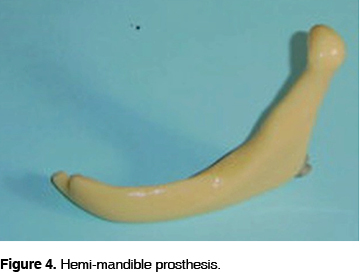
25 year old male patient was admitted in February 2002 at the Oncology Service of the Mexico General Hospital. The patient referred having for two weeks noticed presence of progressive volume increase in the parapharyngeal space. The growth was operated upon in April of the same year performing transmandibular approach. Microscopic evaluation revealed a mixed tumor (pleomorphic adenoma). Five months later, the patient attended the Head and Neck Unit of the Oncology Service at the same hospital due to the presence of a volume increase in the left genal region, together with lockjaw, dysphagia, and pain on that side of the face (Figure 1). A request was made for CAT scan and panoramic radiographic projection (Figure 2). The panoramic radiographic projection revealed a radiolucent, poorly defined lesion, spanning from the premolar area up to ascending mandibular angle and ramus. The CAT scan revealed an osteolytic expansive lesion affecting the mandibular body, extending to the oral cavity in the direction of the masticatory space. An incisional biopsy was taken. Diagnosis emitted was malign mixed tumor (malign pleomorphic adenoma). In February 2003, a hemi-mandibulectomy of the compromised area was performed. The resection rim followed a stepped design to confer stability to the prosthesis (Figure 3). To reconstruct the lost bone segment, an immediate prostheses was manufactured with thermosetting methyl, methyl-methacrylate material. This prosthesis was fixated with osteosynthesis wire at approximately 3 and 6 mm at both resection margins above the cortex. The device was designed based on orthopantomographic study as well as lateral skull X-rays (Figures 4 and 5). Histopathological study of the surgical segment revealed a malignant mixed tumor in the medial and lateral borders of the section (soft tissue). In March 2003 a chemo-therapeutic scheme was initiated, with the use of 5-fluorouracil and cisplatin concurrently with radiotherapy with 50 Gy (Grey units) in ten fractions following an accelerated fraction protocol. A thorax CAT scan performed after the surgical event revealed presence of multiple pulmonary metastases. At the 3 month post-operative follow-up appointment, the patient presented acceptable aspect and mandibular function (Figure 6). Nine months after the surgical event, the patient was still unable to afford conventional rehabilitation procedures, nevertheless, no atrophy of soft tissues, malocclusion or masticatory muscle contractures had developed.


DISCUSSION
In general terms, radical alveolar resection of the mandible does not require special procedures for its reconstruction. Nevertheless, in cases when the mandible is resected by segments, appropriate planning is required so as to reconstruct the lost bone fragment. There are functional, anatomical and esthetic considerations which must be considered when selecting the reconstruction modality of a hemi-mandibulectomized patient. The first considerations to take into account are those pertaining to restoration of oral physiology, mastication, occlusion, phonation and deglutition. Next considerations to observe are those pertaining to the restoration of inter-incisive opening, to the distance and alignment of dental arches, as well as replacement of lost soft tissues. Esthetic considerations are those pertaining to facial symmetry recovery, re-establishment of lower facial height, chin projection, and inasmuch as possible , avoiding scars in the facial skin. Anti-esthetic impact of surgical defect in immediate post-operative stages is minimal. Nevertheless, if there are no measures established to reconstruct the affected mandible, soft tissues will generally atrophy, and this situation will become, for the patient, totally inacceptable. Moreover, when the resection compromises the posterior section of the body of the mandible, as well as its ascending ramus, mandibular function is altered due to the loss of action experienced by ipsi-lateral pterigoyd muscles; this causes that the masseter and contra-lateral internal pterigoyd muscles move the remaining portion of the mandible in an upward and medial direction, generating thus a rotation from a fulcrum point located in the molar area. The result of this phenomenon is formation of anomalous occlusion and regional muscle contracture at the site of the surgical defect.
Segmented sectioning of the mandible anterior portion, especially in incisors, canines and chin areas cause inadmissible esthetic and function alterations. Therefore, form and function must be immediately restored.
For mandibular rehabilitation, it is best to use grafts and osseous or osseous myocutaneous grafts stabilized with plates, manufactured of self-transplanted condyles and bone integrated implants. The fact that most patients subjected to hemi-madibulectomies come from less socio-economic favored sections of the population, indicate that cheaper materials must be used, such as Kirschner wire and immediate thermosetting methyl, methyl-methacrylate prostheses. Use of the latter is recommended for being inexpensive and easy to handle, since they avoid mandibular rotation and prevent soft tissue atrophy. Similarly, when there are neoplasm positive section borders, it is advisable to place one of these prosthesis to avoid radiation under-dosage, or to avoid the possibility of having to remove a metallic plate or graft/flap due to the presence of a relapse.
Finally, when a lesion free patient is able, in the future, to afford another type of rehabilitation, the state of remaining tissues will undoubtedly be better.
CONCLUSIONS
Apparently, thermosetting methyl, methyl-methacrylate material is a valid alternative for implanted, immediate prostheses for patients requiring radiotherapy protocols due to border neoplasm or when the risk of relapse precludes optimal reconstruction techniques. Possibility for future reconstruction is preserved since this device has the effect of safeguarding space.
REFERENCES
1. Frías MM, Zeichner GI, Súchil BL, Ochoa CFJ. Epidemiología descriptiva del cáncer de cavidad bucal en el Instituto Nacional de Cancerología (1985-1992). Rev Inst Nal Cancerol 1997; 43 (2): 80-85. [ Links ]
2. Ramírez AV, Esquivel PL, Ochoa CFJ, Cuapio OA, Frías MM, Meneses GA, Sánchez MG. Cancer of the mobile tongue in Mexico. A retrospective Study of 107 Patients. Oral Oncol, Eur J Cancer 1995; 31B (1): 37-40. [ Links ]
3. Sánchez MMP, Díaz VD, Aparicio CG. Frecuencia del carcinoma epidermoide en cavidad bucal en el Hospital Central Militar de 1987 a 1997. Medicina Oral 1999; 1 (1): 20-22. [ Links ]
4. Shah JP, Patel SG. Head and neck surgery and oncology . 3rd Ed. New York: Mosby; 2003: 589-592, 614-631. [ Links ]
5. Cantor R, Curtis TA. Prosthetic management of edentulous mandibulectomy patients. Part I: Anatomic, physiologic and psychologic considerations. J Prosthet Dent 1971; 25: 446. [ Links ]
6. Beumer III J, Curtis TA, Marunick MT. Maxillofacial rehabilitation . Prosthodontic and surgical considerations. St. Louis: Ishiyaku EuroAmerica, Inc.; 1996: 113-223. [ Links ]
7. Taylor TD. Clinical maxillofacial prosthetics . Chicago: Quintessence publishing Co, Inc.; 2000: 205-213. [ Links ]
8. Lee KY, Kore JM, Perry CJ. Use of the Kirschner wire for mandibular reconstruction. Arch Otolaryngol Head Neck Surg 1988; 114 (1): 68-72. [ Links ]
9. Schusterman MA, Harris SW, Raymond AK, Goepfert H. Immediate free flap mandibular reconstruction: significance of adequate surgical margins. Head Neck 1993; 15(3): 204-207. [ Links ]
10. Martin PJ, O'Leary MJ, Hayden RE. Free tissue transfer in oromandibular reconstruction. Necessity or extravagance? Otolaryngol Clin North Am 1994; 27(6): 1141-1150. [ Links ]
11. Moscoso JF, Keller J, Genden E et al. Vascularized bone flaps in oromandibular reconstruction. A comparative anatomic study of bone stock from various donor sites to assess suitability for endosseous dental implants. Arch Otolaryngol Head Neck Surg 1994; 120 (1): 36-43. [ Links ]
12. Freitag V, Hell B, Fischer H. Experience with AO reconstruction plates after partial mandibular resection involving its continuity. J Craniomaxillofac Surg 1991; 19 (5): 191-198. [ Links ]
13. Futran ND, Urken ML, Buchbinder D et al. Rigid fixation of vascularized bone grafts in mandibular reconstruction. Arch Otolaryngol Head Neck Surg 1995; 121 (1): 70-76. [ Links ]
14. Buchbinder D, Urken ML, Vickery C et al. Bone contouring and fixation in functional, primary microvascular mandibular reconstruction. Head Neck 1991; 13 (3): 191-9. [ Links ]
15. Hidalgo DA. Titanium miniplate fixation in free flap mandible reconstruction. Ann Plast Surg 1989; 23 (6): 498-507. [ Links ]
16. Herford AS, Ellis E 3rd: Use of a locking reconstruction bone plate/screw system for mandibular surgery. J Oral Maxillofac Surg 1998; 56 (11): 1261-1265. [ Links ]
17. Cordeiro PG, Hidalgo DA. Soft tissue coverage of mandibular reconstruction plates. Head Neck 1994; 16 (2): 112-115. [ Links ]
18. Foster RD, Anthony JP, Sharma A, Pogrel MA. Vascularized bone flaps versus nonvascularized bone grafts for mandibular reconstruction: an outcome analysis of primary bony union and endosseous implant success. Head Neck 1999; 21 (1): 66-71. [ Links ]
19. Gurtner GC, Evans GR. Advances in head and neck reconstruction. Plast Reconstr Surg 2000; 106 (3): 672-82; quiz 683. [ Links ]
20. Kuriloff DB, Sullivan MJ. Mandibular reconstruction using vascularized bone grafts. Otolaryngol Clin North Am 1991; 24 (6): 1391-1418. [ Links ]
21. Sullivan MJ, Baker SR, Crompton R, Smith-Wheelock M. Free scapular osteocutaneous flap for mandibular reconstruction. Arch Otolaryngol Head Neck Surg 1989; 115 (11): 1334-1340. [ Links ]
22. Boyd JB. The place of the iliac crest in vascularized oromandibular reconstruction. Microsurgery 1994; 15 (4): 250-256. [ Links ]
23. Cordeiro PG, Disa JJ, Hidalgo DA, Hu QY. Reconstruction of the mandible with osseous free flaps: a 10-year experience with 150 consecutive patients. Plast Reconstr Surg 1999; 104 (5): 1314-1320. [ Links ]
24. Yim KK, Wei FC. Fibula osteoseptocutaneous flap for mandible reconstruction. Microsurgery 1994; 15 (4): 245-249. [ Links ]
25. Hidalgo DA. Fibula free flap mandible reconstruction. Microsurgery 1994; 15 (4): 238-244. [ Links ]
26. Jones NF, Monstrey S, Gambier BA. Reliability of the fibular osteocutaneous flap for mandibular reconstruction: anatomical and surgical confirmation. Plast Reconstr Surg 1996; 97 (4): 707-716. [ Links ]
27. Patel A, Maisel R. Condylar prostheses in head and neck cancer reconstruction. Arch Otolaryngol Head Neck Surg 2001; 127 (7): 842-846. [ Links ]
28. Hidalgo DA. Condyle transplantation in free flap mandible reconstruction. Plast Reconstr Surg 1994; 93 (4): 770-781 [ Links ]
29. Gurlek A, Miller MJ, Jacob RF et al. Functional results of dental restoration with osseointegrated implants after mandible reconstruction. Plast Reconstr Surg 1998; 101 (3): 650-655. [ Links ]
Note This article can be read in its full version in the following page: http://www.medigraphic.com/facultadodontologiaunam Mailing address:
René Jiménez Castillo
E-mail: renejimenezc@gmail.com

