


 nueva página del texto (beta)
 Inglés (pdf)
 Artículo en XML
 Referencias del artículo
 Enviar artículo por email
 Citado por SciELO
  Similares en
SciELO
Similares en
SciELO 
 Permalink
Introduction
The nervous system has five forms of communication: mechanical signaling, electrical signaling, short-range signaling, local signaling, and long-range signaling. Mechanical signaling involves adhesion and recognition, whereas electrical signaling is triggered by electrical synapses and gap junctions (GJs). Short-range signaling occurs thanks to chemical synapses and neurotransmitters, local signaling is triggered by hemichannels, and finally, long-range signaling involves neurohormones1,2.
In most animal cells, except isolated moving cells, intracellular channels are formed between the cytoplasm of adjacent cells. These channels ensure the exchange of ions, metabolites, and other messenger molecules, but they also mediate electrical coupling among connected cells3,4. Similarly, specific intracellular channels are formed by multi-protein complexes, commonly known as GJs. Pathologies affecting GJs have adverse effects on vital organs, senses, and bones.
There are three families of unrelated proteins that are involved in electric synapses connexins, pannexins, and innexins (Fig. 1). The family of connexins comprises 20 members which are found only in chordates4, and some of them are expressed in the central nervous system (CNS)5. On the other hand, pannexins are found in vertebrates, and innexins exist only in invertebrates.
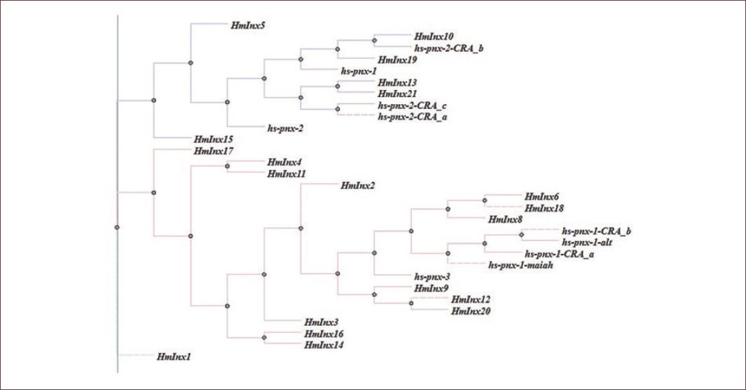
Figure 1 Cladogram showing innexins/pannexins of Hirudo medicinalis and Hirudo sapiens protein sequences.
A GJ channel is formed by two hemichannels, each from one of two neighboring cells, leaving a narrow 2-4 nm gap between them. Furthermore, some hemichannels only serve as a backdoor to important metabolites. Studies on the functional role of innexins in the nervous system of invertebrates generate results that are applicable to all animal species, particularly mammals. Therefore, in this review, we emphasize on the importance of invertebrate animal models, namely, Hirudo medicinalis, to study synaptic function and thus respond to complex questions in neuroscience. Similarly, we present a panorama of neurological disorders, in which synaptic dysfunction is a characteristic. Then, we compare this information to that reported in the literature about human connexins and current treatments of human neurological diseases.
Hirudo development of the nervous system
It is well-known that molecular interactions influence the development of virtually all types of cells, including nerve cells, during embryogenesis6,7. GJs are considered as important mediators of cell communication8. In fact, research has demonstrated that GJs play a key role in the development and preservation of nerve cells: GJ inhibition causes progenitor cells to cease proliferation, thus leading to cell differentiation, or even death9. Cells can affect one another and promote growth. Electrical synapses work both during the development of the nervous system and in adulthood. If an innexin is ectopically expressed, it creates new connections with adjacent neurons, thus forming new communication channels. However, if some innexins are blocked, new channels cannot be formed (Fig 2)9.

Figure 2 Characterization of Hirudo medicinalis adult neurons expressing different innexin transcriptions. A: ventral aspect of a single ganglion6; B: expressions of different innexins family members from quantitative polymerase chain reaction.
H. medicinalis is a well-known species of the genus Hirudo and the family of Hirudinidae; however, it is usually confused with another leech species, Hirudo verbana, mainly due to their physical resemblance and similar behavior (Fig 1). Due to the size of its neurons and the structure of its nervous system, H. medicinalis is a popular model in biology. It is used to study neuron-to-neuron communication, specifically GJs. The nervous system of H. medicinalis comprises 32 ganglia, which is considered as motor brains and contains 400 neurons of approximately 10-50 µ in diameter. These characteristics make H. medicinalis neurons more accessible to neuroscience studies. Furthermore, ganglia six and seven contain the male and female sexual organs of this species, respectively9.
Important cells of the nervous system
GJs take part in electrical coupling and contribute to synapse regeneration after neuronal injury. There is an average of ten glial cells or neuroglia per neuron, which makes them the most abundant cell types in the CNS. Glial cells comprise macroglia (astrocytes and oligodendrocytes), microglia, and ependymal cells. Astrocytes are associated with the majority of neurological disorders and diseases, such as brain ischemia, Alzheimer's disease (AD), and epilepsy. Similarly, GJs can be related to the malignant degree and metastasis of brain tumors, yet the functional role of most of these proteins is still unknown, particularly in the case of diseases of the nervous system9. Oligodendrocytes form the myelin that surrounds and protects axons, whereas microglia are the first and major form of active immune defense in the CNS. Astrocytes also exchange ions and metabolites, such as inositol 1,4,5-triphosphate, and adenosine triphosphate (ATP) through GJs. Similarly, oligodendrocytes maintain myelin using GJs. Furthermore, studies on brain ischemia and epilepsy using mice as biological models have managed to block GJs activity, thus addressing the possibility of neural protection to prevent brain ischemia or metabolic stress. Finally, it has also been demonstrated that macrophages can influence GJ expression in astrocytes, whereas activated microglia communicate using GJs9.
Role of GJs in neuropathological conditions
Diseases introduction
Knowing the functional role of GJs in conditions such as brain ischemia, neurodegenerative diseases, epilepsy, heart arrhythmia, cataracts, and tumors could allow experts to determine and understand the real molecular mechanism of GJ proteins network during disease processes, a practical separation of diseases based on the type of synapse involved, to simplify their study. Consequently, it could also help design treatments to successfully eliminate these conditions. Connexins and pannexins are key proteins to the bone and skeletal muscle development, maintenance, and regeneration. Research has found that connexin channels are present in the bone among osteoblasts, osteoclasts, and osteocytes. Similarly, connexins are important for bone growth and adaptation10.
The permeability of GJ channels is increased by multiple factors, including low pH levels, high cytosolic free calcium levels, and voltage gradient across the GJ. In vertebrate species, embryonic tissues are easily dissociated by treating them with low concentrations of a proteolytic enzyme, such as trypsin, along with a divalent-cation chelator, such as ethylenediaminetetraacetic acid, to disrupt the protein-protein interactions that hold the cells together.
The connection between cancer development and GJs was first reported by Loewenstein and Kanno when they studied liver cancer using electrophysiology techniques in 196611. Twenty-four years later, it was already established that GJ mutations were linked to multiple human genetic diseases12. Nowadays, connexins are known to be related to the development of conditions such as hearing loss, skin disorders, congenital cataracts and other vision disorders, heart arrhythmia, demyelinating diseases, and oculodentodigital dysplasia13.
Although the exact percentage of electrical, chemical, or combined synapse participation is currently unknown, a simplification would help unravel the workings of opening neuronal communication channels during different conditions and would allow progress in these areas. At the molecular level, cellular communication plays a decisive role in the development or treatment of these diseases, and of course, the channels through which these signal molecules travel (Fig 3).
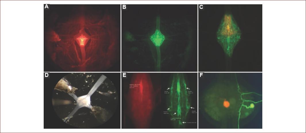
Figure 3 A: internal view of Hirudo medicinalis ganglion through epifluorescence using Lucifer yellow injection, neuropil is illuminated, B: internal view of H. medicinalis ganglion through epifluorescence using Neurobiotin, entire ganglion is illuminated, C: superposition of previous images, showing neuropil location in ganglion, D: ventral view of H. medicinalis ganglion - upper side corresponds to the organism's anterior connective tissue, E: control ganglion, red: TMR-dextran, green: Neurobiotin, F: visualization of the structure of a touch-sensitive neuron (T neuron) in ganglion using Neurobiotin.
Brain ischemia
Cerebrovascular ischemia, or brain ischemia, occurs when there is insufficient blood flow to the brain. It leads to limited oxygen supply and can cause the death of brain cells. The side of the ischemic lesion has the same physiopathological mechanism and local consequences with different clinical manifestations, its severity, and its duration, yet sufferers can risk death. When a person presents symptoms of brain ischemia, they must receive emergency treatment. Even a temporary deficit in oxygen supply can impair the brain, when the brain is damaged as a result of ischemia, the consequences are severe (i.e., physical or mental disability or death). In fact, cerebrovascular diseases such as brain ischemia are a leading cause of death in the USA. Cerebral vascular disease is ranked as the second leading cause of death (9.7%) in Mexico.
The functional role of astrocytes and GJs in brain ischemia is still unclear, yet the two are known to fulfill a potentially neuroprotective function that involves ischemic tolerance and remodeling of neuronal networks by phagocytosis14. Experiments using cell culture techniques have demonstrated that blocking GJs in astrocytes make the cells more vulnerable to glutamate cytotoxicity15. Likewise, using hippocampal slice cultures, it was found that blocking GJs cause neuronal damage in ischemic conditions, such as oxygen and glucose deprivation16. GJs channels are known to remain open during these conditions17.
The most promising strategy for evaluating the functional role of connexins involves blocking them using interfering RNA, drugs such as carbenoxolone (CBX), and mimetic peptides, including Gap 19 and Gap 1617. Yet any state of overregulation can lead to microglial activation and neuroinflammation18. This strategy can help experts identify which connexins are implicated in apoptosis and inflammation. Consequently, it would be possible for scientists to develop new treatment options for brain ischemia. To date, Connexin 43 (Cx43) is the most promising connexin19. Cx43 expression is sensitive to neuronal injury and can be detected as early as 2 h post permanent middle cerebral artery occlusion (pMCAO). These findings underscore Cx43 GJ as a potential early target for therapeutic intervention in ischemic stroke. Cx43 is potentially a useful early marker to delineate and investigate the ischemia progression19.
Epilepsy
Leading causes of epilepsy include infectious diseases and abnormal brain development. Epilepsy is a neurological disorder that develops as a result of abnormal brain function, neural communication anomalies, or imbalanced neurotransmitters - which are responsible for neurotransmission (i.e., communication between neurons). Hence, epilepsy occurs when neurons occasionally send abnormal signals in the brain20.
After a head injury, brain stroke, or any other accident, the brain might try to self-repair; however, this process can accidentally lead to abnormal brain connections, and thus epilepsy. During the epileptic crisis, many neurons send signals at the same time - as many as 500 times/s. This event causes changes in a person's behavior, movements, feelings, and levels of consciousness. Nowadays, epilepsy is referred to as a spectrum of disorders with a myriad of causes, severities, and effects. Some people may experience convulsions or lose consciousness, whereas others may simply stop what they are doing, have a short lapse of awareness, and stare into space for a moment without knowing what is happening around them20.
In general, a person is considered to have epilepsy after two unprovoked seizures; a "seizure" is a paroxysmal alteration of neurologic function caused by the excessive, hypersynchronous discharge of neurons in the brain, separated by at least 24 h. Provoked seizures are caused by known precipitating factors, such as high fever, nervous system infections, acute traumatic brain injury, or fluctuations in blood sugar, or electrolyte levels. To date, nearly 2.3 million adults and 450,000 children and adolescents in the US suffer from epilepsy. In Mexico, there are about 2 million people with epilepsy, according to the Ministry of Health. Anyone can have epilepsy, men and women, regardless of their race, ethnicity, or age.
There are also severe cases of epilepsy, such as Dravet syndrome. This is a rare but lifelong dysfunction of the brain characterized by medically refractory seizures and causing serious learning disabilities. Current medications can control epilepsy in 60% of sufferers, whereas the remaining 40% experience what is commonly known as drug-resistant epilepsy. Nowadays, treatment options for epilepsy include about 20 different types of drugs, special diets, and surgical techniques, and medical specialty like neurosurgery. Common tests for epilepsy diagnosis include imaging and monitoring techniques (e.g., electroencephalography and magnetoencephalography), patient medical history, blood tests, behavioral tests, and neurological exams. Cx43 is related to cryptogenic epilepsy that is defined as a group of focal or generalized epilepsy, which are believed to be symptomatic of a histopathological or cellular occult alteration, but not of a genetic nature21.
Research has found that Cx43 in GJs21 and hemichannels22 adversely affects astrocyte function, thereby causing epileptic seizures, which in turn induce Cx43 expression23. On the other hand, changes in Connexin 36 (Cx36) expression cause cell death24 and neural communication anomalies, thus resulting in epilepsy25. Furthermore, GJs in astrocytes play a crucial role in ionic regulation; particularly, they can change the concentration of potassium ions. Changes in the strength of GJ coupling can cause neuronal hyperexcitability, and consequently, spontaneous epileptic activity26, which ultimately leads to astrocyte uncoupling. Finally, there is evidence that pannexin 1 (Panx1) contributes to the maintenance of epileptic seizures by releasing ATP. The absence of Panx1 in astrocytes potentiates seizure manifestation due to low levels of adenosine kinase, whereas the absence of Panx1 in neurons reduces seizure manifestation27.
GJ blockers to treat epilepsy
In a recent research Scemes et al.28, they studied 13 GJ blockers as treatment options for epilepsy: CBX, quinine, mefloquine, quinidine, anandamide, oleamide, heptanol, octanol, meclofenamic acid, niflumic acid, flufenamic acid, glycyrrhetinic acid, and retinoic acid. In the in vitro experiments, all these compounds demonstrated to have anticonvulsant effects in brain slices. The blockers modified the behavioral parameters related to seizures induced by 4-aminopyridine, pentylenetetrazol, pilocarpine, penicillin, and maximal electroshock. Similar research works suggest that GJ blockers are a future alternative for the treatment of epilepsy. Nevertheless, most of these compounds have been discontinued as treatments due to their side effects, thus implying that further research must be conducted to identify the action mechanisms for neurological disorders such as epilepsy (Table 1).
Table 1 GJ blockers and their side effects
| Blocker | Connexin | Side effects | Reference |
|---|---|---|---|
| Carbenoxolone | Non-selective | Hydroelectrolytic disorders | 58 |
| Quinine | C x 36, C x 45, C x 50 | Serious adverse events tinnitus, deafness, dizziness, vomiting | 59 |
| Mefloquine | C x 36, C x 43, C x 50 | Neurotoxic side effects following large doses | 12 |
| Anandamide | C x 32, C x 43 | Impact on calcium channels Toxicity following large doses | 60 |
| Heptanol | C x 32, C x 43, C x 45 | Unstated | |
| Octanol | C x 43, C x 46, C x 50 | Unstated | |
| Meclofenamic acid | C x 36, C x 43, C x 50 | None | 61 |
| Niflumic acid | C x 43, C x 45, C x 50 | None | 61 |
| Flufenamic acid | C x 26, C x 32, C x 40, C x 43, C x 46, C x 50 | None | 61 |
| Glycyrrhetinic acid | Non-selective | None | 61 |
| Retinoic acid | C x 38 | None | 61 |
| Mimetic peptides | Specific | Reduce connexin function, temporarily | 10 |
| siRNA | Specific | Unknown |
Multiple studies on connexins have been conducted with the aim of understanding and reporting the devastating effects of connexin dysfunctions (Table 2). Connexins are important, since they form synapses. Moreover, GJs are present in all biological cells, except spermatozoids, and some types of blood cells.
Table 2 Tissues and pathologies associated with connexins
| Connexin (Hirudo sapiens) | Tissue or site of expression | Mutation-related pathologies | Reference |
|---|---|---|---|
| C x 26 (GJB2) | Ependymal cells, pinealocytes, breasts, cochlea, placenta, hepatocytes, pancreas, kidneys, intestine, and epidermis | Sensorineural hearing loss, palmoplantar hyperkeratosis | 22, 62, 63, 64 |
| C x 29/C x 30.2-31.3* (GJC3) | Heart, skeletal muscle, liver, myelinating Schwann cells, Bergmann glia, and oligodendrocytes | Hearing impairment with pathological changes in cochlea | 4, 62, 63,64 |
| C x 30 (GJB6) | Epidermis, cochlea, astrocytes, and hippocampal pyramidal neurons | Nonsyndromic hearing loss, hidrotic ectodermal dysplasia | 11, 22,64, 65 |
| C x 31.9 (GJD3) | Smooth muscle cells expressed in the heart | Arrhythmia | 25, 31, 58,67 |
| C x 30.3 (GJB4) | Epidermis and kidneys | Erythrokeratodermia variabilis (EKV) | 29,64,66 |
| C x 31 (GJB3) | Cochlea, auditory nerves, placenta, and epidermis | Hearing impairment, erythrokeratodermia variabilis (EKV) | 4, 22, 31, 36, 58, 63, 64 |
| C x 31.1 (GJB5) | Middle and outer layers of the corneal epithelium and epidermis | Expressed upregulation in corneas affected with Stevens-Johnson syndrome (SJS) | 63,68 |
| C x 32 (GJB1) | Highly expressed in liver, pancreas, and kidneys. Expression in the nervous system is reduced to oligodendrocytes. | Hereditary peripheral neuropathy, Charcot-Marie-Tooth disorder (CMTX) | 4, 5, 31, 58, 64, 65 |
| C x 36 (GJA9/GJD2) | Neurons, microglia, and pancreatic B-cells | Involved in gliomas, astrocytomas, and glioblastomas | 14, 22, 65,69 |
| C x 37 (GJA4) | Endothelial cells, granulosa cells, lungs, and epidermis | Atherosclerosis | 4,13,29,58,66 |
| C x 40.1 (GJD4) | Pancreas, kidneys, skeletal muscle, liver, placenta, myoblasts, and heart | Muscular dystrophy | 3,4,31,58 |
| C x 40 (GJA5) | Cardiomyocytes,endothelial cells, and lungs | Arrhythmia | 4,31,66 |
| C x 43 (GJA1) | Ubiquitous, highly expressed in astrocytes, retinal cells, and basal layers of the corneal epithelium and anterior stroma | Oculodentodigital dysplasia (ODDD), syndactyly type III, vaso atrial heterotaxy, astrocytomas | 6,40,57,58,64,68,70 |
| C x 45 (GJA7/GJC1) | Endothelial cells, neurons, smooth muscle, myoblasts, neurons of the cerebral cortex, claustrum, and olfactory bulb glomeruli | Muscular dystrophy | 3,4,31,58 |
| C x 46 (GJA3) | Highly expressed in heart, placenta, testis, chondrocytes, and ocular lens | Autosomal dominant zonular pulverulent cataract-3 (CZP3) upregulated in breast cancer cells | 7,12,22,69 |
| C x 47 (GJA12/GJC2) | Oligodendrocytes along the surface of internodal myelin | Linked to neuropathies such as Pelizaeus-Merzbacher-like disease 1 | 22,69 |
| C x 50 (GJA8) | Ocular lens | Zonular pulverulent cataract-3 (CZP3) | 21,22 |
Neurodegenerative diseases
AD
AD is the most common degenerative disease, in México, approximately 800,000 people have AD. Clinically, AD is caused by cerebral atrophy of the frontal cortex, along with neurofibrillary tangle and large numbers of senile plaques. The ApoE4 allele is usually seen as a risk factor for AD, yet health experts also look for other indicators. That is, not everybody carrying the ApoE4 gene variant will go onto develop AD as they grow old, although the probabilities are significantly higher. Moreover, some experts state that knowing that a person carries the ApoE4 allele does not benefit at all since none of the current treatment options cures the disease. ApoE gene polymorphic form ApoE4 (ε4) allele is the most commonly associated genetic risk factor linked with the late onset of the AD. In response to injury or neuroinflammation, Apoε4 undergoes neuron-specific proteolysis. Even if it is not clear the relationship between this allele and Cx43, it will be interesting to focus on Cx43 function under this condition28. Thus far, the relationship between AD and GJs remains unclear, yet glial cells are thought to contribute to the aggressive propagation of this disease by inhibiting cell communication and promoting neuroinflammatory responses29. Moreover, scientific evidence suggests a relationship between neurotoxicity of Aβ42 deposits in AD and upregulation of Cx43 as a result of released neurotoxic molecules that cause oxidative stress30.
That said, researchers have found that Cx43 prevents, to some extent, the brain from deteriorating; however, Cx43 overexpression identified in experiments on AD-like pathologies30 seems to severely affect intercellular communication, thus relating Cx43 overexpression with neuroinflammation.
Cx43 also seems to be involved in glutamate excitotoxicity induced by manganese exposure, thus promoting neurodegenerative diseases, such as AD and Parkinson's disease (PD). Moreover, in the relationship between GJ activity in glial cells and glutamate excitotoxicity, the latter is linked to excessive NMDAR activity and neurodegeneration31. That said, two promising treatments of AD include blocking specific GJs for Cx4332 and the Metabolic Enhancement for Neurodegeneration (MEND) approach. MEND is a 36-point therapeutic personalized program including aspects such as medication, comprehensive dietary changes, vitamins, brain stimulation, and exercise, to name but a few. There is a revision that mentions the importance of GJ in AD, their positive and negative role in neurons and other cell types, expressing that there is a need for further investigation in a GJ oriented treatment for AD33.
PD
PD is characterized by the loss of dopaminergic neurons in the part of the brain known as the substantia nigra (SN). There are no exact numbers of Parkinson's patients in Mexico. However, the National Institute of Neurology and Neurosurgery estimates a prevalence of 50 new cases per 100,000 inhabitants/year. Clinical symptoms include shaking, rigidity or tremor, and motor disorders such as bradykinesia which means slowness of movement, and it is one of the cardinal symptoms of Parkinson's (i.e., difficulty with walking). The 1-methyl-4-fenyl-1,2,3,6-tetrahydropyridine (MPTP) chemical model is used to represent the conditions of PD, and it causes permanent symptoms. MPTP is extensively modeled in mice and is characterized by overexpression of Cx43 in both the striatum and the astrocytic hemichannels in the SN, thus contributing to the death of dopaminergic neurons34. In addition, MPTP animal models have shown accelerated neuron loss in the absence of Cx30 as a result of a disruption in astrocyte energy metabolism35.
A breakthrough in research on PD is the study of the inferior olivary nucleus (ION), which is a complex structure forming a bulge in the ventral surface of the medulla oblongata. The ION receives a wide range of sensory and motor afferents, and it is the source of the climbing fibers ascending to the cerebellum. Similarly, the ION is thought to play a key role in the generation of tremor in PD. Nowadays, olivary neurons are coupled by means of GJs, yet further research is necessary to determine their functional role. In other publications they mention the importance of focusing attention on the GJs for PD, for example, they say that GJ blocker treatment combined with inhibition of K+ channels might be a good approach to correct motor dysfunction in PD patients36 and that the use of GJ blockers in PD appears to be a promising treatment, it has been reported to improve motor function in hemiparkinsonian rats since GJ activity elicits beta oscillations in the basal ganglia nuclei, leading to akinesia in PD37.
Brain tumors (glioma)
A glioma is only one type of CNS tumor that originates in the glial cells of the brain or the spine. Gliomas are the most common type of brain tumors - they are space-occupying lesions causing intracranial pressure, vascular occlusion, and cerebral edema. In Mexico, an average of 30,000 new cases of brain cancer is detected annually, according to data provided by the Mexican Council of Neurological Surgery. The literature reports that in cases of severe gliomas, Cx43 is present in low concentrations; however, it is also known that the structure of Cx43 itself encourages glioma proliferation38. In addition, Cx43 suppresses tumor growth, regardless of GJ channels. Namely, it is believed that Cx43 C-terminus directly inhibits tumor metastasis, possibly due to the correlation between Cx43 expression and protein kinase B (Akt)/extracellular signal-regulated kinase39. In other words, Cx43 acts as an inhibitory regulator of the activation of growth factor receptors, usually related to treatments for glioblastomas (GBM).
The study of brain tumor cells entails a deep study of the cell cycle. For instance, preliminary data have shown that Cx43 helps regulate the cell cycle during the S phase by interacting with a protein kinase40. However, Cx43 is also known to promote chemoresistance during GBM treatment by reducing the apoptotic effect of temozolomide41. From this perspective, researchers have been able to identify cancer cell lineages with these characteristics42. In fact, identifying cells with aberrant Cx43 expression allow experts to develop new treatment options for brain tumors and block GJs to prevent chemoresistance43.
New approaches have been developed to identify tumor genesis response mechanisms. For instance, laser scanning microscopy (confocal laser scanning microscopy) and simple molecule localization microscopy are employed for the spatial localization of Cx43 in tumor genesis and the formation of metastasis44. Moreover, the literature reports epigenetic approaches for Cx26, Cx30, and Cx4345. In fact, cell homeostasis can help suppress gliomas, yet it is also theorized that tumor cells affect neighbor cells through GJs46,47, whereas increased GJ regulation decreases the proliferation of glioma cells48.
Autism
Autism spectrum disorder (ASD) is a developmental disorder whose causes are usually unknown. It is influenced by multiple factors across race, ethnicity, and socioeconomic status aspects. According to the literature, in the USA, over 100 million families are affected by ASD. In 2017, around 1% of all children in Mexico, about 400,000 have autism. Recent evidence suggests that alterations of gut microbial-associated epitopes, including GJ alpha 1, can adversely affect the function of Cx4349. Such results associate gastrointestinal problems in ASD with GJs and neuronal loss. In addition, research has found an increase in astrocytic Cx43 expression in a part of the brain's frontal cortex (i.e. Brodmann area 9) of subjects with autism, thus suggesting abnormal glial-neuronal communication in brains of ASD sufferers. Finally, studies on brain connectivity relying on electrophysiological techniques have used H. medicinalis as a key model to reduce the expression of specific connexins. In humans, this could help find a way to reduce overconnectivity in the brains of subjects with autism50.
New studies have emerged to diagnose neuropathologies with different novel approaches. Histochemistry was used to proper identify neurons, neuritic processes and axons, myelin sheaths, neuroglial cells, and connective tissue in the nervous system50. New strategies have been developed to predict ASD. One approach can be using genetic information contained in the Autism Genetic Resource Exchange; this database was used to predict the diagnostic of ASD in combination with an instrument of behavioral evaluation51. The other approach is the use of neuroimaging information which can be useful in the evaluation of psychiatric disorders. For ASD computational techniques were used to process magnetic resonance images of subjects with ASD and control groups52,53. There is no relationship between neuroimaging and GJs. Neuroimaging can be used as alternative information to try to evaluate the condition of a patient. Magnetic resonance procedures can be helpful to see the brain activity of patients with ASD. Moreover, computational techniques can be useful to process that information.
Symptom onset, genetic, and neuropathological data were examined from patients with Lewy body proteins to determine the relationship between those proteins and AD54. In addition, postmortem magnetic resonance was used to examine serial coronal sections, horizontal sections of brainstem, and cerebellum to find neuropathological lesions54,55.
Discussion
The study of both the nervous system and neuropathologies through integrative molecular neuroscience requires not only fully-equipped laboratories and key biological models - such as H. medicinalis - but also extensive training on techniques such as injection of individual neurons, nanoballistics, and confocal microscopy, not to mention neuroimmunology and advanced histological techniques (Fig. 3).
Data show that connexin mimetic peptides can be used as GJ function blockers56,57 to disrupt innexin functions and to analyze the specific function and action of each member of the innexin family in H. medicinalis. In this sense, results from research on the nervous system of H. medicinalis can be extrapolated to understand the functional role of GJs in the nervous systems of other species, including vertebrates.
Conclusion
According to the World Health Organization (OMS), disability is a complex phenomenon that reflects a close and borderline relationship between the characteristics of the human being and the characteristics of the environment where he lives. It is a broad term that contains and encompasses deficiencies, limitations to perform certain activities, and restrictions on participation. According to the National Human Rights Commission (CNDH) in Mexico, disabilities are divided into motor, sensory, cognitive-intellectual, and psychosocial disabilities. If we analyze these disabilities at the cellular-molecular level, we can see a clear lack of cellular communication or defective communication between the neurons of the central and/or peripheral nervous system, a disconnection between different regions of the body.
The CNS plays a central role in the control of our bodily functions, yet failures in this system can cause multiple diseases and disabilities. Any CNS problem diagnosis is devastating. Studying GJs is important when trying to understand neuron-to-neuron communication. In fact, knowing and understanding the functional role of GJs in certain neuropathologies could allow experts to develop new treatment options to tackle such pathologies from the root cause. In this sense, H. medicinalis is a well-known model used in in vivo experiments on neurons, innexins, and neurotransmitters.
Our work demonstrates that cell communication through GJs and electrical/chemical synapses is closely related to neurological conditions. However, evidence suggests that turning off connexins causes collateral damage or side effects that do not benefit humans. Additional and deeper in vivo studies are needed for a medical breakthrough and to find treatment options to cure neurological diseases.

