


 nueva página del texto (beta)
 Inglés (pdf)
 Artículo en XML
 Referencias del artículo
 Enviar artículo por email
 Citado por SciELO
  Similares en
SciELO
Similares en
SciELO 
 Permalink
Introduction
Guillain-Barré syndrome (GBS) is an acute demyelinating polyradiculoneuropathy, of autoimmune origin, with heterogeneous clinical variants1. In most cases, there is a pattern of infection before the clinical manifestations of GBS (acute paralysis, paresthesia, and numbness). Moreover, progressive weakness of lower extremities with subsequent inability to ambulate1,2.
The first cases of GBS were described in 1857 by Landry, specifying that patients with GBS present ascending paralysis of motor predominance, respiratory failure, and death3,4. These clinical characteristics were delimited in 1916 by Guillain et al.4, demonstrating the presence of motor deficit and arreflexia, but with minimal or no sensory involvement. In addition, they established that albuminocytological dissociation is part of the comprehensive diagnosis of stellate ganglion block (SGB)4.
In 1990, after the review of the SGB diagnostic criteria, Asbury and Comblath, they proposed electrodiagnostic criteria, the main characteristic being the delay in the conduction velocity of two or more motor nerves5.
GBS is the most frequent cause of flaccid paralysis in previously healthy children6. Worldwide, the annual incidence is 0.6-2.4 cases per 100,000 inhabitants, in any age group, it affects both genders with an H/ratio. M 1.5:17.
For years worldwide, due to the introduction of vaccination schemes, there was a considerable decrease in the frequency of polio cases, with the eradication of this disease in some countries. In Mexico, the last reported case of poliomyelitis was October 18, 1990, in Jalisco. In April 1995, the wild poliovirus eradication certificate was granted in Mexico8. Therefore, after the reduction of polio cases, GBS disease became the main cause of acute flaccid paralysis (AFP) worldwide at all ages9.
During 1988 and 1996, in Mexico, within the framework of the global eradication of poliomyelitis and through the participation of the epidemiological surveillance system of the AFP, a study was conducted where 3730 cases of AFP were analyzed, specifying that 63% of the cases he had a final diagnosis of GBS, constituting since then the main cause of paralysis in patients under 15 years of age10.
The epidemiology of GBS at the national level is unknown because there is little information available. Depending on the author consulted, the prevalence of GBS is diverse. In a study conducted at the National Institute of Pediatrics, during January 1988 and December 1996, GBS accounted for 77.9% of all acute flaccid paralyzes analyzed by the epidemiology service11. The risk of developing GBS during the course of any patient’s life is <1:100012.
The main infectious agent reported in GBS outbreaks is Campylobacter jejuni13. Other infections associated with GBS are: Cytomegalovirus, Epstein-Barr virus, influenza A virus, Mycoplasma pneumoniae, and Haemophilus influenzae14.
The clinical manifestations of the patient with classic GBS begin 2-4 weeks after an infectious episode (respiratory and/or gastrointestinal), presenting with acute weakness predominantly in the lower extremities, posterior caudocephalic dissemination, and in some cases compromise in bulbar or cranial nerves15.
The diagnosis of GBS is clinical. However, it can be complex in the population of preschool children due to an atypical clinical presentation, so the neurological examination must be thorough16. As mentioned, the clinical diagnosis is based on the latest update of the diagnostic criteria established by Asbury and Cornblath in 19905,17. There are also specific biomarkers but many of these are not positive in some variants of GBS.
The GBS is defined clinically, anatomopathologically, and electrophysiologically as an acute inflammatory demyelinating polyneuropathy (AIDP). According to the characteristics of nerve conduction studies it was observed that GBS is characterized by; slowing of driving speeds, driving blockage, delayed latencies and/or scattered responses; but over time the evidence from several studies indicated that there are several clinical, serological, and electrophysiological characteristics in each of the GBS variants.
The following describes in detail the pathological anatomy, the antibodies present, and the symptomatology of each of the GBS variants:
a. In the acute inflammatory demyelinating type variant; there is involvement of motor roots18, notable segmental demyelination, infiltration of mononuclear cells predominantly T lymphocytes and macrophages in the peripheral nervous system, chains of sympathetic ganglia, and cranial nerves19. In addition to the proliferation of Schwann cells as part of the repair mechanism. There is an antibody cross-reaction against ganglioside GM1, finding axonal epitopes similar to the gangliosides present in C. jejuni (serotypes 19 and 41), whose polysaccharides are similar to the gangliosides located in the nerve, which would explain direct axonal damage and demyelination20. The main characteristic symptom of GBS is symmetric weakness in the lower extremities, decreased or absent deep tendon reflexes (arreflexia) and localized pain in the lower extremities or low back pain, which has been proven in 79% of the reported trials15
b. In the Miller Fisher syndrome (MFS) type variant; the findings found are very similar to those found in the PDIA. The main responsible is the ganglioside GQ1b21, located in the myelin of cranial nerves, constituting the main ganglioside injured by specific antibodies cross-reactive by C. jejuni infections. The ganglioside GQ1b is considered a marker of ophthalmoplegia in SGB22,23. The anti-GT1 antibody is also a compromise marker and translates bulbar cranial nerve injury in SGB24. The classic triad of MFS is: ataxia, areflexia, and ophthalmoplegia in almost 50% of cases, diplopia, and/or facial paresis have been reported as the first clinical sign. In the case of external ophthalmoplegia, the first affected muscle is the superior rectus muscle, followed by paralysis of the lateral rectus muscle and finally the inferior rectus muscle is affected. It is characteristic in patients with MFS to appreciate the clinical phenomenon of bell25
c. In the axonal type variant, no inflammatory changes are seen; only a discrete primary lesion is found at the Ranvier nodes explaining the axonal degeneration. The anti-GD1a antibody is specific in this variant26. The clinical picture is not severe and depends on the extent of axonal injury. Unlike the classic variant of GBS, tendon reflexes are preserved and may even have hyperreflexia. In addition, if there is distal involvement, recovery is rapid and complete25,27. Clarifying that, regardless of the variants of GBS, axons are the main target for autoimmune injury28.
The effect of immunotherapy in GBS has been studied for several years, based on several randomized controlled trials, establishing that the use of intravenous immunoglobulin (IVIG) and plasma exchange (plasmapheresis) is effective29.
The use of IVIG or plasmapheresis should be performed as soon as possible, ideally, start before there is irreversible nerve damage30.
The cornerstone of the treatment of GBS in pediatric patients is based on the use of IVIG. The treatment guidelines are divided according to the dose; 1st guideline (most effective therapy): immunoglobulin dose (2 g/kg of body weight) administered in 2 days at 1 g/kg/day, and the 2nd pattern: dose of immunoglobulin at 0.4 g/kg of body weight administered in 5 days2,31.
The administration of IVIG at 0.4 g/kg in 5 days decreases the risk of cases of side effects32. However, the use of IVIG at a dose of 1 g/kg/dia for 2 days, effectively decreases the subsequent clinical complications with greater limitation of damage neuronal present in GBS, constituting the ideal treatment in pediatric patients33.
The specific indications for the use of IVIG are the following; rapid progression of muscle weakness, respiratory insufficiency or mechanical ventilatory support, bulbar or cranial nerve involvement and inability to ambulate2.
In case of therapeutic use with plasmapheresis, five sessions will be required, each exchange will include 2-3 L of plasma according to the patient’s body weight with a treatment duration of 2 weeks, confirming the therapeutic benefit when treatment is initiated in the first 4 weeks (preferably in the first 2 weeks) from the start of SGB29.
Plasmapheresis has shown the same efficacy as immunoglobulin but is a more invasive treatment, being reserved only for cases of intolerance or poor response to the administration of IVIG32.
Therapy that was previously used based on corticosteroid doses does not show effectiveness in SGB32,34.
The clinical evolution of GBS is usually limited. Symptoms reach their maximum expression in the first 4 weeks after an infectious episode and a recovery period after months or years (secondary to the decrease in the immune response and the period during which the peripheral nerve performs an endogenous repair with limited of the box)30.
The prognosis of GBS in children is generally good. More than 90% of the cases of the acute inflammatory demyelinating variant and all cases of MFS recover completely35. Cases of emergencies are when there is a delay in the diagnosis of GBS especially in young children16.
The severity of the clinical picture is important as a prognostic factor in GBS. About 40% of affected children have the inability to ambulate during the acute phase. In severe cases, approximately 25% of patients will require special support in intensive care units due to the need for support with artificial ventilation secondary to dysautonomias15,36-38.
After the natural evolution of the disease, it has been shown that 20% of patients with GBS will not be able to walk without support after 6 months of starting the clinical picture of GBS39. Therefore, it is important to establish predictive measures clinic, to improve care and establish an opportune treatment in patients with GBS.
In this way, it constitutes the fundamental role of rehabilitation therapy as a coadjutant treatment of patients with GBS. With which, it is allowed to reduce the cases of thrombophlebitis (mobilization and use of elastic bandages) and the subsequent damage of joints (using orthoses and splints). Muscle stimulation is essential to prevent or reduce the degree of muscle atrophy in patients with GBS.
The support established by respiratory and motor physiotherapy will aim to reduce the severity of muscle atrophy due to the prolonged paralysis present in GBS, with the final goal of having an early restoration of motor function with the reintegration of the patient to their autonomy and improve their quality of life12,33.
Because there are currently few studies on GBS in pediatrics, constituting a national public health problem (since it affects thousands of Mexican patients), the present work was aimed at; to describe the clinical severity and associated complications in pediatric patients with GBS in a concentration hospital in Mexico City.
Methods
The study carried out is descriptive and retrospective. Data were collected from the clinical files of patients admitted to Legaria Pediatric Hospital with a diagnosis of GBS, in a period of 3 years (January 2015-December 2017).
Considering as inclusion criteria
a. Complete clinical records of pediatric patients of male and female sex, with an age between 1 and 18 years of age
b. Previously healthy pediatric patients with a history of gastrointestinal and/or previous respiratory infection (2 weeks-1 month) before the onset of neurological symptoms (according to the clinical criteria of GBS established by Asbury)
c. Pediatric patients with clinical criteria characteristic of GBS (clinical criteria of GBS established by Asbury) and comprehensive assessment by the pediatric neurology service
d. Pediatric patients with an integral approach to GBS (laboratory and/or cabinet studies).
Considering as exclusion criteria
a. Complete clinical records of pediatric patients with previous neuropathy or lower motor neuron lesion, not compatible with GBS
b. Pediatric patients who do not have an adequate comprehensive approach to GBS (incomplete clinical file, laboratory studies, and/or incomplete cabinet).
Finally, the elimination criteria
Complete clinical records of patients who moved to another hospital unit during their hospitalization.
Pediatric patients who met the inclusion criteria on admission to the emergency department were evaluated by a pediatric neurologist, verifying that they met the clinical criteria of Asbury and Cornblath to be diagnosed with GBS.
All pediatric patients were requested to enter the hospital unit, as part of the comprehensive diagnostic protocol, the following laboratory studies; complete blood count, serum electrolytes (Na, K, Cl, Ca, Mg, and P), creatine kinase (CK) and CK-MB levels, liver function tests (alanine aminotransferase, aspartate aminotransferase, and lactic acid dehydrogenase [LHD]), general urinalysis, and cerebrospinal fluid (CSF) study during your inpatient stay.
Complementary electrophysiological studies (neuroconduction studies) were also requested to classify each of the present clinical variants of GBS: acute inflammatory demyelinating, motor axonal, motor and sensitive, plus some axonal pattern as established by the International Federation Standards of Clinical Neurophysiology.
The evaluation of motor conduction was performed in the median, ulnar, tibial, and peroneal nerves, including the F wave analysis. Sensory antidromic conduction studies were performed on the median, ulnar, and sural nerves. The patients were classified into three categories according to the electrophysiological criteria of Asbury and Cornblath: (1) AIDP; (2) acute motor axonal neuropathy (AMAN), when in the absence of demyelination parameters, amplitudes of distal composite muscle action potentials < 80% of the lower normal limit in two or more motor nerves were recorded; and (3) acute motor-sensory axonal neuropathy, when with the AMAN pattern there was also a decrease in the amplitude of the sensory nerve action potentials < 50% of the lower normal limit in two or more nerves.
During the study, each of the following variables were analyzed: age, sex, preceding factors (previous gastrointestinal and/or respiratory infections, surgery, toxins, and vaccination), and time elapsed since the event or previous pathology and the onset of symptoms, manifestations clinics, analysis of laboratory studies, CSF study, ventilatory mechanical support and duration of the same, length of in-hospital stay, degree of severity at admission and discharge, clinical variant of the disease, in-hospital clinical evolution, and medical treatment established (steroids, immunoglobulin, plasmapheresis, etc.).
Subsequently each of the pediatric patients were examined and classified according to the functional disability scale for GBS of Winer and Hughes (0: normal; 1: minor signs or symptoms, able to run; 2: can walk 5 m without help, independently; 3: can walk 5 m with a walker or similar support; 4: cannot walk, stays in bed, or wheelchair, 5: requires mechanical ventilation, and 6: death). CSF analysis was performed, with determination of cells, glucose, total proteins, and presence of protein-cytological dissociation.
The descriptive statistical analysis was carried out, where media and standard deviation (SD) are used for the quantitative variables (days of stay), and for the qualitative ones (assisted mechanical ventilation) frequencies and percentages are used.
In the inferential statistical analysis, the Chi-square was determined to establish whether there is an association between the degree of clinical severity of GBS and the support of mechanical assisted ventilation. The percentage of patients was determined according to the association between the degree of clinical severity and the support of mechanical ventilation.
The student’s t-test was applied to compare the means of the continuous quantitative variables of normal distribution and to determine the relationship between the degree of clinical severity of GBS on admission with respect to the degree of clinical severity of GBS at discharge.
Regarding the continuous quantitative variables, they will be described as arithmetic mean and SD, as well as the rank that corresponded to a normal distribution or a non-parametric distribution, respectively.
Contingency tables were made to determine the association between the degree of clinical severity of GBS on admission and discharge with respect to the ideal medical treatment (IVIG 1 g/kg/day for 2 days).
All p-values for comparisons were calculated to two tails and considered significant when p < 0.05. The statistical package SPSS v 20.0 was used in all calculations.
Results
During a period of 3 years (January 2015-December 2017) in the Legaria Pediatric Hospital of Mexico City, a referral hospital for neurological pathologies, 24 patients met the inclusion criteria established for this research work (criteria based on in guidelines and/or international protocols for the diagnostic and therapeutic approach of GBS in pediatrics).
Of the 24 cases that met the inclusion criteria, the frequency according to sex was 18 cases of the male gender and six of the female gender. The percentage according to sex was 75% of the male sex and 25% of the female sex. The average age was 7.33 years. The age range was 15 years, the youngest patient was 1-year-old, and the largest patient was 16-years-old.
The group of patients most affected with GBS according to age was that of school children (5-11 years) with a frequency of 11 patients and an average of 45.8%.
Of the 24 cases, 15 (62.5%) had a history of respiratory infection; 5 (20.8%) had gastrointestinal infection, 4 (16.7%) had no history of the previous infection, and 0 (0%) received previous vaccination (immunization).
The average time between the preceding factors (respiratory, gastrointestinal infection, no previous infection, or vaccination) and the onset of the clinical symptoms of GBS was 8.71 days (range of 27 days, minimum 1 day, and maximum 28 days).
In patients who had a previous respiratory infection, the period of time elapsed at the onset of symptoms of GBS was 10.13 days (range 27 days, minimum 1 day, and maximum 28 days).
Patients with a history of gastrointestinal infection, during the period of time elapsed at the onset of symptoms of GBS was 9.60 days (range 7 days, minimum 7 days, and maximum 14 days).
In the case of patients with no history of the previous infection, the period of time elapsed at the onset of symptoms of GBS was 2.25 days (range 5 days, minimum 1 day, and maximum 6 days).
The main clinical signs that appeared in the patients on admission to the hospital unit were: weakness in the lower extremities 22/24 (91.66%) and diminished tendon reflexes in the lower extremities 22/24 (91.66%).
The time elapsed between the onset of the symptoms of GBS and the clinical diagnosis of GBS was on average 1.88 days (range 5 days, minimum 1 day, and maximum 6 days).
The most frequent clinical variant of GBS, in our group of patients, was the acute inflammatory demyelinating with 15 cases (62.5%), the axonal motor syndrome variant with 7 cases (29.2%), and finally the MFS with 2 cases (8.3%).
The cranial nerves that suffered the most affection were the III, IV, and VI cranial nerve, occurring in up to two patients (8.32% of cases).
Within the in-hospital clinical evolution, four patients (16.7%) presented with dysautonomies characterized by tachycardia or bradycardia.
The average number of days of in-hospital stay was 16 days (range 50 days, minimum 4 days, and maximum 54 days).
All the patients underwent laboratory studies as part of the comprehensive protocol and approach.
The studies carried out were blood count, blood chemistry, serum electrolytes, liver function tests, CK and CK-MB exhaust enzymes, general urinalysis, and acute phase reactants.
In 100% of the laboratory studies, normal results were reported.
In 50% of the patients, when lumbar puncture was performed, they presented albuminocytological dissociation (hyperproteinorrachia or proteins > 50 mg/dl and/or cells < 10 mm3).
The average days between the beginning of the GBS and the lumbar puncture were with an average of 3.13 days, with a range of 13 days. The earliest lumbar puncture was on the 1st day of hospitalization and the later one at 14 days of hospitalization.
Regarding the neuroconduction study (study of support for the confirmation of the clinical variant of GBS), the average between the time of the initial clinical manifestations of GBS and the performance of the neuroconduction study was on average 9.46 days (range 56 days, minimum 1 day, and maximum 57 days).
According to the neuroconduction study, the most frequent clinical variant of GBS was the acute inflammatory demyelinating type with a frequency of 15 (62.5%), followed by the axonal motor variant with a frequency of 7 (29.2%) and finally the Miller Fisher variant with a frequency of 2 (8.3%).
The degree of clinical severity more frequent in patients on admission to the hospital unit was Grade IV (confined to bed) based on the Hughes scale, with 13 patients being affected (54.2%) (Fig. 1).
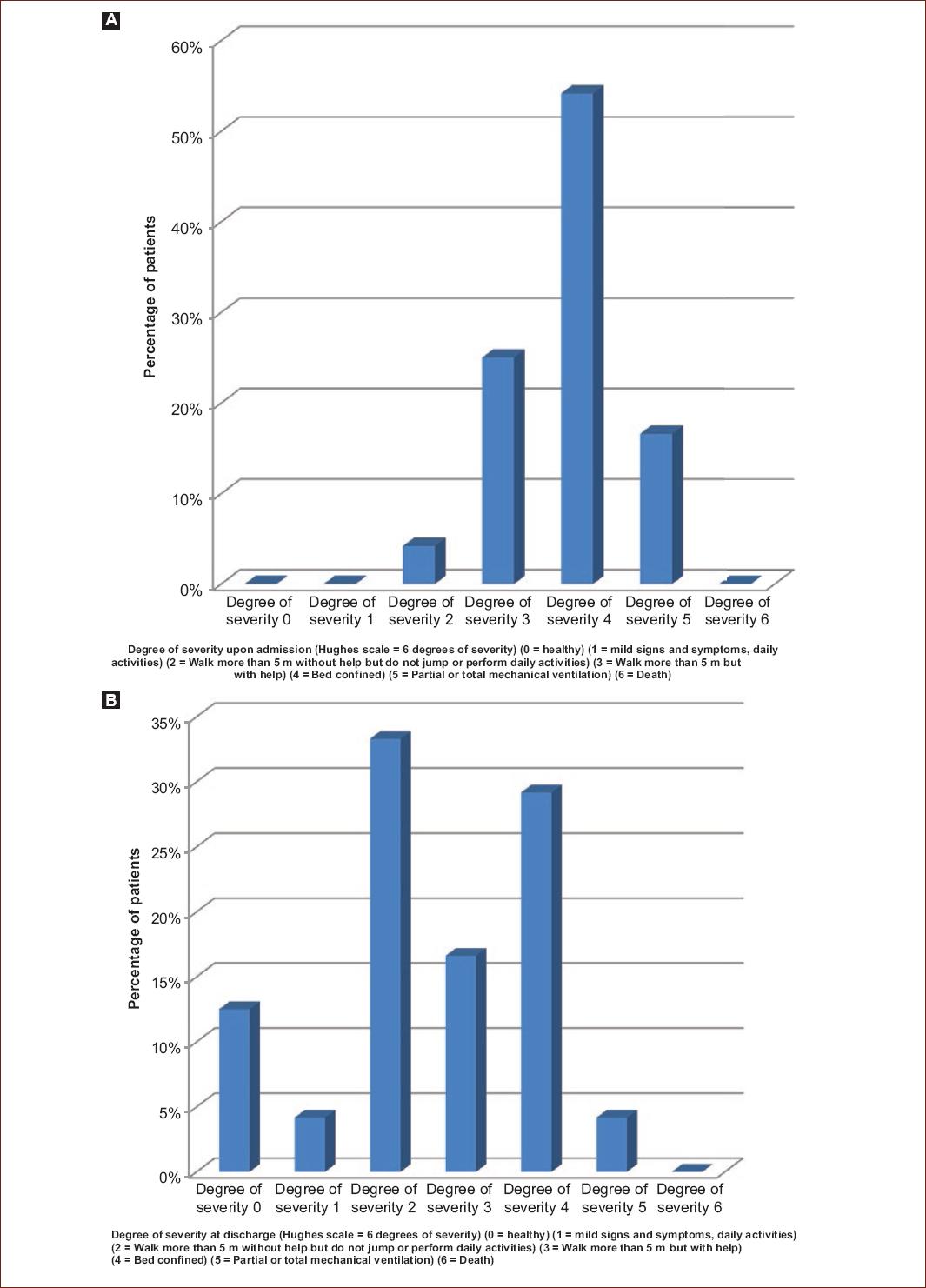
Figure 1 Percentage of patients according to the Hughes severity scale of Guillain-Barré syndrome at hospital admission (A) and hospital discharge (B) (source: Clinical record. Legaria Pediatric Hospital. Secretariat of Health of Mexico City).
The degree of clinical severity more frequent in patients on discharge from the hospital unit was Grade II (walk more than 5 m without help or support but not jump or perform daily activities) based on the Hughes scale, eight patients being affected (33.3%) (Fig. 1).
The average of the different medical treatments used according to availability in the hospital unit were: 1 = steroid (methylprednisolone (dose 30 mg/kg/dia for 3 days) 2/24 (8.3%), 2 = IVIG (dose 1 g/kg/dia for 2 days) 10/24 (41.7%), 3 = IVIG (400 mg/kg/dia dose for 5 days) 3/24 (12.5%), 4 = IVIG (suboptimal dose < 2 kg/kg/dia or < 400 mg/kg/dia) 6/24 (25%), 5 = plasmapheresis 0/24 (0%), and 6 = supports measures 3/24 (12.5%) (Fig. 2).

Figure 2 Percentage of patients according to the medical treatment in the Guillain-Barré syndrome according to availability in the hospital (source: Clinical record. Legaria Pediatric Hospital. Secretariat of Health of Mexico City).
The average time between the onset of clinical manifestations of GBS and the start of hospital treatment with a steroid (methylprednisolone 30 mg/kg/dia for 3 days) was on average 7 days (range 2 days, minimum 6 days, and maximum 8 days).
The average time between the onset of the clinical manifestations of GBS and the start of hospital treatment with IVIG (1 g/kg/dia for 2 days) was on average 4 days (range 10 days, minimum 1 day, and maximum 11 days).
The average time between the onset of clinical manifestations of GBS and the start of hospital treatment with IVIG (400 mg/kg/dia for 5 days) was on average 4 days (range 4 days, minimum 2 days, and maximum 6 days).
The average time between the onset of the clinical manifestations of GBS and the start of inpatient treatment with IVIG (suboptimal dose) was on average 2.5 days (range 6 days, minimum 1 day, and maximum 6 days).
The time between the onset of GBS and the start of hospital treatment with supportive measures was on average 1 day.
According to the clinical evolution during the inpatient stay, 16.7% of the patients (4/24) required assisted mechanical ventilation, reporting an average of 2.25 days between the onset of the clinical manifestations of GBS and the need for support with mechanical ventilation according to the patient’s clinical conditions (range 4 days, minimum 1 day, and maximum 5 days).
Of the four patients who needed assisted mechanical ventilation, the average duration of ventilatory support was 29.75 days (range 13 days, minimum 23 days, and maximum 36 days).
According to the clinical evolution, the four patients that required assisted mechanical ventilation (16.7%) presented respiratory muscle and/or bulbar involvement, this being an absolute indication for it (Phase III mechanical ventilation).
Of the four patients who required mechanical ventilatory support; one-fourth (25% of cases) required orotracheal intubation without requiring tracheostomy and/or gastrostomy due to clinical improvement, presenting adequate suction and swallowing reflex and absence of respiratory distress. One-fourth (25% of the cases) required a tracheostomy but no gastrostomy, presenting an adequate clinical evolution and weaning of mechanical ventilatory support. Two-fourth (50% of cases) required surgical intervention with tracheostomy and gastrostomy due to poor clinical evolution, with subsequent weaning of the ventilator, but in all three cases of patients requiring tracheostomy (75%) all required support of supplemental oxygen at home discharge.
During the hospital stay, five patients (20.8%) developed nosocomial infection characterized by pneumonia associated with health care and 19 patients did not develop intrahospital infection (79.2%).
It was determined whether the degree of clinical severity of GBS constitutes a risk factor for assisted mechanical ventilation.
Of the 24 patients, eight were classified in the group with the highest degree of severity of GBS (Grades IV, V, or VI established by the Hughes severity scale), and 16 patients belonged to the group with the lowest degree of severity of GBS (Grades 0, I, II, and III established by the Hughes severity scale).
Of the eight patients with greater degree of severity of GBS, 4 (50%) required assisted mechanical ventilation, and the remaining 4 (50%) did not require assisted mechanical ventilation (Fig. 3).

Figure 3 Frequency of patients according to the association between the degree of clinical severity of Guillain-Barré syndrome and the support of mechanical assisted ventilation (source: Clinical record. Legaria Pediatric Hospital. Secretariat of Health of Mexico City).
The 16 patients with a lower degree of severity of GBS did not require assisted mechanical ventilation. We obtained a Chi-square 9.60 p < 0.01, establishing that there is a significant difference; there is an association between the degree of clinical severity and the assisted mechanical ventilation (Table 1).
Table 1 Association between the degree of clinical severity of GBS and the support of mechanical assisted ventilation
| Association between degree of severity of GBS and mechanical ventilation | Mechanic ventilation | Total | |
|---|---|---|---|
| No | Yes | ||
| Degrees of severity | |||
| Greater | 4 | 4 | 8 |
| Minor | 16 | 0 | 16 |
| Total | 20 | 4 | 24 |
Chi-square 9.60; p < 0.01, there is significant difference.
The degree of clinical severity of GBS is associated with the need for assisted mechanical ventilation support.
GBS: Guillain-Barré syndrome.
Source: Clinical record. Legaria Pediatric Hospital. Secretariat of Health of Mexico City.
Regarding the degree of clinical severity of GBS at admission related to the degree of clinical severity of GBS at discharge, a Student’s T-test of 5933 was obtained, with p < 0.001; there is a statistically significant difference between the degree of severity at admission versus patient discharge (Table 2 and Fig. 4).

Figure 4 Comparison of the degree of severity at the patient’s admission and discharge. Student’s t = 5.933, with p < 0.01, there is a significant difference (source: Clinical record. Legaria Pediatric Hospital. Secretariat of Health of Mexico City).
Table 2 Relationship between the degree of clinical severity of GBS on admission and hospital discharge (A) and test of matched samples according to the degree of clinical severity of SGB on admission and hospital discharge (B)
| (A) | ||||||||
|---|---|---|---|---|---|---|---|---|
| Clinical severity | Mean | n | Standard deviation | Standard error average | ||||
| Degree of severity on entry | 3.83 | 24 | 0.761 | 0.155 | ||||
| Degree of severity at discharge | 2.58 | 24 | 1.412 | 0.288 | ||||
| (B) | ||||||||
| Clinical severity | Paired differences | T | gl | Sig. (bilateral) | ||||
| Mean | Standard deviation | Standard error average | 95% confidence interval of the difference | |||||
| Lower | Higher | |||||||
| Degree of severity on entry-degree of severity at discharge | 1.250 | 1.032 | 0.211 | 0.814 | 1.686 | 5.933 | 23 | 0.000 |
GBS: Guillain-Barré syndrome.Source: Clinical record. Legaria Pediatric Hospital. Secretariat of Health of Mexico City.
The association between the degree of clinical severity of GBS on admission and the ideal medical treatment (IVIG 1 g/kg/dia for 2 days) was obtained; and the existing association between the degree of clinical severity of GBS at discharge and ideal medical treatment (IVIG 1 g/kg/day for 2 days).
It was determined that there are 3.8 times higher risk of clinical severity in those patients who do not receive the ideal medical treatment (IVIG dose of 1 g/kg/day for 2 days).
Discussion
In the present study, we found predominance of patients with GBS, an inflammatory demyelinating variant, as has been demonstrated in international studies.
Of the 24 patients, according to the clinical evolution during the in-hospital stay, 16.7% (4 patients) required assisted mechanical ventilation due to respiratory and bulbar muscle involvement, reporting an average of 2.25 days between the onset of the manifestations clinics of GBS and the need for support with mechanical ventilation according to the patient’s clinical conditions (range 4 days, minimum 1 day, and maximum 5 days).
Of the four patients who needed assisted mechanical ventilation, the average duration of ventilatory support was 29.75 days (range 13 days, minimum 23 days, and maximum 36 days).
Of the four patients who required mechanical ventilatory support, one patient required orotracheal intubation without requiring tracheostomy and/or gastrostomy due to clinical hospital improvement after administration of IVIG, presenting adequate suction and swallowing reflex and absence of respiratory distress.
Another patient required a tracheostomy due to the respiratory condition but without needing gastrostomy since the clinical evolution was favorable and allowed her to wean from the mechanical ventilatory support. In this way, two patients required surgical intervention with tracheostomy and gastrostomy due to poor clinical hospital evolution, with subsequent weaning of the ventilator after a long inpatient stay. Therefore, of the four patients, only three patients who required a tracheostomy (75%) required additional oxygen support at home.
Nearly 20.8% of pediatric patients with GBS (five patients) developed hospital-acquired infection characterized by pneumonia associated with health care.
The degree of clinical severity of GBS as a risk factor for assisted mechanical ventilation was determined in the study. Of the 24 patients, eight patients (33.3%) were classified in the group of greater degree of severity of GBS (Grades IV, V, or VI established by the Hughes severity scale) and 16 patients (66.6%) belonged to the group of degree of minor severity of SGB (Grades 0, I, II, and III established by the Hughes severity scale).
Of the eight patients with greater severity of GBS, only 50% of them required assisted mechanical ventilation. Being that the 16 patients with a lower degree of severity of GBS, did not require any type of assisted mechanical ventilation. Establishing that there is a significant difference; therefore, there is an association between the degree of clinical severity and assisted mechanical ventilation (Table 1).
In addition, the existing association between the degree of clinical severity of GBS on admission and ideal medical treatment (IVIG 1 g/kg/dia for 2 days) was obtained; and the existing association between the degree of clinical severity of GBS at discharge and ideal medical treatment (IVIG 1 g/kg/day for 2 days). With an estimated risk of 3.8 times greater clinical severity in those patients who do not receive the ideal medical treatment (IVIG dose of 1 g/kg/day for 2 days) in a timely manner and warrants more mechanical ventilatory support and subsequent complications.
The results of the present study could not be analyzed with results of other works performed in pediatric patients with GBS, since in Mexico there are only studies focused on the study of GBS in adult patients, for which we have no previous reference to integrate an opportune analysis.
This study has certain limitations, since it is of a retrospective and observational type, but with the obtained results, it is a matter of encouraging in each one of the health professionals, in the realization of new investigations of the GBS in pediatric patients with a prospective nature. To establish which clinical variant is most prevalent in the Mexican population studied and establish the associated complications, making an opportune diagnosis that influences the prognosis and management of GBS, since the treatments recommended by international guidelines, with plasmapheresis or IVIG, they have a high economic cost and a therapeutic efficiency not yet demonstrated in the Mexican pediatric population.
The present work was carried out with the purpose of describing clinical severity in pediatric patients with GBS, since at the national level; there are no studies that allow us to know an adequate statistics of this disease.
The Legaria Pediatric Hospital in Mexico City is a second-level center for health care and a reference for pediatric patients with neurological diseases.
The GBS is a national health problem that requires costly treatment in addition to generating large sequelae and complications subsequent to the discharge of medical care.
Conclusions
In most hospitals in Mexico, the main limitation is the lack of availability of the ideal treatment in several diseases. Therefore, it would be transcendental to have the necessary resources to offer adequate diagnosis and treatment of GBS, to reduce subsequent complications of the underlying pathology, improving the quality of life and prompt reintegration of patients to their daily activities.

