


 nueva página del texto (beta)
 Inglés (pdf)
 Artículo en XML
 Referencias del artículo
 Enviar artículo por email
 Citado por SciELO
  Similares en
SciELO
Similares en
SciELO 
 Permalink
1. Introduction
In the last decade, there has been interest on the synthesis and characterization of Zinc peroxide (ZnO2) due to its potential applications as biocide, bactericide, inorganic oxidant and so on 1, 2, 3. ZnO2 can be obtained as a powder with a cubic structure and has been utilized as a precursor to obtain Zinc oxide (ZnO). ZnO2 can be considered as an oxygen reservoir since when it is heated at relatively low temperatures oxygen is released.
Various approaches have been utilized to synthetize ZnO2 powders like sol-gel, organometallic precursors, hydrothermal, Laser Ablation of Solids in Liquids (LASL), and so on 4, 5, 6, 7, 8, 9, 10, 11, 12, 13, 14, 15, 16. While the hydrothermal route is one of the most utilized methods to obtain ZnO2, the LASL technique has only been utilized by the Gondal´s group to synthesize zinc peroxide nanoparticles 11-12. Escobedo et al. have been reported a study of the vibrational properties of ZnO2 NPs synthesized by the hydrothermal method. Additionally, they present results of XRD, TEM and by thermal analysis determine the decomposition temperature of their ZnO2 NPs 8. Recently, the Escobedo-Morales's group has reported a green method to prepare ZnO nanostructures employing as starting material ZnO2 (prepared by hydrothermal method) 17.
LASL is a green technique that has been utilized to prepare metal, semiconductor and ceramic nanostructures 18-19. This method is experimentally simple and has been used to obtain metal oxides based colloidal solutions like Zinc oxide, Molybdenum Oxide, Bismuth Oxide, Tin Oxide 11, 12, 20, 21, 22. Gondal et al. have reported the synthesis of ZnO2 NPs colloidal solutions by using the LASL technique. They have performed their experiments with the third harmonic of a Nd:YAG laser emitting at 355 nm, a foil of Zinc as target and 3% hydrogen peroxide (H2O2) solution as liquid medium. Gondal et al. have reported that the band gap of ZnO2 NPs can be within the range 3.0-3.6 eV 12. With the aim to study the properties of ZnO2 NPs obtained by LASL it is necessary to perform experiments with other ablation parameters such as the emitting wavelength (1064 nm) of the Nd:YAG laser and its per pulse laser fluence. To the best of our knowledge, only the work of Gondal et al. has reported the synthesis of ZnO2 by LASL technique.
In this work we present results on the characterization of ZnO2 NPs obtained by LASL technique. A pulsed Nd:YAG laser emitting at 1064 nm, metallic Zinc disk and H2O2 solution were used to prepare the ZnO2 NPs. The samples were characterized by Raman microspectroscopy, calorimetry and TEM. Results show that LALS is a suitable technique to prepare ZnO2 NPs.
2. Experimental
2.1. Sample preparation
Figure 1(a) shows the experimental set-up to obtain ZnO2 NPs by LASL technique. Nanosecond (ns) laser pulses (7 ± 2 ns pulse duration) from a Nd:YAG laser (Minilite II, Continuum) were used to ablate the Zn disk (2.54 cm in diameter, 0.375 cm thick, and 99.99 % purity, Kurt J. Lesker Co.); 10 ml of 30% H2O2 solution was used as liquid medium in each ablation experiment. The laser was operated in its fundamental emission wavelength (1064 nm) at 15 Hz repetition rate. The per pulse laser fluence was varied by adjusting the per pulse energy, and keeping the laser beam cross section on target constant. While the per pulse laser fluences were 8, 13, 19 and 22 J/cm2, the ablation time was fixed at 10 min in each experiment. Figure 1(b) shows 3 vials with, H2O2 solution (left vial), ZnO2 based colloidal peroxide solution obtained at a per pulse laser fluence of 22 J/cm2 by laser ablation (center vial) and the sediment formed at the bottom in the ZnO2 based colloidal peroxide solution (above mentioned), when it was left to rest for one hour (right vial). Figure 2(b) shows a milky solution very similar to that obtained and described by Gondal et al. 11-12. Figure 2(c) shows that ZnO2 NPs are not stable, a sediment at the bottom of the vial was formed.
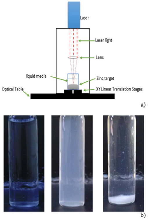
Figure 1. (a) Experimental Set-Up to ablate Zn in H2O2 liquid medium, (b) Photograph of a 30% H2O2 solution (left), ZnO2 based colloidal H2O2 solution obtained at a per pulse laser fluence of 22 J/cm2 by laser ablation during 10 min (center) and ZnO2 based colloidal H2O2 solution left to rest for 1 hour (right).

2.2. Sample characterization
The samples were characterized by Raman microspectroscopy, Calorimetry and TEM. To study the thermal stability of the as-obtained samples a calorimetry equipment (TA Instruments, SDT Q600) was used. Measurements within the range of RT to 550 °C, at a heating rate of 20°C/min under nitrogen atmosphere were performed. Raman spectra were collected by using a microRaman system (Jobin Yvon Horiba, LABRAM HR-800). A 5 mW CW Nd:YAG laser emitting at 532 nm was used to excite the samples. The objective lens of 50 X was used to focus down the laser beam on the sample and at the same time to collect the scattered light. The Raman spectra were the result over 100 acquisitions of 5 s each one. All the samples were prepared in the same manner by drying some drops of the colloidal solution on a glass slide at room temperature. TEM micrographs were obtained with a Transmition Electron Microscope (JEOL-2100). The samples were prepared by putting some drops of colloidal solution on a carbon coated cooper grid.
3. Results and discussion
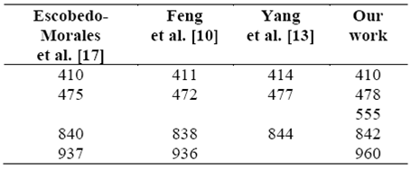
Figure 2(a-d) shows Raman spectra corresponding to the samples prepared at a per pulse laser fluences of 8, 13, 19 and 22 J/cm2. Spectra for the four samples are similar and present peaks at 410, 473 and 842 cm-1. These Raman peaks have been reported by some research groups like Haixin Bai et al. 23 and Guipeng Feng et al. 10 for ZnO2 powder.
Laser fluence might be expected to have an effect on ablation products, as it has been shown by Devaux et al. 24 to be related to the plasma pressure via an experimentally proven analytical model. This relationship is given, assuming constant laser energy I(t)=I 0 throughout the pulse duration, by the following equation:

where P is the plasma pressure (constant) in GPa, I 0 is the laser intensity in GWcm-2, α is a correction factor of P, and Z is related to the shock impedance of each material in gcm-2s-1. Hence laser fluence is related to the square of the pressure, which may have an effect on ablation products via the conditions present during formation. The plasma energy could be further increased because of laser-supported absorption by the plasma plume. The initial experimental trial was conducted by varying laser fluence with fixed short ablation time (10 min) and 30% H2O2 solution. The Raman spectra of ZnO2 nanostructures outlined in figure 2 indicate that laser fluence, indeed, has not some effect on the ablated product. For both, low and high per pulse laser fluences (8 and 22 J/cm2) no particular differences in the Raman spectra can be observed, probably due to an insufficient difference in laser fluence. In the present case, it should be expected that at high reaction rates (i.e., for longer ablation times and/or higher laser fluence) well-controlled ZnO2 NPs could be more readily produced because of a rapid increase in supersaturation and continuous supply of reaction groups over a long period of time.
The TGA-DSC profile of a sample obtained at a per pulse laser fluence of 22 J/cm2 is showed in figure 3. In the range of RT to 500 °C, we have considered three regions for the TG curve as shown in figure 3. In the first region, one can observe a weight decrease of 3.4% from RT to 100 °C, that is due to desorption of physisorbed water in the ZnO2 sample. From 100 to 200 °C a weight loss of 5 % is present. This is related with the elimination of functional groups like OH and CO3. In the third region (200 to 550 °C), it can observe a final weight loss of
Fig. 4(a) shows TEM micrographs as well as a histogram of the size distribution of ZnO2 NPs (inset) obtained at a per pulse laser fluence of 22 J/cm2.
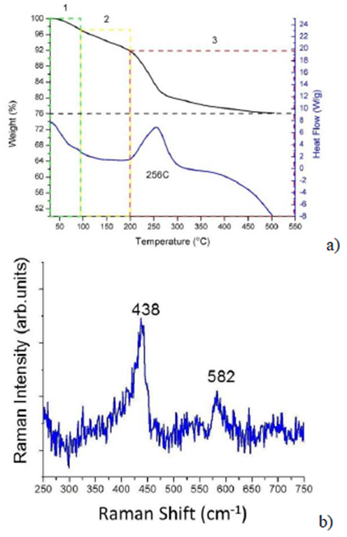
Figure 3. (a) TGA-DSC profiles of ZnO2 obtained by LALS at a per pulse laser fluence of 22 J/cm2. (b) Raman spectrum of the powder thermally treated in the calorimeter.

4. Conclusions
In this work we have characterized ZnO2 NPs obtained by Laser Ablation of Solids in Liquids. We have showed that by using the fundamental wavelength (1064 nm) of a Nd:YAG laser, a Zn disk as target and H2O2 (30 %) as liquid media, it is possible to obtain ZnO2 NPs. Raman results agree well with that reported in the literature. We have obtained NPs 11-14 nm in size. These ZnO2 NPs suffered thermal decomposition in the range of 200-500 °C giving ZnO as resultant material. The work presented and discussed here confirms LASL as an easy, "green", fast and efficient technique for the synthesis of ZnO2 NPs, being (besides chemical methods), another route of synthesis to produce zinc-based nanomaterials.

