


 text new page (beta)
 English (pdf)
 Article in xml format
 Article references
 Send this article by e-mail
 Cited by SciELO
  Similars in
SciELO
Similars in
SciELO 
 Permalink
Introduction
Juvenile systemic sclerosis (JSSc) is a multisystem connective tissue disease of unknown etiology characterized by skin induration and generalized fibrosis of internal organs in patients younger than 16 years of age. The incidence varies between 0.27-0.50 per million children per year in the United Kingdom and Finland, and less than 5% of systemic sclerosis (SS) starts in infancy1,2. Pleuro-pulmonary manifestations in JSSc are rare, but they associate with morbidity and mortality. The most important and fatal pulmonary complications are interstitial lung disease (ILD) and pulmonary arterial hypertension (PAH)3. Interstitial pulmonary fibrosis occurs in approximately one in four children. Knowledge about the pathogenesis, diagnosis, prognosis, and treatment of pediatric ILD and PAH is limited. Laboratory and imaging studies remain essential for early diagnosis, appropriate evaluation, and effective treatment. However, the rarity of JSSc and late diagnosis of its pulmonary complications lead to progressive lung damage4. A few studies on JSSc have comprehensively described pulmonary evaluation5-8. Therefore, this study aimed to describe the clinical aspects, pulmonary function parameters, and imaging findings in JSSc.
Methods
JSSc patients between 5-14 years of age who consecutively arrived at a Pediatric Rheumatology Service in a tertiary referral hospital in 2019 were included in the study and classified based on the preliminary classification criteria for JSSc (Paediatric Rheumatology European Society/American College of Rheumatology/European League Against Rheumatism provisional classification criteria for JSSc)9. Patients with pulmonary disease not associated with JSSc, mixed connective tissue disease, overlap syndrome, or acute cardiopulmonary failure were excluded. All patients underwent a complete physical examination and the following tests: anti-nuclear antibodies, anti-nucleolar and anti-centromere antibodies by indirect immunofluorescence using Hep2 cells as substrate, and anti-Scl-70 (anti-topoisomerase I) antibodies by enzyme-linked immunosorbent assay (ELISA) according to the manufacturers' instructions. In addition, we obtained electrocardiogram (EKG), spirometry (Sibelmed, Datospir 120), chest X-ray, and high-resolution computed tomography (HRCT) (Phillips, Brillance model) using the Wells scale. HRCT was performed by two radiologists blinded to the patients' clinical data (RPJ and MMJM), obtaining a kappa coefficient of interobserver agreement = 0.87. Pulmonary function tests and the 6-minute walk test (6MWT) were also performed according to published guidelines along a 30-meter corridor and standardized normal values for Mexican children10,11. Patients were classified as limited (l-JSSc) or diffuse (d-JSSc) skin subtypes according to the modified Rodnan skin score (maximum total score of 51)12. Heart rate, respiratory rate, and oxygen saturation were measured with pulse oximetry (minicort oximeter model LR78469) using the Fan and Kozinetz severity-of-illness score13.
Interstitial lung disease was considered when chest radiography showed diffuse opacity, confirmed by chest HRCT by Wells scale or restrictive pattern on spirometry according to what has been reported in Mexican students between 8 and 20 years of age14 or oxygen saturation reduction by pulse oximetry on 6MWT. The World Health Organization functional class for PAH is not explicitly designed for children, although it correlates with 6MWT and hemodynamic parameters15. PAH was observed with values ≥ 30 mmHg by echocardiogram: mild PAH between 36-50 mmHg, moderate PAH between 50-100 mmHg, and severe PAH with ≥100 mmHg16. Evaluation of spirometry parameters included forced vital capacity (FVC) and forced expiratory volume in 1 second (FEV1) in a standing position.
The study was conducted following the Declaration of Helsinki (revised in Brazil 2013) and approved by the local Ethics Committee of the Hospital. All parents provided a written informed consent form.
Statistical analysis
Descriptive analysis was performed. Data were presented as mean ± standard deviation or median and interquartile range according to the distribution of variables. Analyses were performed using Statistical Package for the Social Sciences software, SPSS Inc, Chicago, Illinois for Windows (version 20.0).
Results
Clinical characteristics of the patients
We studied 15 patients with an age of disease onset of 7.6 ± 2.7 years and a median follow-up of 12 months. The median time from JSSc diagnosis to the appearance of pulmonary disease was 2 years. Three patients informed a family history of autoimmune disease: a grandmother with systemic lupus erythematosus, a mother with rheumatoid arthritis, and a cousin with SS. Clinical characteristics are shown in Tables 1 and 2.
Table 1 Characteristics of patients with juvenile systemic sclerosis
| Demographic characteristics | Median (n = 15) | Range |
|---|---|---|
| Age (years) | 11 | 4-14 |
| Sex (female/male) | 13/2 | Ratio 9:1 |
| Juvenile systemic sclerosis | Diffuse n (%) | Limited n (%) |
| Clinical subtype | 6 (40) | 9 (60) |
| Female | 6 (46) | 7 (54) |
| Male | 0 | 2 (100) |
| Disease progression | Median | Range |
| Time since initial symptoms (years) | 6 | 3-11 |
| Time of follow-up (months) | 12 | 2-72 |
| Clinical manifestations | n (%) | |
| Cutaneous | ||
| Sclerodactyly | 12 (80) | |
| Calcinosis | 4 (26.7) | |
| Salt and pepper spots | 3 (20) | |
| Peripheral vascular | ||
| Raynaud's phenomenon | 13 (86.7) | |
| Telangiectasias | 7 (46.7) | |
| Digital ulcers | 7 (46) | |
| Musculoskeletal | ||
| Arthritis | 4 (26.7) | |
| Serologic | ||
| Anti-topoisomerase I (Scl 70) | 4 (26.7) | |
| ANA anticentromere | 4 (26.7) | |
| Modified Rodnan skin score for skin thickening | ||
| Normal | 0 | |
| Mild (0-14) | 7 (46.7) | |
| Moderate (15-29) | 7 (46.7) | |
| Severe (30-39) | 1 (6.7) |
ANA, anti-nuclear antibodies.
Table 2 Parameters of each one of the studied patients with juvenile systemic sclerosis
| Patient | Age (years) | Clinical subtype | Time of progression (years) | Rodnan skin score | ANA | Anti-Scl-70-ENA | Pulmonary symptoms | Spirometry FVC<80% | Echo-cardiography | HRCT characteristics | 6MWT |
|---|---|---|---|---|---|---|---|---|---|---|---|
| 1 | 9 | Diffuse | 0.25 | Moderate | Negative | Negative | Chest tightness | Yes | Normal | Abnormal | Normal |
| 2 | 12 | Limited | 1 | Mild | Negative | Negative | Cough | No | Normal | Normal | Normal |
| 3 | 11 | Limited | 1 | Mild | Negative | Negative | Asymptomatic | No | Normal | Normal | Normal |
| 4 | 13 | Diffuse | 7 | Severe | Positive | Positive | Dyspnea | No | Normal | Abnormal | Normal |
| 5 | 11 | Limited | 7 | Moderate | Negative | Negative | Dyspnea | No | Normal | Abnormal | Normal |
| 6 | 11 | Limited | 3 | Moderate | Negative | Negative | Stridor | Yes | Normal | Abnormal | Abnormal |
| 7 | 14 | Diffuse | 3 | Moderate | Positive | Positive | Dyspnea | Yes | Normal | Normal | Abnormal |
| 8 | 13 | Limited | 2 | Moderate | Negative | Negative | Cough | No | PAH | Abnormal | Normal |
| 9 | 10 | Diffuse | 3 | Mild | Negative | Negative | Dyspnea/Stridor | No | Normal | Abnormal | Normal |
| 10 | 11 | Limited | 2 | Mild | Negative | Negative | Asymptomatic | No | Normal | Normal | Normal |
| 11 | 12 | Limited | 8 | Moderate | Negative | Negative | Dyspnea | No | Normal | Abnormal | Normal |
| 12 | 11 | Diffuse | 1 | Moderate | Positive | Positive | Dyspnea | No | Normal | Abnormal | Abnormal |
| 13 | 13 | Limited | 3 | Mild | Negative | Negative | Asymptomatic | No | Normal | Normal | Normal |
| 14 | 12 | Diffuse | 2 | Mild | Positive | Positive | Asymptomatic | No | Normal | Normal | Abnormal |
| 15 | 4 | Limited | 0.5 | Mild | Negative | Negative | Dyspnea | Yes | Normal | Normal | Normal |
ANA, anti-nuclear antibodies; HRCT, high-resolution computed tomography; Anti-Scl-70, anti-topoisomerase antibodies; FVC, forced vital capacity; 6MWT, 6-minute walk test; PAH, pulmonary arterial hypertension.
Pulmonary findings
Pulmonary disease was detected in 11/15 patients (73%). ILD was the most common condition in 10/11 (90%); one patient showed asymptomatic PAH not associated with ILD (sPAP 44 mmHg). Consent for cardiac catheterization was not obtained in the patient with PAH. Respiratory symptoms such as dyspnea, chronic cough, stridor, and chest tightness occurred in 11 patients (73.3%), with dyspnea and chronic cough being the most prominent symptoms. Pulmonary findings, spirometry changes, Fan severity scale, and HRCT findings are shown in Tables 3 and 4. Spirometry reported restrictive pattern in four (26.6%) and obstructive pattern in five patients (33.3%) (FEV1 and FVC < 80%). The 6MWT was abnormal in four patients (26.6%), and both tests were abnormal in only one patient. Interstitial lung disease was present in 10/15 patients (67%), according to the Fan scale.
Table 3 Pulmonary involvement in patients with juvenile systemic sclerosis
| Pulmonary involvement | n (%) |
|---|---|
| Interstitial lung disease | 10 (67) |
| Pulmonary arterial hypertension | 1 (6.6) |
| Lung disease (symptoms) | n (%) |
| Chronic cough | 4 (26.7) |
| Asymptomatic | 2 (13.3) |
| Dyspnea | 7 (46.7) |
| Stridor | 2 (13.3) |
| Chest tightness | 2 (13.3) |
| Changes in spirometry | Abnormal test n (%) |
| PAP, median (min-max) | 28 (20-44) |
| FVC, median (min-max) | 88 (63-115) |
| FVC < 80% | 4 (26.6) |
| 6MWT+ | 4 (26.6) |
| FVC+ 6MWT+ | 1 (7) |
| Fan severity scale (oxygen saturation) | n (%) |
| a Grade I | 4 (26.7) |
| b Grade II | 7 (46.7) |
| c Grade III | 3 (20) |
| d Grade IV | 0 |
| e Grade V | 1 (6.7) |
| FVC, forced vital capacity; 6MWT, 6-minute walk test; PAP, pulmonary artery pressure. |
Fan severity scale in children with interstitial lung disease (ILD):
aAsymptomatic;
bSymptomatic with normal oxygen saturation;
cSymptomatic with exercise desaturation (< 90%);
dSymptomatic with resting oxygen desaturation;
eSymptomatic with pulmonary arterial hypertension.
Table 4 Imaging studies in patients with juvenile systemic sclerosis
| Radiographic findings | n (%) |
|---|---|
| Abnormal chest X-ray | 10 (67) |
| Interstitial infiltrates | 8 (53.3) |
| Lung overdistension | 7( 47) |
| Pulmonary artery dilation | 9 (60) |
| Bronchiectasis | 3 (20) |
| HRCT (Wells' score) | n (%) |
| Abnormal HRCT | 10 (67) |
| Ground glass opacity | 7 (46.7) |
| Grade 0 | 0 (0) |
| Grade 1 | 6 (40) |
| Grade 2 | 1 (6.7) |
| Grade 3 | 0 (0) |
| Honeycombing | 1 (6.7) |
| Fine fibrosis | 3 (20) |
| Grade 0 | 0 (0) |
| Grade 1 | 5 (33) |
| Grade 2 | 1 (7) |
| Grade 3 | 0 (0) |
| Macrocystic pattern (air spaces > 4mm) | 7 (46.6) |
| Microcystic pattern (air spaces < 4mm) | 1 (7) |
| Grade 1 | 0 (0) |
| Grade 2 | 1(7) |
| Grade 3 | 0 (0) |
| Grade 4 | 0 (0) |
HRCT, high-resolution computed tomography of the chest.
HRCT revealed ground-glass images in almost half of the patients. Only two patients were asymptomatic with no imaging abnormalities. Figure 1 shows the chest HRCT of a patient with JSSc and pulmonary involvement with interstitial thickening, typical of ILD. Four patients (26.7%) with the diffuse variety were positive to anti-topoisomerase I antibodies.
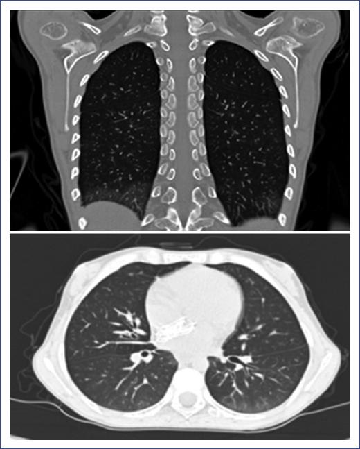
Figure 1 High-resolution computed tomography (HRCT) of a patient with juvenile systemic sclerosis (JSSc) and pulmonary involvement showing interstitial thickening typical of interstitial lung disease (ILD).
Some patients with the d-JSSc variety with severe cutaneous involvement were already receiving cyclophosphamide when the study was being conducted. When pulmonary involvement was found, they continued with their treatment regimen, and supplemental oxygen or immunosuppressive treatment was added in those who had no previous treatment.
Discussion
In the present study, ILD was present in a high proportion of JSSc patients, whereas PAH was present in only one patient. These findings were observed soon after the diagnosis of JSSc in patients with mild clinical findings. Only a few patients showed abnormal pulmonary function tests (PFTs) and 6MWT, regardless of the presence of ILD. It should be considered that ILD and PAH, or a combination of both, are the leading causes of death in JSSc17,18. In general, survival rates of JSSc are higher than those in adults, probably due to the involvement of internal organs and extension of the disease in older patients.
Furthermore, management during childhood differs from that of adulthood. Classically, respiratory involvement is defined by the following factors: pulmonary fibrosis (on chest X-ray or HRCT), reduced diffusing capacity of the lungs for carbon monoxide (DLCO, in carbon monoxide diffusion test) < 80% and FVC < 80%, or PAH (assessed by echocardiography)3. The ILD prevalence is frequently underestimated, depending on the methodology used to identify fibrotic changes. Almost a quarter of the patients in this study showed no pulmonary involvement. Furthermore, patients with pulmonary involvement are usually diagnosed late. It is important to mention that pulmonary fibrosis can develop silently. Therefore, when clinical symptoms become detectable, structural remodeling is already established and often advanced. The most common symptoms are chronic cough and dyspnea. The diagnosis of pulmonary disease in JSSc is often established in asymptomatic patients because a few patients present with dyspnea or persistent cough. A high proportion of pulmonary compromise is identified in children by HRCT, pulmonary function tests, or chest radiography19. Previous studies demonstrated that HRCT is the most sensitive imaging modality for lung involvement in SSc and is considered the gold standard for specific pulmonary abnormalities20.
Moreover, chest X-rays are normal until late in the course of the disease, and the presence of fibrosis in ILD is an independent predictor of death19,21. Another study showed alterations in both HRCT and chest radiography at the diagnosis and during follow-up but in a lower percentage. The most common HRCT finding in JSSc includes ground-glass opacities and irregular linear opacity, similar to our results21. Imaging evaluation plays an essential role in the diagnosis, prognosis, and treatment of JSSc22. Regarding the autoantibody profile, the pulmonary condition most associated with SSc is ILD and is related to anti-Scl-70 antibodies. Positive ANA antibodies, but not anti-Scl-70 antibodies, have been observed in more than one-third of JSSc. Although no correlation with clinical features has been shown, the absence of anti-Scl-70 antibodies has been reported despite having ILD17. In our study, we observed a low percentage of these antibodies in children with ILD.
Regarding the frequency of pulmonary impairment, our results showed a higher prevalence than those reported in asymptomatic patients without pulmonary alterations according to HRCT and chest radiography18,22. Martini et al.23 reported a much lower prevalence of pulmonary fibrosis at diagnosis in patients with JSSc (20%). At the initial evaluation, Panigada et al.21 found pulmonary disease in 58% of patients and increasing pulmonary disease after an 8-year follow-up. In our study, the median follow-up was 12 months, and we observed progression of the pulmonary disease in four patients.
FVC and DLCO tests are important parameters of lung function in the evaluation of JSSc. Low FVC and DLCO have been observed in SSc and ILD, for which they may be indicators of PAH. As only a few patients presented restrictive pulmonary patternmanifested as FVC < 80% independently of ILDand DLCO was not performed, we could not demonstrate this association. One study found that patients with SSc and significant ILD evaluated by HRCT had an average FVC value, with a high false-negative rate reported. In addition, patients with average FVC values show diffuse SSc more frequently despite evidence of fibrosis on HRCT24. Our patients presented mild symptomsa few of them with spirometry alterationsbut with changes in HRCT. Therefore, both pulmonary function tests and HRCT are recommended to increase the possibility of early detection of pulmonary disease.
Patients with mild ILD may be asymptomatic in the early stages of the disease. Subclinical lung disease should be specifically investigated and adequately monitored, mainly during the first years after diagnosis and in the case of cardiopulmonary symptoms. Interestingly, clinical or radiological evidence of severe or progressive pulmonary fibrosis on chest X-rays is a strong predictor of death at presentation and is associated with increased mortality23,25. Six-minute walk test is a noninvasive indicator of cardiopulmonary function that proved to be the most useful in PAH, with a poor correlation between distance traveled and measures of lung function26,27. The interpretation of 6MWT can be confusing in patients with SSc, as they have a cardiopulmonary or musculoskeletal disease and present combinations of cardiac involvement, even in the presence of normal echocardiogram, pulmonary disease, skin fibrosis, muscle damage, and joint disease27. An abnormal 6MWT was identified in a few patients of the present study. We recommend the follow-up of patients with cardiac and respiratory symptoms every 3 to 6 months and annually in the absence of clinical changes. Although optimal radiographic imaging could represent a challenge in infants, it is essential for an accurate diagnosis.
We observed a higher prevalence of l-JSSc, which indicates a mild condition as reported in the literature16. Although l-JSSc is more common in children, some studies found d-JSSc to be more prevalent28. Similar to our study, Morel et al.29 described the clinical characteristics of children with JSSc in our country and found pulmonary involvement in 100% of patients, with a predominance of the l-JSSc subtype. However, since it was not the study's primary objective, they did not describe the detailed characteristics of the pulmonary involvement.
Similar findings have been described regarding the progression of the disease, initial symptoms, diagnosis, and evolution: cardiopulmonary disease is the most frequent visceral involvement, leading to increased morbidity and death7,30. For PAH detection, transthoracic echocardiography has been proposed as a useful diagnostic tool among noninvasive tests, mainly because of its high specificity. However, due to the low sensitivity of noninvasive tests, right heart catheterization remains the gold standard for the confirmatory diagnosis of PAH, although it cannot always be performed16.
In adults, the PAH risk is ≥ 50% for every 10 years at the onset of scleroderma. The follow-up in our patients is still short, so it is advisable to continue it through adulthood. In this context, the most common cause of death in JSSc is heart failure followed by pulmonary involvement31. The main results in cohorts, case reports, and cross-sectional studies in patients with JSSc and pulmonary involvement are summarized in Table 5.
Table 5 Clinical and imaging findings reported on juvenile systemic sclerosis with pulmonary involvement
| A. Cohort studies | |||||||||
|---|---|---|---|---|---|---|---|---|---|
| Author (year) | Type of study Number of patients | PAH n (%) | ILD n (%) | PF | Mean age (min-max) at the start (years) | Follow-up | PFTs | CXR | HRCT |
| Céspedes-Cruz et al. (2020) | Cohort JSSc n = 15 | 1 (6.65) | 10 (67%) | + | 3 (1-11) | 12 (2-72) months | FVC<80%: 4 (26.6%) 6MWT+: 4 (26.6 FVC+ 6MWT+: 17% | Abnormal CXR: 60% Interstitial infiltrates: 8 (53%) Lung overdistension: 7 (47%) Pulmonary artery dilation: 9 (60%) Bronchiectasis: 3 (20%) |
Abnormal: 10 (66%) Ground glass opacification: 7 (46.7%) Honeycomb image: 1 (6.7%) Fine fibrosis: 3 (20%) Macrocystic pattern: 7 (46.6%) Microcystic pattern: 1 (7%) |
| Adrovic et al. (2015)18 | Cohort JSSc n = 35 | 0 | NR | NR | 9.5 (2-18) | Median: 3 (0.5-14.5) years | Normal FVC Normal DLCO |
NR | NR |
| Foeldvari et al. (2012)22 | Cohort EUSTAR JSSc n = 60 | 8 (13.3) | 14 (23.3) | NR | 12.4 (2-15.9) | 7 | NR | NR | NR |
| Misra et al. (2007)28 | Retrospective cohor n = 23 | PAH severe 1 (4.3) | Total 15 (65%) d-JSSc 6 (26%) l-JSSc 9 (39%) | NR | d-JSSc 13* (8-16) l-JSSc 10* (5-16) | Mean: 34 months (0-156 months) | NR | NR | NR |
| Scalapino et al. (2006)8 | Retrospective cohort n = 111 | 4 | 3 | 58/105 (55.2%) | 2.8 | 7 years | PF FVC < 50% of predicted in 10 patients (9%) | NR | NR |
| Zulian et al. (2005)31 | Retrospective cohort n = 750 | NR | NR | NR | 7 | 4 years | Dyspnea or persistent cough Restrictive pattern ↓ DLCO 5 (0.7%) |
Basal infiltrates: 2 (0.26%) |
Basal infiltrates: 2 (0.26%) |
| B. Case reports and cross-sectional studies | |||||||||
| Panigada et al. (2009)21 | Cross-sectional n = 17 | 0 | 10 (58.8) | + | 8.9 (3.9-16) | 4.3 (1-10) years | ↓ FEV1 ↓ FVC ↓ DLCO Restrictive pattern Normal FEV1/FVC ratio |
NR | Good correlation with severity changes on HRCT and PFTs
FEV (r= -0.75; p=0.02) FVC (r= -0.685; p=0.035) DLCO (r= -0.71; p=0.03) |
| Russo et al. (2007)30 | Cross-sectional n = 23 | 2 (8.6) | 9 (39%) | + | 6 (1-14) | 5 (1-11) years | Abnormal: 15 (65%) Dyspnea: 6 (26%) ↓ DLCO (≤ 80% of
predicted value): 1 (3%) FVC ≤ 80: (65%) ↓ FEF (25-75%): 3 (13%) |
Pulmonary fibrosis: 9 (39%) (bilateral basal interstitial infiltrates) |
Abnormal: 6/11 Reticular pulmonary fibrosis Ground-glass opacification |
| Aoyama et al. (2007)6 | Case report n = 3 | NR | 0 | NR | 9 (8-10) | NR 18 years23 years | Normal | Normal | Normal |
| Murata et al. (2005)5 | Case report n = 5 | NR | NR | 1 | 7 (0.5-13) | 5 (1-9) | NR | NR | Pulmonary fibrosis: 1 |
| Foeldvari et al. (2012)7 | Cross-sectional multinational n = 135 | NR | NR | 68/135 (50.4%) | 8.8* (1.5-15.8) | NR | NR | NR | NR |
CXR, chest X-ray; DLCO, diffusing capacity of the lungs for carbon monoxide; FEF, forced expiratory flow; FEV1, forced expiratory volume first second; FVC, forced vital capacity; HRCT, high-resolution computed tomography; ILD, interstitial lung disease; JSSc, juvenile systemic sclerosis; 6MWT, 6-minute walk test; NR, not reported; PAH, pulmonary arterial hypertension; PF, pulmonary fibrosis; PFTs, pulmonary function tests.
Our study has limitations such as the cross-sectional design, the absence of specific details on antibodies, and the lack of other cardiopulmonary parameters, including pro-BNP and DLCO testing. Although the sample size is small, a strength of our study is that it is an adequate number of patients in a single hospital center, considering that JSSc is an uncommon disorder during childhood.
As conclusions, we found that a high proportion of patients with JSSc with limited subtype presentation showed ILD, which occurs in patients with mild clinical findings. Therefore, early detection of JSSc and routine pulmonary monitoring once diagnosed are mandatory to avoid further complications.

