SciELO - Scientific Electronic Library Online

 
vol.90 número3Defecto cardíaco congénito y agrandamiento de la vena cavaMiocarditis aguda como simuladora de infarto con ST elevado índice de autoresíndice de materiabúsqueda de artículos
Home Pagelista alfabética de revistas  

Servicios Personalizados

Revista

Articulo

Indicadores

Links relacionados

  • No hay artículos similaresSimilares en SciELO

Compartir


Archivos de cardiología de México

versión On-line ISSN 1665-1731versión impresa ISSN 1405-9940

Arch. Cardiol. Méx. vol.90 no.3 Ciudad de México jul./sep. 2020  Epub 23-Oct-2020

https://doi.org/10.24875/acm.20000065 

CARTA CIENTÍFICA

“Tiger stripes” sign in acute coronary syndrome

Signo “manchas de tigre” en síndrome coronario agudo

Jorge A. Cervantes-Nieto1 

Daniel A. Rivera-Silverio2 

Rodrigo Gopar-Nieto1 

Francisco Azar-Manzur3 

Maria E. Ruiz-Esparza2 

Grecia I.M. Raymundo-Martínez1  * 

1Cardiology Fellow in Training. National Institute of Cardiology Ignacio Chávez, Mexico City, Mexico

2Department of Echocardiography. National Institute of Cardiology Ignacio Chávez, Mexico City, Mexico

3Department of Teaching. National Institute of Cardiology Ignacio Chávez, Mexico City, Mexico


Abstract

The incidence of mitral regurgitation in acute myocardial syndromes is variable. Echocardiographic evaluation is fundamental in making a proper diagnosis of mechanical complications and to offer timely treatment. We present a case of a 64-year-old male who was admitted to the ER in acute pulmonary edema. The electrocardiogram showed negative ST-segment deviation from V4-V6, positive ST-segment deviation in aVR. Multivessel disease with severe mitral regurgitation was seen in catheterization. A transthoracic echocardiogram revealed important mitral regurgitation showing the “tiger stripes” sign, seen in the presence of intracardial oscillating structures, in this case, suspected papillary muscle rupture. Echocardiographic evaluation is necessary in every case of myocardial infarction who present with new-onset mitral regurgitation. Treatment is complex and must be determined with an interdisciplinary group.

A 64-year-old male with no medical background was admitted to the ER after 1 week of epigastric pain. At his arrival was found in acute pulmonary edema, electrocardiogram revealed sinus tachycardia, negative ST-segment deviation in V4-V6, and positive deviation in aVR. He developed cardiogenic shock. Coronary angiography showed multivessel disease with the left circumflex artery as the culprit artery. A transthoracic echocardiogram was performed, reporting a left ventricle ejection fraction of 48%, hypokinesia of inferolateral wall in the basal segment, the mitral valve showed normal leaflets but with closure limitation conditioning important mitral regurgitation with suspected rupture of the papillary muscle. On continuous Doppler interrogation of the mitral regurgitant jet, the spectral signal had a peculiar sound such as goose croak and a linear fragmentation of the image on the Fourier register resembling “tiger stripes” suggesting an oscillating intracardiac structure, in this case, posteromedial papillary muscle rupture (Fig. 1), which was confirmed with a transesophageal echocardiogram (Fig. 2).

Figure 1 Transthoracic echocardiography showing continuous Doppler interrogation of the mitral regurgitant jet showing fragmentation on the Fourier Register. 

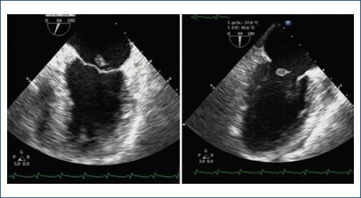
Figure 2 Transesophagic echocardiogram in midesophageal two chamber view showing rupture of the posteromedial papillary muscle. 

The causes of mitral regurgitation after acute myocardial infarction include ischemic papillary muscle dysfunction, left ventricular dilatation or true aneurysm, and papillary muscle or chord rupture1-2. Transesophageal echocardiography is essential for confirmation. These band-like signals appear to be associated with intracardiac oscillating structures with the first band (lowest frequency on the Doppler recording) representing its fundamental frequency. These structures vibrate with a single frequency with several harmonic overtones. Differential diagnosis of “tiger stripes” includes valve regurgitation, flail prosthetic valve leaflet, and possibly Lambl’s excrescences as previously reported1-3.

Acknowledgments

None.

Ethical disclosures

Protection of human and animal subjects. The authors declare that no experiments were performed on humans or animals for this study.

Confidentiality of data. The authors declare that no patient data appear in this article.

Right to privacy and informed consent. The authors declare that no patient data appear in this article.

References

1. Davogustto G, Fernando RR, Loghin C. Lambl's excrescence, migrainous headaches, and “tiger stripes“:puzzling findings in one patient. Tex Heart Inst J. 2015;42:70-2. [ Links ]

2. Sahu AK, Kapoor A. 'Striped TR signal':an intriguing Doppler echocardiography artefact or a pathological correlate?Heart Asia. 2016;8:45. [ Links ]

3. Goyal KK, Mukund DE, Sajeev CG. Tiger stripes sign. J Indian Acad Echocardiogr Cardiovasc Imaging. 2018;2:139-40. [ Links ]

FundingNone.

Received: February 12, 2020; Accepted: April 19, 2020

* Correspondence: Grecia I.M. Raymundo-Martínez E-mail: graymundo87@gmail.com

Conflicts of interest

None.

Creative Commons License Instituto Nacional de Cardiología Ignacio Chávez. Published by Permanyer. This is an open ccess article under the CC BY-NC-ND license