


 nueva página del texto (beta)
 Inglés (pdf)
 Artículo en XML
 Referencias del artículo
 Enviar artículo por email
 Citado por SciELO
  Similares en
SciELO
Similares en
SciELO 
 Permalink
A 30-year-old man with a history of innocent heart murmur diagnosed in childhood presented recurrent high fever that did not go away with antibiotics or self-administered antipyretic treatment. In addition, he developed the consumptive syndrome. Therefore, an echocardiography was performed, which showed the presence of a fistula between the right coronary aortic sinus of Valsalva and the right atria. Infective endocarditis was diagnosed and treated with intravenous Vancomycin-gentamicin, so the fever resolved. Then, he was transferred to our hospital to surgical treatment.
Laboratory test revealed a normal white blood cell count and negative blood cultures.
Transthoracic echocardiography showed right atria and left cavities enlarged due to chronic fistula with preserved left ventricular systolic function (Fig. 1A). Transesophageal echocardiography (TEE) demonstrated high flow fistulous tract (8 mm) between the right coronary aortic sinus of Valsalva and the right atria (adjacent septal tricuspid valve) with small hyperechogenic images that may correspond to vegetation (Fig. 1B-C; Video. S1-S2).
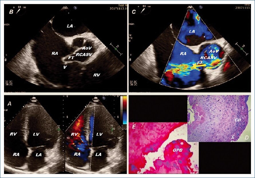
Figure 1 AoV: aortic valve; RCASV: right coronary aortic sinus of Vasalva; RA: right atrium; LA: left atrium; RV: right ventricle; LV: left ventricle; FT: fistulous tract; V: vegetation; GPB: colonies of Gram-positive bacteria; Arrow (shunt left-right); CII: chronic inflammatory infiltrate.
Surgery confirmed the echocardiographic findings, so surgical closure of the defect with a pericardial patch was performed. Then, intra-operative TEE by color flow showed severe tricuspid regurgitation and no residual fistula at the site of repair, so a tricuspid annuloplasty was performed. The patient was admitted to the intensive care unit, under antibiotic treatment, with favorable evolution. Hematoxylin and eosin staining showed vegetation constituted by fibrin with scarce chronic inflammatory infiltrate, multiple areas of dystrophic calcification, and multiple bacterial colonies (Fig. 1D). No acute inflammatory infiltrate was identified. A gram stain revealed colonies of Gram-positive bacteria (Fig. 1E).

