


 nueva página del texto (beta)
 Inglés (pdf)
 Artículo en XML
 Referencias del artículo
 Enviar artículo por email
 Citado por SciELO
  Similares en
SciELO
Similares en
SciELO 
 Permalink
A 25-year-old woman presented to our echocardiography laboratory for a follow-up imaging study. She had a history of muscular ventricular septal defect (VSD) closure by Amplatzer Muscular VSD Occluder (St. Jude Medical, USA) 4 years before this visit. Since this repair, she has remained asymptomatic and was on no medication. In transthoracic echocardiography, the double disc device was visualized in apical four-chamber view (Fig. 1) with no residual VSD flow. Interestingly, in off-axis view of parasternal long-axis imaging, the device was visualized in the form of two side-by-side figure of 8 “Clover-leaf” appearances (Figs. 2 and 3).
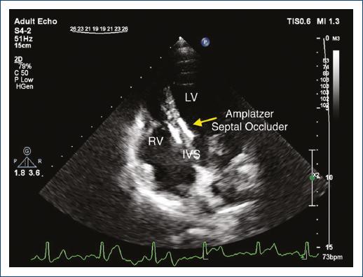
Figure 1/Video 1 Apical four-chamber echocardiogram showing normal biventricular function. The double-disc occluder device is visualized in the mid part of the interventricular septum.
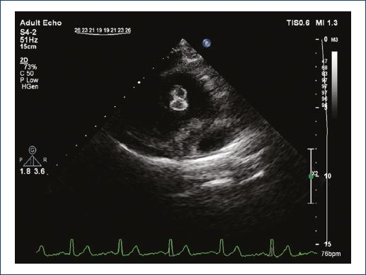
Figure 2/Video 2 Off-axis parasternal long-axis view showing the figure of 8 image artifact of the occluder device.

Figure 3/Video 3 The occluder device resembles a clover-leaf in this off-axis parasternal long-axis view.
Percutaneous disc occluders due to their specific “epitrochoidal” mesh configuration and the interaction with ultrasound waves; form this image artifact when viewed from a coronal imaging position1. Transthoracic echocardiography is the main method of follow-up for post-procedural device position, and accurate interpretation of views is of utmost importance2.
By increasing utilization of percutaneous closure devices for various indications including atrial and VSD, left atrial appendage, patent foramen ovale, recognition of this particular pattern as a normal imaging artifact of a deployed double-disc device when visualized from a coronal imaging position is necessary to avoid misinterpretations of studies such as malposition or displaced device.
Ethical disclosures
Protection of human and animal subjects. The authors declare that no experiments were performed on humans or animals for this study.
Confidentiality of data. The authors declare that they have followed the protocols of their work center on the publication of patient data.
Right to privacy and informed consent. The authors declare that no patient data appear in this article.

