


 nueva página del texto (beta)
 Inglés (pdf)
 Artículo en XML
 Referencias del artículo
 Enviar artículo por email
 Citado por SciELO
  Similares en
SciELO
Similares en
SciELO 
 Permalink
Coronary artery ectasia is observed in 0.3---5% of diagnostic angiograms; >50% of cases are related to coronary atherosclerosis, 20---30% are congenital and up to 20% are related to connective tissue or inflammatory diseases. The impact of strenuous exercise on the left ventricle’s structure has been demonstrated. Studies with ECGs and echocardiography discovered a high prevalence of increased cardiac voltage, suggestive of left ventricle enlargement and hyper- trophy respectively.1 Coronary artery anomalies have been described in 14% of athletes.2 A 37-year-old man, a retired professional football soccer player with history of syncope upon exertion, presented to the ED with ischemic acute chest pain at rest. On admission, his pulse was 70 beats/min, blood pressure 146/85 mmHg. Physical examination was normal. ECG revealed ST dynamic changes and negative deep T waves on anterior-lateral region (Fig. 1A and B). Laboratory tests revealed normal values of cardiac biomarkers.
Immediate diagnostic coronary angiography showed evidence of left main and proximal anterior descending coronary artery ectasia (12 mm) with intracoronary con- trast reflux phenomenon, no obstructive disease or thrombus were observed (Fig. 1C). Transthoracic echocardiography showed mild bilateral atrial dilation, left ventricular hyper- trophy without wall motion abnormalities and ejection fraction 56%. Computed tomography angiography shows coronary ectasia; aorta evaluation with no evidence of dilation, hematoma or dissection (Fig. 1D). Therapy was started with non-fractioned heparin infusion. No abnormalities were found on 24-h Holter monitoring. Clinical and ECG improve- ment was observed at 48 h. The patient was discharged with oral anticoagulation. This case establishes the relevance of coronary angiography as a diagnostic tool, in those cases in which the diagnosis of ST-elevation myocardial infarction is uncertain.
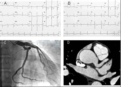
Figure 1: (A) ECG shows isolated increase of QRS amplitude with normal QRS axis and normal atrial activation; V2 to V6 with negative deep T waves and V3 to V6 with ST depression. (B) ECG 48 h after shows improvement of ST depression. (C and D) Coronary angiogram and computed tomography angiography showed left main (12 mm) and proximal left anterior descending coronary ectasia.

