


 nueva página del texto (beta)
 Inglés (pdf)
 Artículo en XML
 Referencias del artículo
 Enviar artículo por email
 Citado por SciELO
  Similares en
SciELO
Similares en
SciELO 
 Permalink
Localized aneurysms of the sinus of Valsalva are extremely rare. They may be congenital or acquired (as a consequence of trauma, degeneration, inflammation or infection).1
A 74-year-old man with hypertension, type 2 diabetes mellitus and dyslipidemia, was admitted in the emergency room after an episode of retrosternal chest pain and shortness of breath. Physical exam was unremarkable. The ECG showed ischemic T waves from V1 to V5 and the peak troponin I level was 0.5 ng/ml. He was referred for coronary angiography, which demonstrated as unique pathologic finding left main extrinsic compression from an ovoid-shaped structure with turbulent flow of dye inside (Fig. 1; SVA — sinus of Valsalva aneurysm, LM — left main). Magnetic resonance imaging confirmed the presence of a left Valsalva sinus unruptured aneurysm below the left main, causing extrinsic compression (Fig. 2; AV — aortic valve). The ascending aorta was dilated and the aortic valve was bicuspid with mild aortic insufficiency. To avoid future life-threatening ischemic events and the possibility of enlargement and sudden rupture, cardiac surgery was performed. The operative findings revealed a 2.5 cm diameter left aortic sinus aneurysm, just below the left main (Fig. 3). Repair was performed with aortic valve substitution by a bioprothesis and ascending aorta replacement by a dacron graft, with coronary ostium reimplantation. The postsurgical evolution was unremarkable.
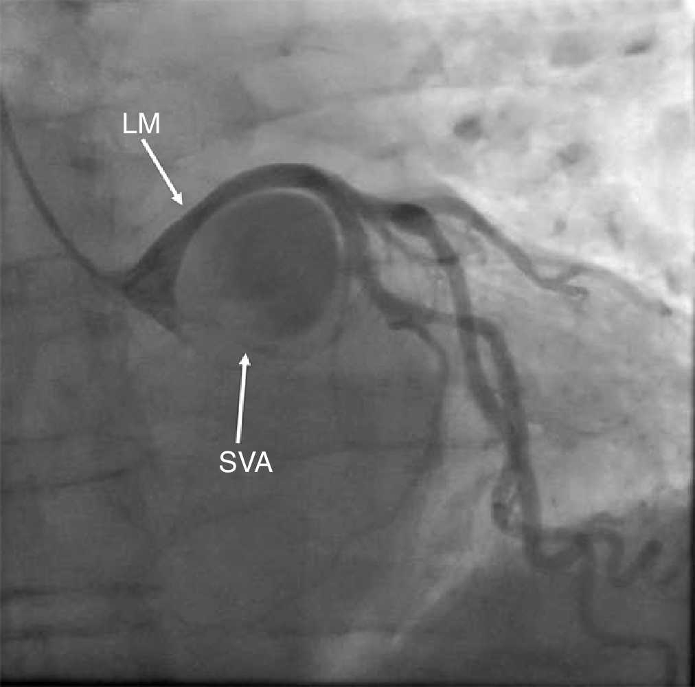
Figure 1 Coronary angiography showing left main extrinsic compression from an ovoid-shaped structure with turbulent flow of dye inside (LM — left main coronary artery; SVA — sinus of Valsalva aneurysm).
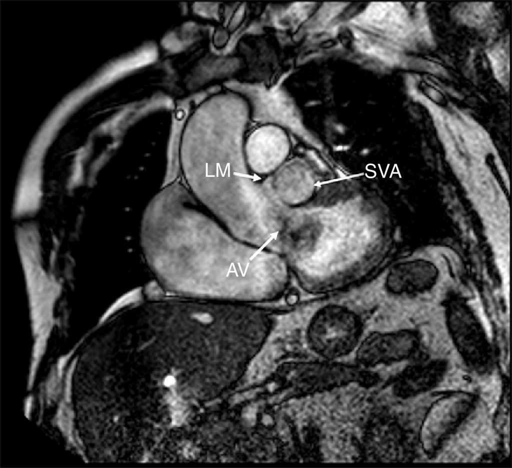
Figure 2 Magnetic resonance imaging confirming the presence of a left Valsalva sinus unruptured aneurysm below the left main, causing extrinsic compression (LM — left main coronary artery; SVA — sinus of Valsalva aneurysm; AV - aortic valve).
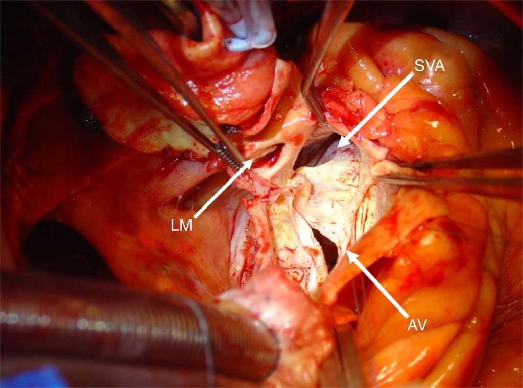
Figure 3 Operative findings revealing a 2.5cm diameter left aortic sinus aneurysm, just below the left main (LM — left main coronary artery; SVA — sinus of Valsalva aneurysm; AV — aortic valve).
Sinus of Valsalva aneurysms may imply high morbidity since they are prone to rupture.2 We report a clinical case of spontaneous aneurysm with unusual clinical presentation (NSTEMI), which had good outcome as a result of prompt diagnosis and surgery.

