


Servicios Personalizados
Revista
Articulo
 Inglés (pdf)
 Artículo en XML
 Referencias del artículo
 Enviar artículo por email
Indicadores
-
 Citado por SciELO
 -
 Accesos
Links relacionados
-
 Similares en
SciELO
Similares en
SciELO
Compartir

 Permalink
Archivos de cardiología de México
versión On-line ISSN 1665-1731versión impresa ISSN 1405-9940
Arch. Cardiol. Méx. vol.81 no.1 Ciudad de México ene./mar. 2011
Comunicaciones breves
Intracardiac ultrasound–guided percutaneous mitral valvuloplasty
Valvuloplastía mitral percutánea guiada por ultrasonido intracardiaco
Marco Antonio Peña–Duque, Carlos Zabal–Cerdeira, José Alejandro Amado–de León, José Antonio–García Montes, Marco Antonio Martínez–Ríos
Departamento of Hemodinámica del Instituto Nacional de Cardiología Ignacio Chávez.
Corresponding author:
Marco Antonio Martínez Ríos.
Hemodynamic Service. Instituto Nacional de Cardiología.
E–mail: mtzrios@cardiologia.org.mx
Received on December 24, 2009;
Accepted on February 16, 2010.
Abstract
Intracardiac echocardiography (ICE) is a relatively new method of ultrasound images useful during cardiac percutaneous interventional procedures. The first human experience with this method was published in 2000 and, since then, several original trials have enrolled this useful method, proving similar and more useful than transesophageal echocardiography in percutaneous treatment of several congenital cardiopathies, like interatrial communication, permeable oval foramen, mitral stenosis, and atrial fibrillation ablation. In this presentation, we publish a single case report of percutaneous mitral valvuloplasty under ICE guidance.
Keywords: Mitral stenosis; Percutaneous mitral valvuloplasty; Intracardiac echocardiography; Mexico.
Resumen
La ecocardiografía intracardiaca (EIC) es un método relativamente nuevo de imágenes por ultrasonido que utilizamos principalmente durante procedimientos de cateterismo intervencionista. La primera experiencia en seres humanos con este método fue reportada en 2000 y, desde entonces, se han publicado diversos artículos sobre su utilidad como procedimiento de control sustituto de la ecocardiografía transesofágica, en el tratamiento por cateterismo de la comunicación interatrial, comunicación interventricular, foramen oval permeable, estenosis mitral y ablación de fibrilación auricular. En esta ocasión presentamos un caso de estenosis mitral y valvuloplastía percutánea, guiado por EIC.
Palabras clave: Estenosis mitral; Valvuloplastía mitral percutánea; Ecocardiografía intracardiaca; México.
Case presentation
A 65–year–old woman with high blood pressure and past medical history positive for rheumatic fever during childhood on ambulatory medical treatment with spironolactone, enalapril, and furosemide presented at our institution, the Instituto Nacional de Cardiologia Ignacio Chavez, with a history of progressive worsening of her functional class over the last 6 years. She was referred to our Institute after detecting a cardiac murmur. A trans–thoracic echocardiogram showed evidence of inactive rheumatic heart disease, pure mitral valve stenosis with a mean trans–mitral valve pressure gradient of 15 mmHg, and dilatation of the left atrium of 47 mm. In addition, there was evidence of mild mitral valve, tricuspid valve, and aortic valve regurgitation, as well as pulmonary artery hypertension with a pulmonary artery systolic pressure calculated at 62 mmHg. Left ventricular ejection fraction was normal (70%). Echocardiography Wilkins score was calculated at 6 and the patient was considered for percutaneous mitral valvuloplasty with the Inoue balloon.
Valvuloplasty technique
Using the modified Seldinger technique, introducer sheaths were placed in the right (8 Fr) end left (11 Fr) femoral veins, as well as the left femoral artery (6 Fr). Left and right cardiac catheterization was performed, measuring baseline pressures at main pulmonary artery, pulmonary capillary wedge, and left ventricle; as well as simultaneous trans–mitral pressure gradient. In addition, a left ventriculogram was performed in the RAO projection.
After baseline hemodynamic assessment, percutaneous mitral valvuloplasty was performed, for which a Mullins catheter was advanced with the Brockenbrough needle through the right femoral vein. At this point, an AcuNav (Acuson, Mountain View, CA) intracardiac echocardiogram (ICE) catheter was advanced through the left femoral vein. This catheter system consists of an element located at the tip, which collects two dimensional ultrasound images at 7 and 8.5 MHz resolution, and a handle with three rotator controls, two to position the tip of the catheter and the other to lock it in position.
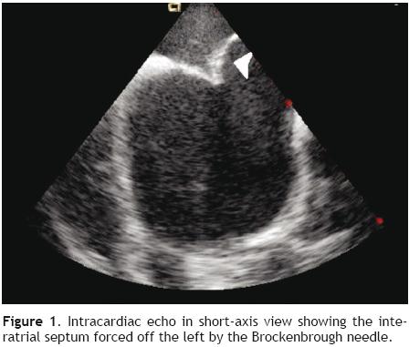
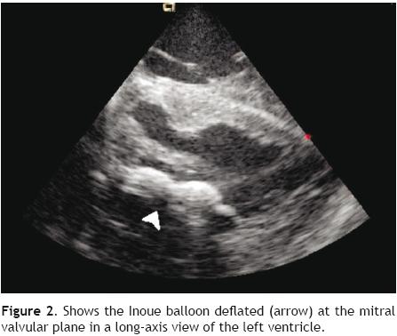
The transeptal puncture was performed under direct ultrasound guidance (Figure 1). After hemodynamic confirmation of left atrial curve morphology, the Mullins catheter was advanced and exchanged for a spiral guide–wire. Interatrial septum was dilated with the usual technique and an Inoue 28 balloon was advanced through the mitral valve into the left ventricle under direct ultrasound guidance (Figure 2). Mitral valvuloplasty at the level of the valve (Figure 3) was performed by a single inflation with the Inoue balloon with 28 ml of normal saline. ICE analysis across the mitral valve showed a significant drop of mean transmitral valve gradient and an increase of the mitral valve area, without an increase of mitral regurgitation.

Conclusions
Intracardiac echocardiography is a relatively new ultrasound imaging modality that has been progressively used for guidance of interventional cardiac procedures.1 The first experience in humans with this method was reported in 2000 presenting two cases of mitral valvuloplasty with distorted anatomy of the interatiral septum due to bulking of the fossa ovalis within the atrium;2 since then, several reports have been published confirming its usefulness in the guidance of several interventional cardiac procedures, and it is currently replacing transesophageal echocardiography in the guidance of transcatheter treatment of interatrial and interventricular septal defects, patent foramen ovale, as well as in the guidance of transeptal puncture procedures for mitral stenosis and radiofrequency ablation of atrial fibrillation.3,4 At the Instituto Nacional de Cardiologia Ignacio Chavez, this method was started to be used around the year 2002 for guidance of the interventionist treatment of congenital structural heart disease.5 This is the first case at our Institute, however, of ICE guidance of a percutaneous mitral valvuloplasty with Inoue balloon. Its potential advantages would be that it provides guidance for a safe and rapid transeptal puncture, shortens fluoroscopy exposure time, and provides immediate on–line assessment of the result of the mitral valvuloplasty by both two–dimensional ultrasound and Doppler technique.5,6,7 We hope that the ICE guidance for mitral valvuloplasty will help the low–volume interventionist to obtain similar results as those performing lots of procedures per year in a high–volume reference center. This can be achieved by reducing the potential complications of trans–septal puncture during the Inoue balloon mitral valvuloplasty.
References
1. Koenig P, Cao Gl, Heitschmidt M, et al. Role of intracardiac echocardiographic guidance in transcatheter closure of atrial septal defects and patent foramen ovale using the Amplatzer device. J Interv Cardiol 2003;16: 51–62. [ Links ]
2. Cafri C, de la Guardia B, Barasch E, et al. Transseptal puncture guided by intracardiac echocardiography during percutaneous transvenous mitral commissurotomy in patients with distorted anatomy of the fossa ovalis Catheter Cardiovasc Interv 2000;50:463–467. [ Links ]
3. Packer DL, Stevens CL, Curley MG, et al. Intracardiac phasedarray imaging: methods and initial clinical experience with high resolution, under blood visualization. J Am Coll Cardiol 2002;39:509–516. [ Links ]
4. Ren JF, Marchlinski FE, Callans DJ, et al. Clinical use of AcuNav diagnostic ultrasound catheter imaging during left heart radiofrequency ablation and transcatheter closure procedures. J Am Soc Echocardiogr 2002;5:1301–1308. [ Links ]
5. Zabal C. Tratamiento con oclusores Amplatzer de los defectos septales. Arch Cardiol Mex 2003;73:S158–162. [ Links ]
6. Hijazi Z, Wang Z, Cao Q, et al. Transcatheter closure of atrial septal defects and patent foramen ovale under intracardiac echocardiographic guidance: feasibility and comparison with transesophageal echocardiography. Catheter Cardiovasc Interv 2001;52:194–199. [ Links ]
7. Hijazi ZM, Shivkumar K, Sahn DJ. Intracardiac echocardiography during interventional and electrophysiological cardiac catheterization. Circulation 2009;119:587–596. [ Links ]

