


Servicios Personalizados
Revista
Articulo
 Inglés (pdf)
 Artículo en XML
 Referencias del artículo
 Enviar artículo por email
Indicadores
-
 Citado por SciELO
 -
 Accesos
Links relacionados
-
 Similares en
SciELO
Similares en
SciELO
Compartir

 Permalink
Archivos de cardiología de México
versión On-line ISSN 1665-1731versión impresa ISSN 1405-9940
Arch. Cardiol. Méx. vol.80 no.1 Ciudad de México ene./mar. 2010
Comunicaciones breves
Usefulness of integrated dual–source multislice computed tomography and cardiac SPECT in a patient with previous myocardial infarction
Utilidad de la evaluación integral con tomografía multicorte de doble–tubo de rayos X y SPECT cardiaco en una paciente con antecedentes de infarto
Enrique Vallejo,1 Hermes Ilarraza,2 Oscar Quiroz,3 Carlos Rodríguez,3 José Luis Ramírez–Arias.3
1 Department of Nuclear Cardiology and Cardiac Hospital Ángeles del Pedregal.
2 Department of Cardiac Rehabilitation, Instituto Nacional de Cardiología Ignacio Chávez.
3 Department of Radiology, Hospital Ángeles del Pedregal, Mexico City.
Corresponding author:
Enrique Vallejo.
Nuclear Cardiology and Cardiac CT,
Hospital Ángeles del Pedregal.
Camino a Santa Teresa 1055–C No. 645,
Col. Héroes de Padierna, C.P. 10700,
México, D.F., México.
Phone and Fax number: 5255 5135 2984.
Email: vallejo. enrique@gmail.com
Received: March 20, 2009.
Accepted: August 20, 2009
Abstract
We present the case of a 62–year–old woman with chest pain and a history of an inferior myocardial infarction for which she underwent stenting in the right coronary artery. Patient was evaluated by cardiac SPECT and the recently introduced dual–source computed tomography (DSCT) system (equipped with two X–ray tubes and two corresponding detectors), in order to detect ischemia associated to stent restenosis. In this case, DSCT demonstrated a very high diagnostic performance to exclude in–stent restenosis, using a dual–energy protocol, and clearly showed subendocardial distribution of the myocardial perfusion defect, in contrast with the transmural defect seen in the SPECT images. As a result, the integration of a dual–energy CT protocol for the evaluation of myocardial blood pool during the assessment of coronary anatomy in this patient, may redefine the diagnostic power of DSCT.
Key words: Cardiac SPECT; CT Angiography; Dual–Source CT.
Resumen
Se presenta el caso de una mujer de 62 años de edad con dolor torácico en estudio y antecedente de infarto de la pared inferior, por lo cual se le colocó una prótesis endovascular (stent) en arteria coronaria derecha.
La paciente fue evaluada con tomografía por emisión de positrones (SPECT) cardiaco y con tomografía multicorte de dos tubos de rayos X (DSCT) con el fin de descartar isquemia asociada a reestenosis en la prótesis. En esta investigación se demostró que la DSCT permite gran precisión diagnóstica para excluir reestenosis en la prótesis cuando se aplica un protocolo de doble energía y también para demostrar la distribución subendocárdica del infarto previo, en comparación con el defecto transmural detectado en el estudio SPECT. En consecuencia, integrar las imágenes de la anatomía coronaria con las de perfusión miocárdica obtenidas mediante DSCT aumenta el valor diagnóstico de la tomografía multicorte.
Palabras clave: SPECT cardiaco; Angiotomografía Coronaria; Tomografía con Dos Tubos de Rayos X.
Case history. A 62–year–old woman was evaluated at the outpatient clinic for shortness of breath and mild substernal heaviness, which increased in severity with moderate exertion. The patient had a history of hypertension and coronary artery disease (CAD), and had suffered an acute inferior myocardial infarction six years earlier, for which she underwent bare–metal stenting in the distal right coronary artery (RCA). Her physical examination was normal and the ECG revealed normal sinus rhythm at 75 beats per minute, with Q waves and T–wave inversion in leads II, III and aVF. Suspicion for in–stent restenosis was raised and she was scheduled for a coronary computed tomography angiogram (coronary CT angiogram) after consultation with a cardiologist.
The patient underwent a cardiac CT with a dual–source CT system (DSCT: two X–ray sources and 64 detectors with temporal resolution of 83 ms; SOMATOM Definition, Siemens Medical Solutions, Forchheim, Germany) in dual–energy mode for performing coronary CT angiogram , and myocardial perfusion evaluation, as recently described.1–2 From the dual–energy scan, 3 different image reconstructions were obtained using the automated dual–energy image reconstruction software of Syngo MultiModality Workplace (Siemens) and compared to previous reports.1 The DSCT system delivers the lowest possible radiation dose in cardiac CT because DSCT images the heart two times faster; furthermore, adaptative ECG–pulsing delivers the necessary radiation amount for cardiac imaging in less than half the time required by the most dose–efficient single source CT scanners. The protocol described is completed under 10 mSv, which is lower than the dose of a single day SPECT scan.2 No pharmacological heart rate control was used.
Figure 1 coronary CT angiogram images revealed right dominant circulation with normal left main coronary artery and non–significant stenosis in the left anterior descending and circumflex arteries territory. Analysis of the RCA showed two high–density calcified plaques with an intermediate stenosis in the middle segment of the RCA (Figures 1B and 1C). Multiplanar images with a special post–processing B46f reconstruction filter showed a patent stent with a very mild neointimal hyperplasia (thin dark line inside the stent lumen) (Figure 1D). Figure 2 coronary CT angiogram images demonstrated a suben–docardial perfusion defect in the right coronary territory, within the inferior and inferolateral wall of the left ventricle (area that appears as a black thick line at the su–bendocardial region) (Figures 2B, 2C and 2D).

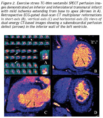
Due to clinical history and the findings of coronary CT angiogram (RCA with two intermediate stenoses), the patient was scheduled for SPECT myocardial perfusion imaging (SPECT MPI). Exercise–stress Tc–99m sestamibi SPECT MPI demonstrated an inferior and inferolateral transmural infarct extended from base to apex with mild ischemia (Figure 2A). Gated SPECT images showed inferior and inferolateral akinesia with normal left ventricular ejection fraction. The patient was managed medically without symptoms during follow–up evaluations.
Discussion
Cardiac imaging has become the cornerstone in the wor–kup of patients with suspected or known CAD. The integration of multimodality imaging represents a natural extension of current imaging paradigms for diagnosing CAD, assessing risk and guiding therapeutic decision–making. The latest generation of multidetector CT scanners and the software configuration of 64–section CT scanners provide an appealing alternative for noninvasive luminal assessment in patients with chest pain.3,4 However, a major limitation of MSCT is that only anatomic information is obtained, whereas no information on the hemodynamic significance of CAD is evaluated.5,6 As a result, a wide discrepancy may be present between anatomic and functional testing results.
Recently, it has been published that single dual–energy CT provides morphological information on coronary artery luminal integrity and, using the different imaging spectra contained within the same scan, allows the reproducible differentiation of iodine distribution within the myocardium to delineate myocardial perfusion defects, in good correlation with standard techniques.1 In our case, DS–MSCT demonstrated a very high diagnostic performance to exclude in–stent restenosis when using a dual–energy protocol. Furthermore, due to the higher spatial resolution of DS–MSCT, compared to SPECT, dual–energy CT images clearly demonstrated subendocardial distribution of the myocardial perfusion defect, in contrast to the transmural defect seen within the SPECT images. As a result, the integration of a dual–energy CT protocol imaging to evaluate myocardial blood pool during the assessment of coronary anatomy in this patient, may redefine the diagnostic power of DS–MSCT.
However, myocardial blood deficits demonstrated with DS–MSCT cannot differentiate between reversible ischemia and scar with current imaging protocols, because coronary CT angiogram is acquired only at rest; this is true for any relative or absolute evaluation of myocardial perfusion reserve (MPR). Recently, it has been described an experimental protocol using DS–MSCT to assess both stress and rest myocardial perfusion, in order to identify areas of infarcted or ischemic myocardium.7 However, disadvantages of CT perfusion imaging protocols include:
a) radiation exposure, b) administration of a large volume of iodinated contrast, c) poor contrast resolution, d) artifacts that can result in false positive images, e) unable to quantify myocardial blood flow, and f) they are highly dependent on appropriate bolus timing. Therefore, to further define the sequelae of coronary atherosclerosis, functional testing for assessment of MPR is ideally performed by nuclear perfusion imaging. Advantages of cardiac SPECT imaging over other cardiovascular imaging modalities (cardiovascular magnetic resonance, contrast echocardiography and computed tomography) include an extensive literature supporting efficacy and prognostic value, standardized protocols for performing studies, published user guidelines and appropriated criteria, as well as a proven cost effectiveness for diagnosis, management and risk assessment.8
Thus, rather than being competitive, MSCT and SPECT imaging should be considered to be complementary for both diagnostic and a prognostic perspective,9 as it has been shown in this case. The use of the latest generation of multidetector CT scanners and developing novel imaging protocols, combined SPECT and MSCT cardiac imaging, will play a prominent role to detect, quantify, and characterize both clinical and subclinical atherosclerosis, with potential reduction of radiation burden.
References
1. Ruzsics B, Lee H, Zwerner PL, et al. Dual–energy CT of the heart for diagnosing coronary artery stenosis and myocardial ischemia–initial experience. Eur Radiol 2008;18:2114–2124. [ Links ]
2. Achenbach S, Anders K, Kalender WA. Dual–source cardiac computed tomography image quality and dose considerations. Eur Radiol 2008;18:1188–1198. [ Links ]
3. Leber AW, Johnson T, Becker A, et al. Diagnostic accuracy of dual–source multi–slice CT–coronary angiography in patients with an intermediate pretest likelihood for coronary artery disease. Eur Heart J 2007;28:2354–2360. [ Links ]
4. Heuschmid M, Brugstahler C, Reimann A, et al. Usefulness of noninvasive cardiac imaging using dual–source computed tomography in an unselected population with high prevalence of coronary artery disease. Am J Cardiol 2007;100:587–592. [ Links ]
5. Tio RA, Dabeshlim A, Siebelink HMJ, et al. Left ventricular function and myocardial perfusion reserve in patients with is–chemic heart disease. J Nucl Med 2009;50:214–219. [ Links ]
6. Nissen SE. Limitations of computed tomography coronary angiography J Am Coll Cardiol 2008;52:2145–2147. [ Links ]
7. Blankstein R, Okada DR, Rocha–Filho JA, et al. Cardiac myocar–dial perfusion imaging using dual source computed tomography. Int J Cardiovasc Imaging 2009 (in press). doi:10.1007/s10554–009–9438–1. [ Links ]
8. Mahmarian JJ. Combining myocardial perfusion imaging with computed tomography for diagnosis of coronary artery disease. Curr Opin Cardiol 2007;22:413–421. [ Links ]
9. Santana CA, Garcia EV, Faber TL, et al. Diagnostic performance of fusion of myocardial perfusion imaging (MPI) and computed tomography coronary angiography. J Nucl Cardiol 2009;16:201–211. [ Links ]

