Services on Demand
Journal
Article
Indicators
-
 Cited by SciELO
 -
 Access statistics
Related links
-
 Similars in
SciELO
Share
Archivos de cardiología de México
On-line version ISSN 1665-1731Print version ISSN 1405-9940
Arch. Cardiol. Méx. vol.72 n.1 Ciudad de México Jan./Mar. 2002
Comunicaciones Breves
Detection of right ventricular myocardial perfusion and contractil reserve by contrast echocardiography and low dose dobutamine in myocardial infarction after successful right coronary angioplasty
Estudio de la perfusión miocárdica y de la reserva contráctil del ventrículo derecho con ecocardiografía
Jesús Vargas-Barrón,* Marco Peña-Duque,* Francisco-Javier Roldan,* Ángel Romero-Cárdenas,* Nilda Espinola-Zavaleta,* Sergio Férez-Santander*
* From the Instituto Nacional de Cardiología "Ignacio Chávez", México D.F.
Correspondence:
Jesús Vargas-Barrón,
M.D. Department of Echocardiography
Instituto Nacional de Cardiología "Ignacio Chávez"
(INCICH. Juan Badiano No. 1,
Col. Sección XVI Del. Tlalpan, 14080 México, D.F.).
México. Telephone (52-55) 5573-2911
Ext. 212 Fax (52-55) 5573-0994.
E-mail: eco_vargas@terra.com.mx
Recibido: 14 de septiembre de 2001
Aceptado: 6 de noviembre de 2001
Summary
We assessed right ventricular myocardial reperfusion through intracoronary contrast and transesophageal echocardiography after right coronary angioplasty. Low dose dobutamine was used to demonstrate contractile reserve in areas with microvascular integrity.
Key words: Ultrasound contrast. Transesophageal echocardiography. Right ventricular infarction. Right atrial infarction. Contractile reserve.
Resumen
Valoramos el éxito de la reperfusión miocárdica en un paciente sometido a angioplastia coronaria mediante ecocardiografía transesofágica y la inyección intracoronaria de un ecorealzador. En las áreas en las que el ecorealzador mostraba integridad de la microcirculación, se pudo demostrar mediante la administración de Dobutamina a dosis bajas la presencia de reserva contráctil.
Palabras clave: Ultrasonido de contraste. Ecocardiografía transesofágica. Infarto del ventrículo derecho. Infarto de la aurícula derecha. Reserva contráctil.
Introduction
In the field of ischemic heart disease, contrast echocardiography is useful in the ventricular opacification and enhancement of endocardial border definition in patients with technically suboptimal studies in resting and stress echocardiography. In the same way, myocardial perfusion in the infarcted area has been assessed by myocardial contrast echocardiography (MCE) a validated technique that has been used to assess microvascular integrity in the setting of acute and subacute myocardial infarction. This evaluation is complemented using low dose dobutamine to study the contractile reserve.1
The assessment of myocardial reperfusion by intravenous or intracoronary contrast echocardiography after primary percutaneous transluminal coronary angioplasty (PTCA) or thrombolysis has been limited to the left ventricle.2,3 The purpose of this report is to show the utility of contrast and dobutamine echocardiography in the reinstauration of myocardial perfusion and in the study of the contractile reserve of the right ventricle in a patient with acute ischemic heart disease subjected to angioplasty of the right coronary artery.
Case report
Forty nine year old male with positive smoking and hypertension history, there was no previous angina. He was admitted to a public hospital with an acute myocardial infarction and no reperfusion therapy was provided.
After 10 days of evolution he was sent to the National Heart Institute "Ignacio Chávez" where he was diagnosed with an inferior infarction extended to the right ventricle. The echocardiogram showed an almost normal left ventricle, there was slight hypokinesis of the inferior wall and the ejection fraction was of 50%. Light enlargement of the right ventricle with marked hypokinesis of its inferior and lateral walls was also registered. During the right ventricle filling the presence of the "A"wave on Doppler indicative of the right atrium mechanical activity was corroborated.
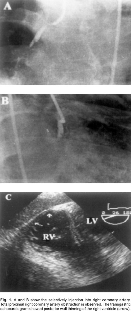
At the following day coronary angiography was performed which did reveal non significant obstruction of the left anterior descending artery, the circumflex artery did not show any obstruction neither. Right coronary artery had a big thrombus with minimal plaque which occupied all the proximal segment until the coronary ostium; the middle third of the artery was completely obstructed. Grade II collateral circulation coming from the anterior descending artery was identified.
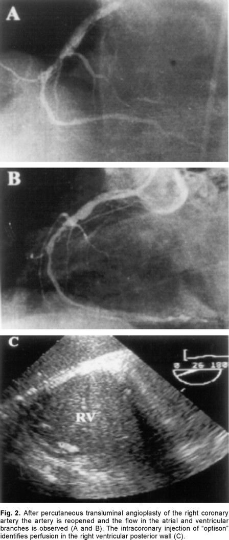
During the following 10 days the patient received heparin intravenously until a second coronary angiography was performed, this study demonstrated persistence of the thrombi in the right coronary artery, total oclusion at the middle third and flow TIMI 0. Therefore, previous canulation with a guide catheter of the artery a coronary angioplasty of the proximal segment and middle third was performed. After finishing the procedure large and difuse lesions of the distal segment just at the origin of the posterior descending artery were observed therefore angioplasty with a balloon of all the distal segments was performed obtaining recanalization of the vessel with minor residual lesions less than 50%, visualizing the posterior descending artery and TIMI 3 distal flow. The original lesion of the mid-third was reduced to a minor stenosis of 40% with TIMI 3 flow.
In the catheterization room, previous informed consent, transesophageal echocardiogram (TEE) with local anesthesia of the oropharynx and slight sedation with midazolam was performed. In the transgastric transverse plane, dilation of the right ventricle was corroborated and hypokinesis of the inferior wall of both ventricles was observed. Before the angioplasty a bolus of "Optison" (0.6 mL) was inyected in the ostium of the right coronary artery without observing any changes in the right ventricular myocardium (Fig. 1). Due to technical difficulties it was not possible to inyect the contrast in the left coronary artery. After recanalized infarct-related right coronary artery "Optison" (0.5 mL) was injected again, demonstrating perfusion in the inferior wall of the right ventricle (Fig. 2). In both injections the echocardiographic study of myocardial perfusion was obtained with continuous imaging.


Three weeks after angioplasty, as outpatient, TEE was repeated. In basal conditions, residual hypokinesis of the inferior wall of the right ventricle was observed; with the administration of 5 and 10 gammas/kg/min of dobutamine and without changes in the gain controls there was increase in the thickness of the same segment of the right ventricular inferior wall (Figs. 3A and 3B).
Discussion
The demonstration of microvascular integrity by MCE performed immediately after primary PTCA has diagnostic value in predicting salvaged myocardium. In theses patients subjected to a reperfusion process the recent advances in ultrasound technology and contrast agents have enabled the detection with transthoracic echocardiography of myocardial perfusion after intravenous injection of transpulmonary contrast agents, valuable for the study of the left ventricle.
Nevertheless, the analysis of all the wall segments of the right ventricle with transthoracic echocardiography is reduced, because apical segments frequently can not be evaluated. In recent years it has been demonstrated that TEE is a simple and safe procedure which allows to characterize the infarction extended to the right atrium and ventricle.4,5 In the same way with this technique, complemented with dobutamine, it is possible to recognize myocardial viability in the right myocardium at risk.6
In this study we demonstrated for the first time that the microvascular integrity of the right ventricle can be observed with intracoronary ultrasound contrast and transesophageal echocardiography. This information was complemented a few weeks later with low dose dobutamine echocardiography, which allowed to demonstrate the late functional recovery or contractile reserve.
Unlike left ventricular function, right ventricular function has not been widely studied after PTCA. Based upon the findings in this patient, we think that the recovery of right ventricular myocardial perfusion in myocardial infarction after successful balloon angioplasty could be demonstrated with contrast and transesophageal echocardiography; the use of low dose dobutamine also permits to demonstrate the contractile reserve.
References
1. BROCHET E, CZITROM D, KARILA-COHEN D, ET AL: Early changes in myocardial perfusion patterns after myocardial infarction: Relation with contractile reserve and functional recovery. J Am Coll Cardiol 1998; 32: 2011-2017. [ Links ]
2. PORTER TR, LI S, OSTER R, ET AL: The clinical implications of no reflow demonstrated with intravenous perflurocarbon containing microbubbles following restoration of thrombolysis in myocardial infarction (TIMI) 3 flow in patients with acute myocardial infarction. Am J Cardiol 1998; 82: 1173-1177. [ Links ]
3. LEPPER W, HOFMANN R, KAMP O, ET AL: Assessment of myocardial reperfusion by intravenous myocardial contrast echocardiography and coronary flow reserve after primary percutaneous transluminal coronary angioplasty in patients with acute myocardial infarction. Circulation 2000; 101: 2368-2374. [ Links ]
4. VARGAS-BARRÓN J, ROMERO CÁRDENAS A, SANCHEZ-UGARTE T, ET AL: Transesophageal echocardiography and right atrial infarction. J Am Soc Echocardiogr 1993; 6: 543-547. [ Links ]
5. GÓMEZ-VILLALOBOS MJ, VARGAS-BARRÓN J, ROMERO-CÁRDENAS A, ET AL: Right atrial and ventricular infarction: Evaluation with transesophageal echocardiography. Echocardiography 1995; 12: 129-137. [ Links ]
6. D'CRUZ IA: Right heart ischemic involvement. TEE-Dobutamine stress assessment. Echocardiography 1998;15:169-170. [ Links ]


