


 nueva página del texto (beta)
 Inglés (pdf)
 Artículo en XML
 Referencias del artículo
 Enviar artículo por email
 Citado por SciELO
  Similares en
SciELO
Similares en
SciELO 
 Permalink
Introduction
Cancer is one of the most important diseases around the world and a major cause of mortality in children and adults. The effectiveness of chemo- and radiotherapies is limited and represents a high economic cost; furthermore, their strong side effects harm the patient both physically and emotionally. For this reason, the constant search for alternative methods that do not cause these side effects is necessary (Abhishek, Rohini & Malleshappa, 2011).
Natural products have played an important role in the discovery and development of new drugs, which are used in a wide variety of diseases. Particularly, plants have been used for a diverse array of purposes including medicine, nutrition, flavorings, beverages, dyeing, repellents, fragrances, cosmetics, charms, smoking and industrial uses. They are an attractive source for discovering new therapeutic drugs because they contain compounds with a wide chemical diversity and potential antitumor activity. Approximately 60% of the drugs used in cancer treatment come from a natural source (Abhishek et al., 2011; Butler, Robertson & Cooper, 2014).
In Sonora, Mexico, there are many varieties of plants that are used in alternative medicine. An example of such plants comes from the Verbenaceae family, Aloysia sonorensis, also known as “Mariola”. This perennial plant is common on the southern coastal plain of Sonora and is endemic to coastal thorn scrub. It is important to mention that some species of the Aloysia genus have shown anti-proliferative and antibacterial activity (Hister, Laughinghouse, Da Silva, Do Canto Dorrow & Tedesco, 2009; Vandresen et al., 2010). The Aloysia gratisima essential oil and infusions are responsible for inducing apoptosis on Allium cepa cells (Hister et al., 2009), while an Aloysia citriodora essential oil showed antiproliferative activity against P815, MCF7 y VERO cancerous cell lines with IC50 values of 6, 34 and 32 µg/mL, respectively (Oukerrou et al., 2017). Based on these studies “Mariola” could have a potential anti-proliferative activity against cancer cell lines, however, this plant has only been used as a treatment for cold, fever, and stomach aches (Yetman & Denver, 2002). Due to this, the aim of this work was to determine the Aloysia sonoresis potential anti-proliferative activity on cancer cell lines and the identification of the compounds responsible for such activity.
Material and methods
Plant collection and extraction
The aerial parts of Aloysia sonorensis were collected in Guaymas, Sonora (N 28° 06.9331 W 111° 04.3621), during the fall. The specimen was authenticated (catalog No. 12417) by Jesús Sánchez Escalante and stored at the herbarium of the Universidad de Sonora, Mexico.
The aerial parts of the plant were dried at room temperature and then powdered. Then, 1,500 g of the dried plant were macerated with methanol (1:10 w/v) for 10 days. After filtration, the methanol (MeOH) was evaporated to dryness using a rotatory evaporator Yamato RE 300 under reduced pressure, to obtain 96 g of methanol extract. This extract was then consecutively fractionated with hexane (Hex), ethyl acetate (AcOEt), and ethanol (EtOH); (3 × 300 mL each solvent). The Hex, AcOEt and EtOH fractions were concentrated to dryness under reduced pressure to give 7.8 g of Hex, 2.2 g of AcOEt, and 15.5 g of EtOH fractions. The part not dissolved in any of the solvents was named residual fraction (70.5 g). The extract and fractions were stored in amber glass vials at -4 °C.
Open and low pressure column chromatographies were carried out on silica gel 60 (230-400 mesh [Merck]). Preparative TLC was performed on precoated silica gel 60 F254 plates (Merck).
The hexane fraction was subjected to an open column chromatography using hexane, mixtures of Hex:AcOEt, at increasing polarity (50:50, 30:70, 10:90, 5:95), AcOEt, mixtures of AcOEt:MeOH, at increasing polarity (95:5, 90:10, 70:30, 50:50), and MeOH as elution solvents.
Subfractions FH12 and FH22 were subjected to low pressure column chromatography using mixtures of Hex:AcOEt (60:40, 50:50), and AcOEt as elution solvents for FH12. For subfraction FH22, mixtures of AcOEt:MeOH (90:10, 70:30) and MeOH were used as elution solvents.
The ethyl acetate fraction was subjected to low pressure column chromatography using mixtures of Hex:AcOEt, at increasing polarity (90:10, 60:40, 50:50), and MeOH as elution solvents.
Spectroscopic analysis
All compounds were characterized by 1H NMR and 13C NMR, DEPT, COSY, HSQC spectroscopy and mass spectrometry, and data were compared to those found in the literature. All spectra were recorded using a Varian Unity 400 spectrometer at 400 MHz for 1H NMR, and 100 MHz for 13C NMR at 25°C in deuterated chloroform as solvent. The chemical shifts were recorded in ppm referenced to tetramethylsilane.
Cell lines
The murine cancerous cell line M12.C3.F6 was provided by Dr. Emil R. Unanue (Department of Pathology and Immunology, Washington University in St. Louis, MO, USA). The cell lines NCTC clone L-929 (normal subcutaneous connective tissue), RAW 264.7 (macrophage transformed by Abelson murine leukemia virus) and HeLa (human cervix carcinoma) were purchased from the American Type Culture Collection (ATCC; Rockville, MD, USA). All cell lines were cultured at 37 °C under a 5% CO2 atmosphere in Dulbecco’s modified Eagle’s medium (DMEM) supplemented with 5% heat-inactivated fetal calf serum.
Cell proliferation assay
Cell proliferation was determined using the 3-(4,5-dimethylthiazol-2-yl)-2,5-diphenyltetrazolium bromide (MTT) assay (Mosmann, 1983) with some modifications (Hernandez et al., 2007). Briefly, cells (10,000/well in 50 µL) were placed in each well of a 96-well plate. After 24 h incubation at 37 ºC in a 5% CO2 atmosphere, aliquots (50 µL) of medium containing different concentrations of “Mariola” extracts and fractions were added and then incubated further for 48 h. All experiments were conducted in parallel with controls (0.5% DMSO). Finally, 10 µL of MTT solution (5 mg/mL) were added to each well and incubated for 4 h. The formazan crystals were dissolved with acidic isopropyl alcohol (100 µL). The absorbance of the samples was measured with an ELISA plate reader (Multiskan EX, ThermoLabSystem) using a test wavelength of 570 nm and a reference wavelength of 630 nm. Plates were normally read within 10 min after adding isopropanol. The anti-proliferative activity of fractions, or subfractions, was reported as IC50 values (IC50 was defined as the concentration of fractions, or subfractions, required to inhibit cell proliferation by 50%) and doxorubicin used as positive control.
Statistical analysis
The results are presented as mean ± standard deviation (SD) of triplicate observations from at least three independent experiments. The significant difference among various treatments was analyzed with the SPSS software and the Tukey test. A p value less than 0.05 was considered to be statistically significant. The IC50 was determined by linear sample and nonlinear regression using the Graph Pad Prism program (Intuitive Software for Science, San Diego, CA).
Results
The methanolic extract of “Mariola” and its four fractions (Hex, AcOEt, EtOH, and residual) were first screened for anti-proliferative activity against the cancerous cell line, M12.C3.F6, and the normal cell line, L-929, using the MTT colorimetric assay.
The anti-proliferative activity of the methanolic extract and fractions are summarized in Table I, expressed as IC50 values (mg/mL). It is worth noting that the Hex and AcOEt fractions presented the highest anti-proliferative activity against the M12.C3.F6 cancerous cell line, with IC50 values of 41 and 39 µg/mL, respectively. Importantly, the Hex and AcOEt fractions showed low cytotoxicity (IC50 ~130 µg/mL) against normal L-929 cells. These two fractions were considered to undergo a further bioactivity-guided fractionation, and antiproliferative activity evaluation.
Table I Anti-proliferative activity (IC50)1 of “Mariola” extract and fractions.
| Extract/Fraction | IC50 values1 55. 56. | |
|---|---|---|
| M12.C3.F6 | L-929 | |
| MeOH | 69.40 ± 0.14 | 164.50 ± 0.70 |
| Hex | 41.50 ± 7.20 | 129.60 ± 8.80 |
| AcOEt | 39.70 ± 11.60 | 128.60 ± 8.70 |
| EtOH | 98.80 ± 10.50 | >200* |
| Residual | >200* | >200* |
1IC50 values of fractions (µg/mL) are shown as the mean ± SD from three independent experiments. The asterisk (*) indicates the highest concentration tested that did not reach IC50 values.
After the chromatographic separation of the most active fractions, Hex and AcOEt, the resulting subfractions (24 hexane and 6 AcOEt) were screened for their anti-proliferative activity against the cancerous cell line, M12.C3.F6. Figure 1 shows that the most active hexane-derived subfractions were FH11 to FH15, and FH21 to FH23 which elicited almost complete inhibition of cellular proliferation at the concentration tested (12.5 µg/mL). From the AcOEt fraction, only one subfraction, FAc1, showed anti-proliferative activity at 12.5 µg/mL.
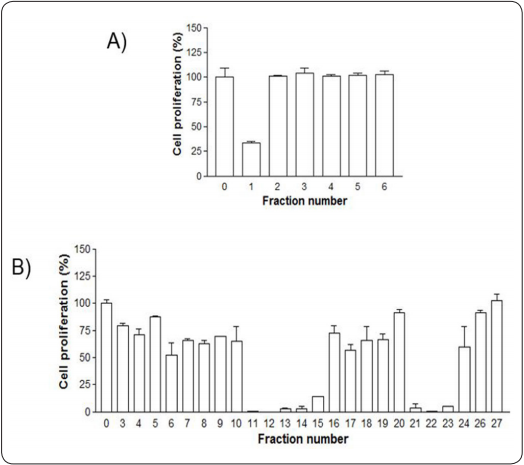
Figure 1 Anti-proliferative activity screening of (A) AcOEt - and (B) hexane -derived subfractions from Aloysia sonorensis against the M12. C3.F6 cancerous cell line. A concentration of 0 means untreated cells (0.05% DMSO) used as negative control. Each percent value is shown as the mean ± SD of three independent experiments.
All active subfractions were screened for their anti-proliferative activity against other cancerous cell lines (RAW 264.7, HeLa). The anti-proliferative activity of these subfractions is summarized in Table II. Most subfractions showed low cytotoxicity against the normal cell line L-929, but selective cytotoxicity against different cancerous cell lines. For example, the FH12 subfraction displayed selective cytotoxicity with IC50 values of 2.84 and 12.12 µg/mL against RAW 264.7 and HeLa cancerous cell lines, respectively. Similarly, FH11 subfraction showed IC50 values of 5.13 and 5.44 µg/mL against RAW 264.7 and M12.C3.F6 cancerous cell lines, respectively. Moreover, the FAc1 displayed cytotoxicity with an IC50 value of 9.42 and 8.93 µg/mL against RAW 264.7 and M12.C3.F6 cancerous cell lines, respectively.
Table II Anti-proliferative activity (IC50)1 of Aloysia sonorensis subfractions.
| IC50 values2 | ||||
|---|---|---|---|---|
| Fractions | RAW 264.7 | M12.C3.F6 | HeLa | L-929 |
| FH11 | 5.13 ± 0.53a | 5.44 ± 0.15a | 16.18 ± 3.37a, b | 12.69 ± 2.40a |
| FH12 | 2.84 ± 0.77 | 10.65 ± 0.33c | 12.12 ± 0.32a | 14.31 ± 3.27a, b |
| FH13 | 5.95 ± 0.27a, b | 7.26 ± 1.40b, d | 31.82 ± 6.07 | 33.34 ± 8.66c |
| FH14 | 6.76 ± 0.13b | 7.33 ± 0.46b, d | 18.73 ± 1.69b | 20.16 ± 5.71a, d |
| FH21 | 7.95 ± 0.65c | 7.60 ± 1.59b, d | 16.50 ± 3.79a, b | 22.79 ± 3.63d |
| FH22 | 14.10 ± 2.50 | 6.32 ± 0.85a, b | 13.87 ± 1.95a, b | 22.23 ± 1.48b, d |
| FAc1 | 9.42 ± 0.04c | 8.93 ± 2.50c, d | 31.48 ± 6.95 | 37.99 ± 8.38c |
1IC50 values of fractions (µg/mL) are shown as the mean±SD from three independent experiments.
2Values in one column, with the same letter, are not significantly different (p ≤ 0.05).
In order to determine if a further purification could improve the anti-proliferative activity of the most active subfractions, FH12 and FH22 were submitted to a bioactivity-guided chromatographic fractionation. The anti-proliferative activity of the different subfractions (evaluated at 12.5 µg/mL) obtained from FH12 and FH22 are shown in Figure 2. The FH12-4, FH12-5, and FH22-3 displayed selective cytotoxicity against RAW 264.7 cancerous cell line with IC50 values of 6.9, 2.1, and 7.27 µg/mL, respectively. Similarly, the FH12-5 subfraction showed IC50 values of 5.44 and 6.64 µg/mL against M12.C3.F6 and HeLa cancerous cell lines, respectively (Table III).
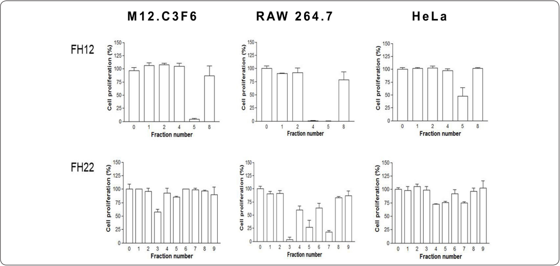
Figure 2 Anti-proliferative activity screening of subfractions FH12 and FH22 from Aloysia sonorensis against M12.C3.F6, RAW 264.7 and HeLa cancerous cell lines. A concentration of 0 means untreated cells (0.05% DMSO) used as negative control. Each percent value is shown as the mean ± SD of three independent experiments.
Table III Anti-proliferative activity (IC50)1 of FH12- and FH22-derived subfractions from “Mariola”.
| IC50 values2 76. 77. | ||||
|---|---|---|---|---|
| Sub-fractions | RAW 264.7 | M12.C3.F6 | HeLa | L-929 |
| FH12-4 | 6.91 ± 0.32a | ND | ND | 59.73 ± 13.27 |
| FH12-5 | 2.10 ± 0.49 | 5.44 ± 0.01 | 6.64 ± 1.25 | 4.82 ± 0.05 |
| FH22-3 | 7.27 ± 0.39a | 12.50 ± 2.16 | ND | 14.70 ± 2.36 |
| FH22-4 | 14.90 ± 0.82b | ND | 24.80 ± 1.07 | 105.31 ± 19.30 |
| FH22-5 | 12.68 ± 1.35b | ND | 27.77 ± 0.12a | 29.40 ± 12.13b |
| FH22-6 | 19.14 ± 3.16 | ND | ND | 31.86 ± 9.85b |
| FH22-7 | 12.36 ± 0.07b | ND | 27.12 ± 2.17a | 30.60 ± 5.69b |
| Doxorubicin | 0.8 ± 0.20 | 0.06 ± 0.01 | 2.3 ± 0.03 | > 60 |
1IC50 values of fractions (µg/mL) are shown as the mean ± SD from three independent experiments.
2Values, in one column, with the same letter are not significantly different (p ≤ 0.05). ND: not determined.
During the anti-proliferative activity study, it was evident that different subfractions induced similar morphological changes on all cancerous cell lines, after 48 h after exposure to the subfractions. Such changes consisted in the formation of apoptotic bodies and membrane blebbing, mainly by FH12-5 and a vacuolation effect by FAc1 (Figure 3), indicating that the possible mechanism of cellular death induced by those subfractions could be apoptosis and oncosis, respectively (Kumar, Abbas, Fausto & Aster, 2005).
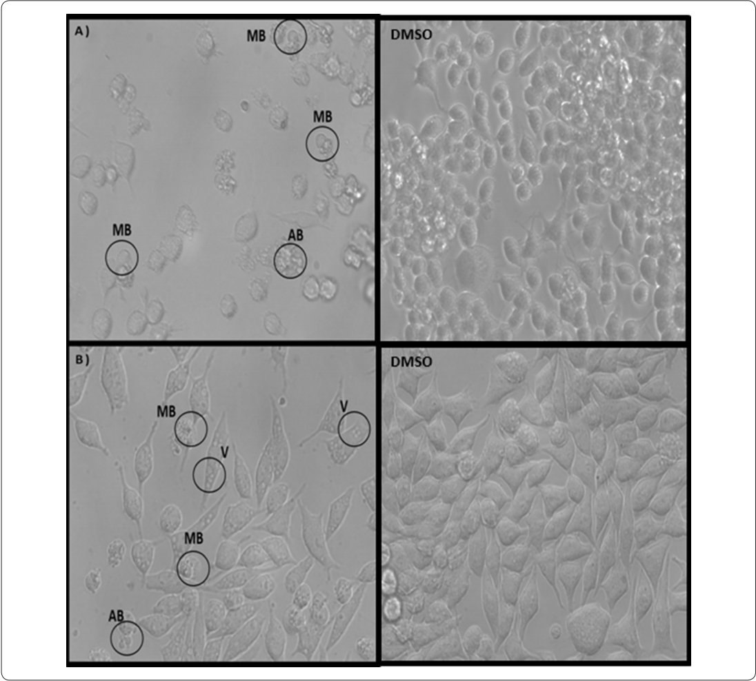
Figure 3 Photomicrographs of morphological changes induced by active sub-fractions FH12-5 and FAc1 at 12.5 µg/mL on cancerous cell lines, after a 24 h treatment. A) RAW 264.7 after treatment with FH12-5. B) M12.C3.F6 after treatment with FAc1. DMSO) negative control. Membrane Blebbing (MB), Apoptotic Body (AB) and Vacuolation (V). All images are magnified at 40X in inverted microscope. Images shown are representative of at least four fields per sample and three independent experiments.
During the bioactivity-guided fractionation it was possible to isolate, from the AcOEt fraction, a compound, that based on the 1H-NMR, 13C-NMR, DEPT, COSY, HSQC spectra and the comparison with data obtained in the literature, was identified as the 3-methyl-6-propan-2-ylidene-cyclohex-2-en-1-one, a monoterpene commonly known as Piperitenone (Figure 4).

Discussion
In this work the anti-proliferative activity of Aloysia sonorensis, known as “Mariola”, against different cancerous cell lines was investigated. It is important to keep in mind that the United States National Cancer Institute, catalogs plant extracts as active, middle-active, and non-active if the IC50 values are less than 30, 30-50, and more than 50 µg/mL, respectively (Suffness & Pezzuto, 1990).
In general, the extract and fractions showed low or no cytotoxicity against the normal cell line, but selective cytotoxicity against the cancerous cell lines. These results are important because one of the goals in the search of new anticancer drugs is to find compounds that act selectively on cancerous cells without exerting cytotoxicity on normal cells. Only the FH12-5 subfraction showed cytotoxicity against every cell line tested.
The bioactivity-guided fractionation of the “Mariola” methanolic extract showed an improvement in the anti-proliferative activity observed in the MeOH extract. For example, starting from an IC50 of 69.40 µg/mL for the methanolic extract against M12. C3.F6 cell line, the subfraction FH12-5 showed an IC50 of 5.44 µg/mL, an almost 13-fold anti-proliferative activity increment against the same cell line.
Comparing the anti-proliferative activity showed by “Mariola” with other species of the Aloysia genus, we found that the aqueous, methanolic, and hexane extracts from Aloysia wrightii known as “oreganillo”, displays a 7.2%, 36.7%, and 61.6%, respectively, anti-proliferative activity against the HeLa cell line at 200 µg/mL (Donalson & Cates, 2004). This anti-proliferative activity is clearly lower than that shown by Aloysia sonorensis against HeLa cell line where the highest IC50 value was around 32 µg/mL. Similarly, the most active fractions in both plants corresponded to those less polar, and they could be the source of anti-proliferative compounds; these fractions are mainly composed by hydrophobic compounds similar to essential oils, which have shown different biological properties (Gautam, Mantha & Mittal, 2014). In the Aloysia genus, the most common essential oils found are limonene, β-caryophyllene, p-cymene, linalool, citral, α-pinene, and 1.8-cineole, and these are thought to be responsible of the biological activity displayed by those plants (Crabas, Marongiu, Piras, Pivetta & Porcedda, 2003; Gil, van Baren, Di Leo Lira & Bandoni, 2007; Oukerrou et al., 2017). Actually, the 1,8-cineole and citral are two monoterpenes, which have shown antimicrobial, anti-inflammatory and anti-cancer activities (Moteki et al., 2002; Katsukawa et al., 2010; Xia et al., 2013; Li et al., 2014).
The cytotoxicity of some of those essential oils against the HeLa cancerous cell line was studied. The authors found that linalool (IC50 > 200 µg/mL) and (-) limonene (IC50=22.1 µg/mL) were the less and most active compounds, respectively (Zapata, Durán, Stashenko, Correa-Royero & BetancurGalvis, 2009). The citral showed EC50 values of 7, 3.7 and 1.3 µg/mL against HepG2, Caco2, MCF7 cancerous cell lines, respectively (Fitsiou et al., 2018). In our study, subfractions FH12 and FH12-5 from “Mariola” displayed IC50 values of
12.12 and 6.64 µg/mL, respectively, against the HeLa cell line, showing similar or higher anti-proliferative activity than those pure essential oils. Preliminary characterization of the constituents of subfractions FH12 and FH12-5 from “Mariola”, has detected the presence of 1,8-cineole and citral in these active fractions. However, it is necessary to continue the phytochemical characterization on “Mariola” to relate the presence of these and other compounds with the antiproliferative activity found in this plant.
It has been proposed that cytotoxicity shown by compounds or extracts can be the result of a particular cell death mechanism induced by those, which can be related to certain morphological changes induced after a cell is exposed to them (Leyva-Peralta et al., 2015). In this sense, during the study of anti-proliferative activity, certain subfractions, such as FH12-5, induced the formation of apoptotic bodies, vacuolation and membrane blebbing in cancerous cells after 48 h of exposure. These changes have been associated with apoptosis mediated by the caspase family as a mechanism of cell death, induced by terpene type compounds such as β-terpineol, terpinen4-ol, lynalyl acetate, linalool and 1,8-cineole (Estanislao et al., 2016; Laghezza et al., 2020). In the case of AcOEt subfractions, FAc-1, induced vacuolization in cancerous cells, which has been associated to a mechanism of cell death similar to necrosis known as oncosis (Shubin, Demidyuk, Komissarov, Rafieva, Kostrov, 2016). That type of mechanism of cell death has been observed in essential oils such as atractylon, hinesol and p-eudesmol (Chen et al., 2013).
During the bioactivity-guided fractionation of “Mariola” extract, a cyclic ketone, the 3-methyl-6-propan-2-ylidenecyclohex-2-en-1-one, commonly known as Piperitenone, was isolated. Piperitenone has been previously identified in the Tagetes, Mentha and Micromeria genus and some biological activities, such as antifungal and antibacterial, have been proven (Marinkovié et al., 2003; Romagnoli et al., 2005; Foganholi et al., 2015). However, no data have been published on anti-proliferative activity, neither on its presence in the genus Aloysia. This compound was detected in AcOEt fraction that displayed selective cytotoxicity with IC50 values of 9.42 and 8.93 µg/mL against RAW 264.7 and M12. Ak.C3.F6 cancerous cells, respectively. A related compound, the piperitenone oxide, was found as the main component in the AcOEt fraction of the ethanolic extract of Mentha suaveolens, showing IC50 values between 5 and 7 µg/mL against liver carcinoma cell line (Božović, Pirolli, Ragno, 2015). Based on this finding, the Piperitenone present in the subfractions obtained from the AcOEt fraction of “Mariola” could contribute to the anti-proliferative activity displayed by those subfractions.
Conclusion
Different fractions from the methanolic extract of “Mariola” have been screened against cancerous cell lines, and they have shown selective anti-proliferative activity against different cancerous cell lines. All subfractions from Hex and AcOEt showed IC50 values lower than 30 µg/mL against the cancerous cell lines tested, and the most active showed anti-proliferative activity from 2.1-9.4 µg/mL. The active subfractions induced morphological changes in the cancerous cell lines that are related with apoptosis and oncosis as mechanism of cellular death. During the bioactivity guided fractionation the monoterpene Piperitenone was isolated in one of the active subfractions. This study provides the basis for further research regarding phytochemical characterization and isolation of antiproliferative compounds present in Aloysia sonorensis.

