


 nova página do texto(beta)
 Inglês (pdf)
 Artigo em XML
 Referências do artigo
 Enviar este artigo por email
 Citado por SciELO
  Similares em
SciELO
Similares em
SciELO 
 Permalink
Introduction
It is well known that plants biosynthesize a diversity of bioactive molecules, which can be mainly used for the discovery and development of new drugs (chemotherapeutics). However, the food industry is increasingly interested in edible plants containing health-promoting molecules, particularly those plants rich in phenols because of their antioxidant properties (Mubashar Sabir, Khan, Rocha, Boligon & Athayde, 2015). Polyphenolic compounds like acid-phenols, flavonoids, tannins, catechins, and anthocyanins are a wide group of natural antioxidant molecules that have the ability to neutralize free-radicals and prevent the cellular damage caused by oxidation processes (Rotta, Windson, Haminiuk, Maldaner & VIssentainer, 2017).Therefore, these polyphenols or phenol-rich plant extracts could be added as active ingredients to biofunctional foods to reduce the risk of oxidative-stress associated diseases.
Thus, new plant antioxidants sources rich in phenols, including edible and nonedible plants, are targeted by researchers looking for food additives, nutraceuticals, and diet supplements (Ćetković, Djilas, Cannadanović-Brunet & Tubas, 2004; Rotta, Windson, Haminiuk, Maldaner & Vissentainer, 2017). Previous studies have demonstrated that plant phenols act as free-radical scavengers, reduce Fe+3/Fe+2, chelate metals, quench singlet and triplet oxygen, inhibit peroxidation and have an anti-inflammatory effect by inhibiting pro-inflammatory cytokine expression (Farahpour, 2014; Girola et al., 2015).
Calendula officinalis L./Asteraceae is a medicinal and edible herb used as spice, tea, and therapeutic agent (Yassen, Habib, Zaghloul & Khaled, 2010). It biosynthesizes polyphenols as flavonoids and acid-phenols (Girola et al., 2015; Ćetković, Djilas, Cannadanović-Brunet & Tubas, 2004), which possess pharmacological properties against cancer (Cabrera & Mach, 2012; Girola et al., 2015), a multifactorial disease that has been related with oxidative stress and inflammatory processes (Lobo, Patil, Phatak & Chandra, 2010). This suggests its use first as a bio functional food due to its high content of phenolic compounds with antioxidant capacity, and perhaps as the base of a new treatments.
Particularly, Calendula officinalis flowers are a rich source of phenolic compounds (Hamzawy, El-denshary, Ezzeldein, Hassan, Mannaa & Abdel-Wahhab, 2013) and their aqueous and alcoholic extracts have shown antioxidant properties assayed by ABTS, DPPH, and hydroxyl and lipid peroxyl radicals (Ćetković, Djilas, Cannadanović-Brunet & Tubas, 2004). Furthermore, Calendula officinalis extract from flowers has been reported to protect the skin against UVB-radiation. It behaves as, hepatotoxic, as well as neuroprotective, given that it reduced oxidative stress and produces proinflammatory cytokines (Hamzawy, El-denshary, Ezzeldein, Hassan, Mannaa & Abdel-Wahhab, 2013).
Therefore, for a better understanding of the antioxidant and anticancer properties of Calendula officinalis petals, this work analyzes the antioxidant capacity (Trolox equivalents in ABTS and DPPH) of the hydro-alcoholic extract obtained from Calendula officinalis petals. We also studied: The in vitro antioxidant efficiency in ferric reducing power, copper (II) and Iron (II) chelation and hydroxyl radicals. In addition, biological activities in human cancer derived cell lines: breast (MDA-MB-231), uterine cervix (HeLa), lung (A-549) and colon (HT-29 and Caco-2). Then, in order establish a possible correlation between polyphenol concentration and growth inhibition or cell death in human cancer cells a polyphenols profile was determined using TLC and HPLC-UV-DAD.
Materials and methods
Preparation of hydro-alcoholic extracts Calendula officinalis flowers, physicochemical analysis
Calendula officinalis fresh flowers were harvested a greenhouse at the Ecatepec Valley State University (March, 2014, Mexico). The physicochemical analysis of the lyophilized petals was carried out according to standard methods (AOAC, 1995). Properties like, moisture/925.09b, protein contents/960.52, ash/923.03, fat/920.39, fiber/991.43 were determined and total carbohydrates estimated by the difference from a 100%. All the determinations were done by triplicate and the statistical averages were reported.
The collected petals were lyophilized at -50 ºC -50 °C and 145 x 10-3 mbar (FreeZone 4.5 L, Labconco, USA). Then, 25 g of lyophilized petals were extracted with 40, 60 and 70% ethanol (500 mL), and shaken at room temperature by 48 h. Later, extracts were filtered and concentrated using an in-vacuum rotatory evaporator at 37 °C and 175 mbar (Buchi R-3, Germany). The extract was dissolved in ethanol for the tests of ABTS, DPPH, HPLC-UV DAD as well as antioxidant activity. For cell culture tests the extract was dissolved in DMEM culture medium supplemented with 2% SFB.
Antioxidant capacity screening by trolox equivalent (TEAC test)
The antioxidant capacity in millimole of trolox equivalents per liter (mM TEAC L-1) of Calendula extracts was assessed by antiradical capacity (Rigane, Chtourou, Ben Mahmoud, Medhioub & Ammar, (2015). ABTS (2,2′-azino-bis-3-ethylbenzthiazoline-6-sulphonic acid) (Braham, Mighri, Jannet, Matthew & Abreu, 2005; Song et al., 2010) and DPPH (1,1-diphenyl-2-picrylhydrazyl). Later, TEAC curves for ABTS (50-250 µM mL-1) and DPPH (100-800 µM mL1) were obtained: 𝑦ABTS = 0.3812𝑥 - 2.64 (r2 = 0.991, p < 0.05) and 𝑦DPPH = 0.000922738𝑥 − 0.0521072 (r2 = 0.988, p < 0.05).
Polyphenols identification and quantitation by TLC and HPLC-UV-DAD
The polyphenols from the 70% hydro-alcoholic extract of Calendula petals were quantified by the Folin- Ciocalteu method (Blainski, Lopes & Palazo de Mello, 2013) and expressed as milligrams of gallic acid equivalent per 100 g (mg GAE g-1). Then, a GAE curve was obtained: 𝑦 GAE = 4.4658𝑥 + 0.0052 (r2 = 0.999, p < 0.05). Polyphenols screening by TLC (25 x 200 mm), silica gel ATLC 60F254, Merck, Germany) was carried out. Samples and standards applied (10 µL) and the mobile phase was methanol:acetone (1:1). The standards (1 mg mL-1) used were caffeic acid, p-coumaric acid, ferulic acid, gallic acid, vanillin, catequin, quercetin and rutin. Spots were revealed by UV light exposure (254 nm). Later, the retention factor for each compound was calculated (Li et al., 2016).
Quantitation of polyphenols by HPLC-UV-DAD (1100 Agilent technologies, USA) was done using a C18 column (4.6 x 150 mm, 5 µm particle size, Zorbax) and readings were obtained at 280 nm. The mobile phase was water-acetic acid (phase A, 98:2) and water-acetonitrile-acetic acid (phase B, 68:30:2) using a linear gradient from B to A (10-100%) and 1.5 mL min-1 flow during 30 min. The sample and standards (1 mg mL-1) were injected (10 µL). Then, a concentration-absorbance relationship for each standard was established: 𝑦caffeic acid = 160.88x + 146 (r2 = 0.9973, p < 0.05); 𝑦catechin = 21.135x + 172.2 (r2 = 0.9742, p < 0.05); 𝑦 p -coumaric acid = 172.63x + 53.286 (r2 = 0.9971, p < 0.05); 𝑦ferulic acid = 115.11x + 254.13 (r2 = 0.9962, p < 0.05); 𝑦gallic acid = 24.104x + 12.176 (r2 = 0.9978, p < 0.05); 𝑦quercetin = 47.057x + 160.04 (r2 = 0.9986, p < 0.05); 𝑦rutin = 45.773x + 12.266 (r2 = 0.9982, p < 0.05); 𝑦vanillin = 116.11x + 12.176 (r2 = 0.9998, p < 0.05). Final identification was obtained by comparison of the retention time and the quantitation expressed in mg mL-1 (Cassani et al., 2015).
Antioxidant efficiency in vitro profile
The antioxidant abilities of the 70% hydro-alcoholic extract from Calendula petals were determined (0.104-3.35 µg mL-1) by Fe3+/Fe2+ reducing power, Cu2+ and Fe2+ chelation and hydroxyl radical scavenging (Oyaizu, 1986; Saiga, Tanabe & Nishimura (2003). The hydroxyl radical (•OH) scavenging activity was assessed according to Lin et al. (2008) Quercetin, gallic acid and caffeic acid were used as standards (1 µg mL-1).
In vitro cytotoxic activity on human cancer cell
The inhibitory concentration (IC50) of Calendula officinalis hydro-alcoholic extract was determined on several human cell lines. The 70% alcoholic extract was select due to its in vitro cytotoxic activity on human cancer cells, and since this extract showed the best antioxidant capacity by ABTS (233.71±0.001) and DPPH (280.44±0.013) tests.
The cytotoxicity of Calendula officinalis petals 70% (5-20 µg mL-1) hydro-alcoholic extract was estimated by the MTT assay (Mosmann, 1983). Taxol was used as control of cell death and the 50% inhibitory concentration (IC50) was estimated by the Hill equation obtaining Hill slope (-1.227±0.065, r2 =0.9639).Obtaining the following: breast (MDA-MB-231) (-2.28 ± 0.24, r2 = 0.915), uterine cervix (HeLa) (-2.36 ± 0.22, r2 = 0.935), lung (A-549) (-2.72 ± 0.28, r2 = 0.931), colon (HT-29) (-2.40 ± 0.28, r2 = 0.898), and (Caco-2) (2.33 ± 0.33, r2 = 0.848). Finally, the relationship between concentration-absorbance relationship was established to obtain the percentage of left over viability of the treatments (Schreml, Lehle, Birnbaum & Preuner, 2007; Kim et al., 2010; Milian-Vázquez, Morales-Ojeda, Vázquez-Montero, Martín-Álvarez & Quiroz-Enríquez, 2010; Dizdarevic et al., 2014).
Statistical analysis
Antioxidant efficiency tests were analyzed by One-way ANOVA and Tukey’s test
(p < 0.05). Phenols quantitation, TEAC test and other
correlations were calculated by linear regression analysis (
Results and discussion
Physicochemical analysis of Calendula officinalis petals
The composition of Calendula officinalis petals has been previously reported as rich in carbohydrates and low in protein content, fat, and ash (Miguel et al., 2016) while Yassen, Habib, Zaghloul & Khaled (2010) reported high fiber and proteins and but low fat. Nevertheless, our physicochemical analysis of lyophilized Calendula officinalis petals showed low content of moisture (6.96 % ± 0.37), carbohydrates (3.27 % ± nd), and ash (6.78 % ± 0.32), and high content of fiber (59.20 % ± 1.70), proteins (14.30 % ± 0.40), and fat (13.78 % ± 0.07), compared to those from edible plants. That perhaps let us to propose that the petals used in this study represent a rich nutritional source. Although differences in nutritional composition were reported between this and previous studies, those may be easily attributed to differences in cultivar, soil-type, harvest season, agro-climatic characteristics, and other variations of environmental conditions.
Antioxidant capacity by TEAC of Calendula officinalis hydro-alcoholic extracts
The results of Calendula officinalis hydro-alcoholic extracts obtained by Trolox equivalents of antioxidant capacity (TEAC) are shown in Table I. The percentage of free radicals inhibition at 70% for DPPH radical neutralization was greater than the ones found at in the 60% and 40% hydro-alcoholic extracts. The antiradical effect observed may be in correlation with the elevated content of polyphenols (Ćetković, Djilas, Cannadanović-Brunet & Tubas., 2004; Hamzawy, El-denshary, Ezzeldein, Hassan, Mannaa & Abdel-Wahhab, 2013; Rigane, Chtourou, Ben Mahmoud, Medhioub & Ammar, 2015).
Table I: Screening of antioxidant capacity by TEAC of hydro-alcoholic extracts from Calendula officinalis petals.
| Calendula officinalis H2O-EtOH extract | Folin mg GAE g-1 | Inhibition (%) ABTS | mM TEACABTS L-1 | Inhibition (%) DPPH | mM TEACDPPH L-1 |
|---|---|---|---|---|---|
| 70% | 37.01±0.015 a | 86.45±1.09a | 233.71±0.001a | 81.12±2.09a | 280.44±0.013a |
| 60% | 13.06±0.032 b | 19.78±0.40b | 58.81±0.002b | 76.64±0.09b | 278.45±0.001a |
| 40% | 9.68±0.009 b | 10.25±0.87c | 33.81±0.005b | 76.13±1.3b | 233.66±0.022b |
TEAC= Trolox equivalent antioxidant capacity; value are mean, represent mean ±SD, n= 3. ANOVA One-way, Tukey´s test p <0.05. Letters a, b, c are statistically significant values.
According to these preliminary results (Table I), the 70% hydro-alcoholic extract of Calendula officinalis was selected due to its in vitro antioxidant efficiency as well as the a possible antiproliferative or rather cytotoxic effect.
Identification and quantitation of polyphenols by TLC and HPLC-UV-DAD
Total phenolic compounds content in Calendula officinalis petals 70% hydro-alcoholic extract
The content of polyphenols of Calendula officinalis flowers was 37.01±0.015 g GAE g-1. Other authors have been reported values of 109.27, 51.62, and 26.5 mg GAE g-1 dry weight in hydro-methanol (Rigane, Chtourou, Ben Mahmoud, Medhioub & Ammar, 2015) and hydro-alcoholic extracts (Farahpour, 2014) from Calendula officinalis flowers the estimated content of polyphenols was 37.01±0.015 g GAE g-1. The differences may be attributed to plant variety (Olennikov & Kashchenko, 2014) or perhaps the use of hydrophobic solvents involved in the extract preparation, such as methanol, hexane and acetone (Rigane, Chtourou, Ben Mahmoud, Medhioub & Ammar, 2015). It would be relevant to highlight that the content of polyphenols shown by 70% hydro-alcoholic extract from Calendula officinalis in this study also high antioxidant efficiency.
Identification and quantification of polyphenols by TLC and HPLC-UV-DAD respectively
Eight phenolic compounds were identified by TLC screening, according to their Rf values, as follows: ferulic acid, coumaric acid, caffeic acid, gallic acid, vanillin, catechin, quercetin and rutin. The polyphenolic acids above were also revealed by HPLC-UV-DAD (Table II); where the content of flavonoids (rutin> quercetin> catechin) was greater than those of acid-phenols (ferulic acid> cumaric acid> vainillin> gallic acid> caffeic acid); being rutin the most abundant polyphenol present in the extract (58.7%).
Table II: Polyphenols profile from petals of Calendula officinalis 70% hydro-alcoholic extract.
| Polyphenol-Type | Compound | Rf | [mg mL-1] |
|---|---|---|---|
| Acid-phenols | Caffeic acid | 0.76 | 2.41 ± 0.20 |
| p-Coumaric acid | 0.73 | 5.80 ± 0.64 | |
| Ferulic acid | 0.70 | 19.40 ± 0.52 | |
| Gallic acid | 0.78 | 3.22 ± 0.22 | |
| Vanillin | 0.84 | 5.67 ± 1.49 | |
| Flavonoids | Catechin | 0.87 | 2.88 ± 0.00 |
| Quercetin | 0.79 | 13.97 ± 0.09 | |
| Rutin | 0.87 | 75.84 ± 16.1 | |
| Total polyphenols | 129.19 | ||
Previously, Fonseca et al, (2010) reported rutin as an important glycosylated flavonoid in a Calendula officinalis hydro-alcoholic extract among several medicinal plants. Miguel et al., (2016) mainly determined glycosylated isorhamnetin from infusions and a methanolic extract prepared from air-dried Calendula officinalis flowers. This study reports a rich profile in phenolic compounds from petals of Calendula officinalis 70% hydro-alcoholic extract, whose efficient antiradical activities contribute to its important antioxidant activity.
Because antioxidants are important to minimize inappropriate oxidation processes, the 70% hydro-alcoholic extract Calendula officinalis petals represent a potential resource of natural antioxidants that may play a crucial role in protection of human health. It is known that the antioxidant activity of phenolic acids is mainly due to (i) the number of substituents, (ii) its relative position (-para> -metha > -ortho), and (iii) substitution of hydroxyl groups by carboxylate.
For instance, molecules of the kind of the hydroxycinnamic acid, such as p-coumaric, caffeic, and ferulic acids (Table II) are considered strong antioxidants, better that the caffeic acid (3,4-hydroxy cinnamic acid), even more the reducing properties of p-coumaric acid (4-hydroxy cinnamic acid), include the neutralization of inappropriately elevated oxidant activity associated with to reactive oxygen species (ROS) (Re et al., 1999).
Calendula officinalis 70% hydro-alcoholic extract from this study, contains phenolic compounds, such as ferulic acid, with potent antioxidant activity. The mechanism of those compounds is mainly equilibrate an anomalously activated ROS response by stimulation of antioxidant enzymatic activity, as the glutathione S-transferase and quinone reductase (Zang et al., 2000). In this study, Calendula officinalis has also been found to be an important source of flavonoids such as caffeic and p-coumaric acids.
On the other hand, the antionxidant capacity of hydroxyl-benzoic acid and gallic acid (a, 3,4,5- trihydroxy benzoic acid) (Song et al., 2010) is improved due to the number of OH substituents in the benzoic ring on the contrary, in molecules like hydroxyl-benzaldehydes, the carboxylate group is reduced to aldehyde, and their effectiveness as antioxidants is reduced (Bountagkidou, Ordoudi & Tsimidou, 2010); for instance, this happens in vanillin (3-methoxy-4-hydroxy benzaldehyde) when it is compared to its acid counterpart (vanillic acid). Finally, the antioxidant mechanism of flavonoids such as catechin, quercetin and rutin (Table II) has also been suggested to be dependent of (i) the number of hydroxyl groups of the C-ring, especially 3-OH, (ii) methoxy substituents, (iii) unsaturation on C-ring and (iv) position of hydroxyl groups on B ring (Seyoum, Asres & El-Fiky, 2006; Song et al., 2010).
Chemical structures of flavanoids identified here as quercetin and as well as the flavan-3-ol catechin, display identical arrangement of the five hydroxyl groups except for its glycosylated derivative rutin, which has a glycosylated group in the position 3 of the C-ring. This glycosylation actually reduces the antioxidant effect, when compared to the corresponding aglycone (quercetin). Furthermore, quercetin has a 2,3-double bond on the C-ring and a substitution 3´4´-ortho function on B-ring, this structural change means an advantage increasing the molecule´s antioxidant ability when compared to the saturated heterocyclic ring of catechin (Seyoum, Asres & El-Fiky, 2006; Song et al., 2010).
Antioxidant efficiency in vitro profile
Oxidation processes are complex in vivo and therefore, the antioxidant efficiency of natural products must be analyzed by different methods to obtain a reliable antioxidant profile (Pérez-Jiménez, 2006). In pursuing this goal, the antioxidant efficiency profile of the petals of Calendula officinalis 70% hydro-alcoholic extract was evaluated in vitro by several methods.
When evaluating its Fe3+/Fe2+ reducing capacity, the Calendula officinalis hydro-alcoholic extract proved to be highly reducing (69.9-73.4%; 0.104-3.350 µg mL-1); (Figure 1a). The antioxidant capacity of the extract was lower than the one observed in the positive controls using the concentration of 1 µg mL-1: quercetin (87.2%), gallic acid (75.1%) and caffeic acid (71.5%). The variated content of polyphenolic compounds identified in the extract is probably showing a synergistic effect as increased antioxidant capacity, in comparison with the molecules tested separately. The Calendula officinalis hydro-alcoholic extract showed a hydroxyl antiradical effect (1-52.5%; 0.104-3.350 µg mL-1) dependent on concentration (Figure 1b). This •OH radical scavenging effect of Calendula officinalis extract could be particularly attributed to its content of flavonoids like quercetin, which displayed the highest hydroxyl antiradical effect (68.9%) in comparison with phenolic acids (gallic acid, 35.1% and caffeic acid, 8.7%).
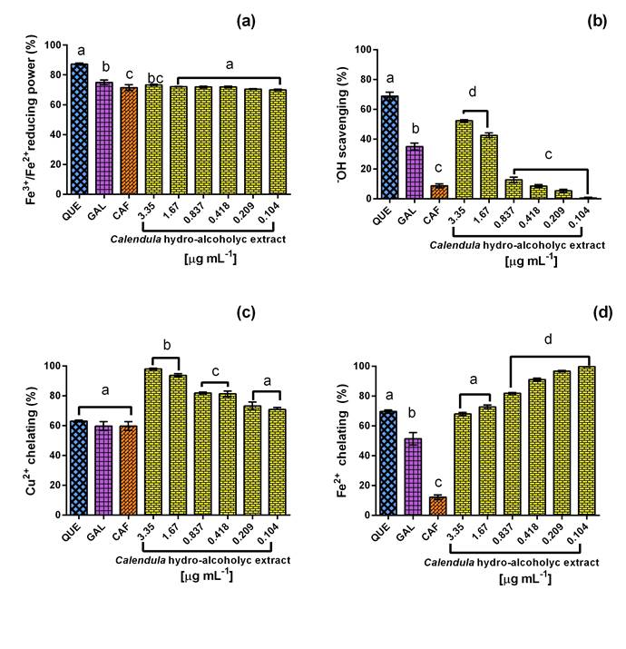
Figure 1: In vitro antioxidant efficiency profile of 70% hydro-alcoholic extract from Calendula officinalis petals. (a) Ferric-reducing power, (b) Hydroxyl-radical scavenging, (c) Cooper (II) and (d) Iron (II) chelation. QUE, Quercetin; GAL, Gallic acid; and CAF, Caffeic acid (1 µg mL-1). Bars represent mean ± SD, n = 3. ANOVA One-way, Tukey’s test p < 0.05. The letter a, b and c indicate a statistical difference between samples.
The Cu2+ chelation ability (Figure 1c) of the Calendula officinalis hydro-alcoholic extract displayed a concentration-dependent effect (70.8-98.1%; 0.104-3.350 µg mL-1). The chelation ability of quercetin (63.1%), gallic acid (59.8%), and caffeic acid (59.8%) were not higher than the one from the whole extract, even in low concentrations, suggesting that the extract shows an accumulative effect of polyphenol activity.
In the case of the Fe2+ chelation ability (Figure 1d), the extract shows an inverse concentration-dependent activity. This is because the chelation capacity results from the Calendula officinalis hydro-alcoholic extract chelation capacity seem to be higher (99.8-67.9%; 0.104-3.350 µg mL-1) than the one observed for quercetin (69.6%), gallic acid (51.3%) and caffeic acid (12.1%) by themselves (Figure 1).
The extract is highly capable of chelating ions and thus has an effect as a neutralizer of free radicals present in the cell causing cell toxicity. Its antioxidant activity upon metals such as iron and copper may play an important role in preventing those ions from catalyzing oxidative processes. These observations would be in agreement with studies suporting that iron and copper catalyze the formation of free radicals (Preethi, Kuttan & Kuttan, 2006).
Previous reports on the aerial parts of Calendula officinalis supported the antioxidant activity of the extracts (Preethi, Kuttan & Kuttan., 2006), which may be explained by a synergism of the antioxidant abilities of each of their phytochemicals-perhaps mainly polyphenols (Liu, 2004) which can be readily observed from the differences of individual antiradical effects among the molecules used as standards (quercetin, gallic acid, and caffeic acid). The flavonoid quercetin is the greatest antioxidant contributor, when compared against hydroxybenzoic acid (gallic acid) and hydroxycinnamic acid (caffeic acid). The inverse effect was noticed in the Fe2+ chelation of Calendula officinalis extract, which could be explained by the high concentration of flavonoids (Martínez-Flores, González-Gallego, Culebras & Tuñón, 2002) or a dual activity (antioxidant/pro-oxidant) of those flavonoids, probably caused by a weak chelation/stability of Fe-Flavonoid complex.
Therefore, the antioxidant efficiency of the Calendula officinalis extract could be attributed to the polyphenols (phenolic acids and flavonoids) identified, because these compounds (i) are reducing agents, (ii) donate hydrogen, and (iii) chelate metals such as copper and iron (Leopoldini, Russo & Toscano, 2011). The free-radical scavenging observed on in vitro assays here, offer an approximation to explain an antioxidant protective activity of the hydro-alcoholic extract from Calendula officinalis petals. These tests may be good antioxidant estimators to build an antioxidant efficiency profile. Nevertheless, other estimators can also be considered if they enhance the resolution capacity of the antioxidant efficiency.
In vitro cytotoxic effect on human cancer cell lines
Oxidative stress caused by reactive oxygen species (ROS) contributes to the development of chronic-degenerative diseases and cancer (Vauzour, Rodríguez-Mateos, Corona, Oruna-Concha & Spencer, 2010). The free-radical scavenging activity from the polyphenols present in nutritional vegetables, is linked to the required stabilization of free-radicals from ROS, representing an important way to control the induction of cellular damage and for this reason, plant-phenols may offer protection against pathophysiological conditions such as cancer development (Beer, Joubert, Gelderblom & Manley, 2002; Saxena, Saxena & Pradhan, 2012). In diseases like cancer, the inclusion of antioxidants in the diet is suggested to contribute in its prevention. Therefore, one of the objectives of this study was to find out a biologic effect on the growth of cancer cells in vitro, perhaps the high content antioxidants from the extract from Calendula might show cytotoxic activity. To assess this goals, this extract was evaluated using five different human cancer derived cell lines.
In vitro treatments showed that Calendula officinalis hydro-alcoholic extract displayed an apparent growth inhibition activity (IC50 ± SEM; 95% confidence intervals) on the human cancer cell lines (Table III) due to cytotoxicity, as proven by microscopic observations (not shown). The most effective cytotoxicity happened in the colon cancer cell line (HT-29) (10.84 ± 1.53 µg mL-1; 9.91-11.86) assay, followed by the breast cancer cell line (MDA-MB-231) (11.26 µg mL-1± 1.04; 10.41-12.18), the uterine cervix cell line (HeLa) (11.53 µg mL-1 ± 1.03; 10.79-12.32), the colon cancer derived cell line (Caco-2) 11.73 µg mL-1 ± 1.05; 10.60-12.98) and the lung cancer cells line (A-549) (11.96 µg mL-1 ± 1.03; 11.22-12.75). These may be considered interesting results compared towards other authors, since the present assays were performed using up to 20 μg/mL suggesting a stronger cytotoxic activity, because other authors used crude extracts in concentrations of ≤ 50 µg mL-1 yielding less than 50% of survival rate (Wamidh & Mahasheh, 2010).
Table III: IC50 values of different cell lines after 18 h exposure at 70% hydro-alcoholic extract of Calendula officinalis petals.
| Line cell | Tumor type | IC50 µg mL-1 ± SEM | Confidence intervals 95% |
|---|---|---|---|
| HT29 | Colon | 10.84 ± 1.53 | 9.91 ± 11.86 |
| MDA-MB-231 | Breast | 11.26 ± 1.04 | 10.41 ± 12.18 |
| HeLa | Cervical | 11.53 ± 1.03 | 10.79 ± 12.32 |
| Caco-2 | Colorectal | 11.73 ± 1.05 | 10.60 ± 12.98 |
| A-549 | Lung | 11.96 ± 1.03 | 11.22 ± 12.75 |
Each value represents the mean of three experiments.
Both, in vivo and in vitro studies have described the cytotoxic activity of Calendula officinalis extracts (aerial-parts) in tumor cell lines from several origins (Girola et al., 2015), we speculate that the reason could be a relationship between cytotoxicity and the content of polyphenols (Olennikov & Kashchenko, 2014).
Another component, the isorhamentin-3-O-β-glucoside, was studied in vivo and proved to reduce the size and weight of tumors previously induced in mice. The mechanism to explain these results was the activation of the caspase system (Cabrera & Mach, 2012). Regarding cytotoxicity, the molecules present in the Calendula officinalis hydro-alcoholic extract in this study, e.g. flavonoids like quercetin, have been described as activators of the MEK-ERK signaling pathway and regulators of p53.
Moreover, the flavonoid rutin (quercetin-3-O-rutinoside) is an antioxidant and anti-inflammatory molecule whose, activities are suggested to be useful in cancer treatments because they may downregulate the angiogenesis process to some extent, contributing to an antitumor activity described in vivo (Olthof, Hollman, Buijsman, van Amelsvoort & Katan, 2003). Rutin, frequently found in edible plants, has been the subject of investigation of Zhang et al., 2017, who found it neuroprotective and anticancer activities. An in vitro cell viability assay supported that rutin alone had a low cytotoxic effect, but in combination with other treatments it enhances the efficacy of the antitumor drug Temozolomide (TMZ) in a dose-dependent manner. Besides, the in vivo studies showed that experimental tumors significantly diminished their volumes in mice receiving a combined treatment of TMZ/Rutin, in comparison with treatments including only TMZ or only rutin (Zhang et al., 2017).
Furthermore, another study performed in lung cancer patients reported a direct relationship between quercetin administration and protection against this cancer. The authors found an inverse association between quercetin intake and the risk of lung cancer. They proposed that quercetin may be acting as an inhibitor of CYP3A4 and therefore activate programmed cell death, perhaps through 3- and 9- caspases (Le Marchand, Murphy, Hankin, Wilkens & Kolonel, 2000).
Relationship between the Calendula officinalis extract antioxidant efficiency and its cytotoxic activity
Cellular oxidative stress is regulated by antioxidant protection mechanisms that can be enzymatic and non-enzymatic, an abnormal antioxidant/oxidant dysregulation is dangerous because it may trigger extensive damage to important biomolecules, such as DNA and proteins. Oxidation would cause cell injury and, more importantly, it may contribute to the development of chronic-degenerative diseases and, neoplasia (Beer, Joubert, Gelderblom & Manley, 2002; Reuter, Subash & Gupta, 2011). The above reasoning may explain supports the antitumor activity of a plant extract rich in antioxidant molecules, as it is the case of the present study.
For in vitro assays, were performed to establish a relationship between the content of polyphenols in the Calendula officinalis hydro-alcoholic extract and its biological activities (Figure 2). Then a linear regression analysis between the extract of Calendula officinalis and its antioxidant efficiency and another one between the same extract and its cytotoxic activity were performed.
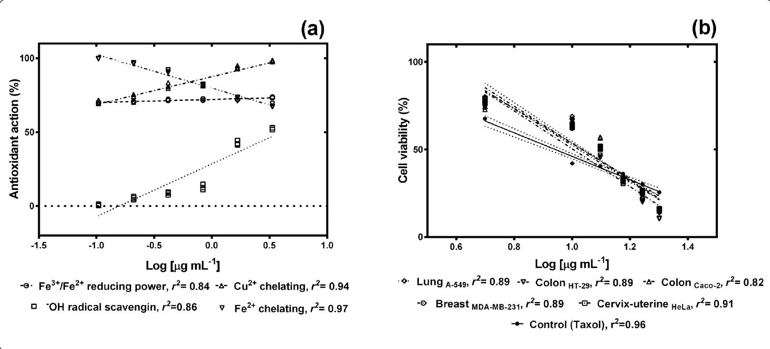
Figure 2: Relationship of Calendula officinalis extract - antioxidant ability (a) - cytotoxicity (b). Determination coefficients (r2), n = 3, p < 0.05.
Direct relationship (p < 0.05) between the content of phenols in the extract and its antioxidant capacity was established, as shown in Figure 2a. Interestingly an exception occurred in the Fe (II) chelation assay, which instead displayed an inverse correlation (ferric reducing power: r 2 = 0.84; •OH scavenging: r 2 = 0.86; Cu2+ chelation: r 2 = 0.94; Fe2+ chelation: r 2 = 0.97).
Several studies indicate that even though flavonoids actually have antioxidant activity, when high doses of them are used, they show a pro-oxidant activity owed to the diminished chelating activity from the flavonoid-iron complex (Martínez-Flores, González-Gallego, Culebras & Tuñón, 2002). Similarly, other studies established a relationship between the content of phenols and the antioxidant capacity of edible and medicinal plant extracts (Zheng & Wang, 2001). Therefore, the results of this study showed high the antioxidant efficacy of 70% hydro-alcoholic extract of Calendula officinalis with a performance (339.2g/kg) in comparison with extracts obtained using lower ethanolic contents (hydro-alcoholic extracts at 60% and 40% (374g/kg and 187.125g/kg respectively). So, the experimental evidence from this study, support that the 70% hydro-alcoholic extract of Calendula officinalis can be considered a new source, rich in antioxidants, mainly of the kind of phenolic compounds.
As shown in Figure 2b, the experimental data from this study helped to established an inverse relationship (p < 0.05) between the concentration of Calendula officinalis hydro-alcoholic extract and the viability of human cancer cells follows: breast (MDA-MB-231), r 2 = 0.896; uterine cervix (HeLa), r 2 = 0.912; lung (A-549), r 2 = 0.892; colon (HT-29), r 2 = 0.889; colon (Caco-2) r 2 = 0.817. Besides, the results from this paper also correlate with a previous study reporting that a Calendula officinalis extract with potent in vitro cytotoxic activity, may be further supported by their experimental results were in vivo models in which rats were treated with 40mg/kg for day in period for 34 days showering reduction diameters of tumors, interpreted as antitumor activity (Barajas Farías et al., 2006).
Extracts from other plant species related to Calendula officinalis have been recently characterized in several studies. For instance, researchers have assessed the cytotoxic activity of Calendula arvenis in cancer cells in vitro. Also, the quantitative content of phytochemical molecules in the species was evaluated; the methanolic and aqueous extracts revealed contents of flavonoids and phenolic acids with considerable antioxidant activities. Both the methanolic and aqueous extracts exhibited cytotoxicity (assessed using the indirect MTT test) in a myeloid cancer cell line (IC50 31 μg/mL) (Khedid, Ansar & Ibrahimi, 2016).
The presents study show clearly that the Calendula officinalis hydro-alcoholic extract from petals of the flower, are a promising antioxidant source. When the appropriate concentrations are used, the extract may show cytotoxic effect in cancer cells in vitro, and therefore the extracts and its molecules, will probably be useful as part of cancer treatments causing cell death, in vitro and perhaps also in vivo.
The experimental results mentioned above, show that the Calendula officinalis hydro-alcoholic extract has a cytotoxic activity, demonstrable in five human cancer cell lines from different lineages, possibly by its high content of polyphenols, through stimulation of a pathway of free-radical scavenging and metal chelation. However molecular biology studies, the pathway of cytotoxicity and the cell death mechanisms involved are necessary. The relationship between antioxidant capacity and the in vitro cytotoxicity, may be related to a probable antitumor in vivo activity justified perhaps by the kinds and levels of antioxidant-enzymes, as well as the participating cell signaling pathways.
Conclusions
Since the results from this study strongly suggest that the 70% hydro-alcoholic extract from petals represents a natural source in antioxidants with potential application, mainly in the pharmaceutical industry because, it was found to be rich in acid phenols and flavonoids, strongly linked to antioxidant efficiency.
From the point of view of the biologic activity, the hydro-alcoholic Calendula officinalis extract shows a powerful cytotoxic effect in vitro in human cancer cells in general, given that five lineages of cancer cell lines were assayed. Since the content of polyphenols and flavonoids is relatively high, it might be associated with the antioxidant efficiency, exhibiting the following biologic activities: high free-radical scavenging capacity (ABTS, DPPH), efficient metal-chelation, moderate ability to neutralize hydroxyl radicals and a strong reducing power.
The linear regression analysis in the present study showed that the concentration of the hydro-alcoholic extract of Calendula officinalis correlated with both the antioxidant efficiency and the cytotoxic effect. Therefore, we consider that the content of polyphenols could also be related to both antioxidant and cytotoxic capacities.

